Search Results for: Covid

COVID-19 and Neurology Update

By B. Jeanne Billioux, MD, and Avindra Nath, MD, and edited by Chandrashekhar Meshram, MD

In this month’s neurology and COVID-19 review, we’ve included several topics that have arisen in the literature and news, including new updates regarding long COVID/PASC and neurologic outcomes of infants born to mothers with COVID-19.

Post-acute sequelae of SARS-CoV-2 (PASC) or long COVID continues to be a puzzling entity. However, numerous new studies are underway to help better understand the nature of this syndrome and the mechanisms involved in its pathogenesis, including neurologic aspects.

This month, the initial findings from the University of San Diego NeuCOVID longitudinal cohort study was published, describing the neurologic manifestations in two cohorts: Patients referred to the neurology department after recovering from COVID-19, and patients with pre-existing neurologic disorders who subsequently were diagnosed with COVID-19.

In this cohort of 56 patients, fatigue was the most commonly reported symptom at baseline (89.3%), followed by headache (80.4%), insomnia (66.1%), and memory impairment (64.3%). Almost all patients had a baseline assessment at least 28 days after onset of neurological symptoms, with a median of 16 weeks from infection. In those who were able to follow up six months later, a small number (n=9) noted resolution of their symptoms, but others reported persistent fatigue (52.2%), memory complaints (68.8%), insomnia (51.3%), headache (45%), and difficulty concentrating (47.6%). However, many of these symptoms had lessened in severity.

MOCA scores generally improved over time in patients who were able to follow up (from average score of 26 to 28/30), although MOCA scores declined in about one quarter or these patients. Interestingly, a small subset of patients (7.1%) displayed a triad of symptoms, including tremor, ataxia, and cognitive dysfunction (Shanley 2022).

Another neurologic post-COVID study from Italy found that PASC patients could usually be grouped into two cohorts of PASC/long-COVID based on their neurologic symptoms. The first cohort typically presented with a constellation of memory issues, headache, psychological issues, anosmia, and ageusia; the second cohort presented with symptoms referrable to the peripheral nervous system (PNS). Upon analysis of risk factors for either cohort, the cohort with PNS-related symptoms was found to be more likely to have had a larger number of comorbidities at onset, a more severe course of COVID-19, as well as a higher number of non-neurologic COVID complications (Grisanti 2022). Longitudinal cohorts such as these will be informative for the natural history of PASC and its neurologic manifestations, as well as the potential long-term socioeconomic impact, and will help guide modes of intervention.

Several studies are ongoing to elucidate the pathogenesis of the neurologic sequelae of COVID-19. A paper published in Cell this month described diffuse microglial cell activation in the brain of patients who had died despite a mild respiratory infection with SARS-CoV-2. This was replicated in a humanized mice model of a mild form of SARS-CoV-2 infection. There was no evidence of SARS-CoV-2 neuroinvasiveness (as evidenced by lack of virus in the CNS), however the mice displayed elevated levels of cytokines in both the serum and CSF at 7 days post-infection. Moreover, these mice were found to have increased levels of microglial/macrophage reactivity in the subcortical white matter, as well as upregulation of inflammatory gene expression in these microglia. They also found that the hippocampi of these mice had microglial/macrophage activation with impairment of neurogenesis that persisted several weeks post infection. This was accompanied by decreased levels of oligodendrocytes and oligodendrocyte precursor cells in the subcortical white matter. These findings suggest that even mild infections with SARS-CoV-2 may lead to persistent neurologic inflammation, myelin dysregulation, and decreased hippocampal neurogenesis, which may lead to the neurologic symptoms currently seen in long COVID/PASC, such as memory impairment and “brain fog” (Fernández-Castañeda 2022).

Another interesting topic in the news this month is on the outcome of infants born to mothers infected with SARS-CoV-2. A cohort study of 7,772 infants, with 222 infants born to mothers who tested positive for SARS-CoV-2 during pregnancy, found that infants born to mothers with COVID-19 during pregnancy had a higher risk of being diagnosed with neurodevelopmental abnormalities over a 12-month time period (OR 2.17), with a higher risk (OR 2.34) associated with infections during the third trimester. This risk was present after adjusting for a variety of factors, including preterm labor (which can be associated with SARS-CoV-2 infection (Edlow 2022). These findings warrant further evaluation, but could reflect potential detrimental effects of SARS-CoV-2-related inflammation on fetal brain development, similar to other known maternal infections. •

Avindra Nath, MD, is chief of the Section of Infections of the Nervous System and Clinical Director, National Institute of Neurological Disorders and Stroke (NINDS), National Institutes of Health in Bethesda, Maryland.

B. Jeanne Billioux, MD, is staff clinician and head of the program in International Neuroinfectious Diseases within NINDS. Her research focus is on emerging infectious diseases and conducting research on the neurological consequences of infections in an International setting.

Chandrashekhar Meshran is co-opted trustee of the WFN.

References

Shanley JE, Valenciano AF, Timmons G, Miner AE, Kakarla V, Rempe T, Yang JH, Gooding A, Norman MA, Banks SJ, Ritter ML, Ellis RJ, Horton L, Graves JS. Longitudinal evaluation of neurologic-post acute sequelae SARS-CoV-2 infection symptoms. Ann Clin Transl Neurol. 2022 Jun 15. doi: 10.1002/acn3.51578. Epub ahead of print. PMID: 35702954. DOI: 10.1002/acn3.51578


Grisanti SG, Garbarino S, Barisione E, Aloè T, Grosso M, Schenone C, Pardini M, Biassoni E, Zaottini F, Picasso R, Morbelli S, Campi C, Pesce G, Massa F, Girtler N, Battaglini D, Cabona C, Bassetti M, Uccelli A, Schenone A, Piana M, Benedetti L. Neurological long-COVID in the outpatient clinic: Two subtypes, two courses. J Neurol Sci. 2022 Jun 3;439:120315. doi: 10.1016/j.jns.2022.120315. Epub ahead of print. PMID: 35717880; PMCID: PMC9212262. DOI: 10.1016/j.jns.2022.120315


Fernández-Castañeda A, Lu P, Geraghty AC, Song E, Lee MH, Wood J, O’Dea MR, Dutton S, Shamardani K, Nwangwu K, Mancusi R, Yalçın B, Taylor KR, Acosta-Alvarez L, Malacon K, Keough MB, Ni L, Woo PJ, Contreras-Esquivel D, Toland AMS, Gehlhausen JR, Klein J, Takahashi T, Silva J, Israelow B, Lucas C, Mao T, Peña-Hernández MA, Tabachnikova A, Homer RJ, Tabacof L, Tosto-Mancuso J, Breyman E, Kontorovich A, McCarthy D, Quezado M, Vogel H, Hefti MM, Perl DP, Liddelow S, Folkerth R, Putrino D, Nath A, Iwasaki A, Monje M. Mild respiratory COVID can cause multi-lineage neural cell and myelin dysregulation. Cell. 2022 Jun 13:S0092-8674(22)00713-9. doi: 10.1016/j.cell.2022.06.008. Epub ahead of print. PMID: 35768006; PMCID: PMC9189143. DOI: 10.1016/j.cell.2022.06.008


Edlow AG, Castro VM, Shook LL, Kaimal AJ, Perlis RH. Neurodevelopmental Outcomes at 1 Year in Infants of Mothers Who Tested Positive for SARS-CoV-2 During Pregnancy. JAMA Netw Open. 2022 Jun 1;5(6):e2215787. doi: 10.1001/jamanetworkopen.2022.15787. PMID: 35679048; PMCID: PMC9185175. DOI: 10.1001/jamanetworkopen.2022.15787

How Do You Diagnose COVID-19 Patients With Neurological Complications?

Call for Survey Respondents

By Dr. Arina Tamborska

Arina Tamborska

Researchers with the Global COVID Neurology Survey are calling on clinicians to help them better understand how neurological complications of COVID-19 are diagnosed around the world.

The project is being led by the University of Liverpool with support from the World Health Organization (WHO) and the World Federation of Neurology (WFN).

“We need clinicians around the world to share their experiences of how they diagnose neurological and neuropsychiatric syndromes and perceive their association with COVID-19,” said Dr. Arina Tamborska, NIHR academic clinical fellow in neurology at the University of Liverpool. “Your responses will play a role in the validation of a prospective WHO clinical record form, which will then be made openly available to everyone following the study’s completion.”

The survey will take up to 30 minutes to complete and involves questions about your clinical experience and several short case scenarios, describing patients with neurological complications as a result of COVID-19.

Participants will be named as a collaborator in any publications arising from the survey. All clinicians who treat patients with COVID-19 are eligible to participate, including trainees.

“Whether you are a neurologist or any other health care professional involved in the care of COVID-19 patients, your help would be greatly appreciated with this study, which we hope will lead to better treatments and outcomes for patients,” said Dr. Benedict Michael, senior clinician scientist fellow at the University of Liverpool.

Visit https://redcap.link/covidneurosurvey to take part. •

Dr. Arina Tamborska is a NIHR academic clinical fellow in neurology, brain infections group at the University of Liverpool, The Walton Centre NHS Foundation Trust, Liverpool, U.K.

An Epidemic of Mucormycosis Strikes India During the COVID-19 Pandemic

By Chandrashekhar Meshram

Chandrashekhar Meshram

Chandrashekhar Meshram

The second wave of the COVID-19 pandemic was devastating in India with a sudden spurt in cases during the second quarter of the year, and with the count of daily cases crossing 400,000. During this pandemic, there was another epidemic of the fungal infection mucormycosis.

This pandemic and epidemic completely exposed the lacunae in the health care system in the country. There was a shortage of medicines, a shortage of hospital beds, a shortage of oxygen supply, and a shortage of health care personnel, resulting in high morbidity and mortality. According to the statement on June 28 by the Union Health Minister, 40,845 cases of mucormycosis were reported in the country. Out of these, 85% patients had COVID-19, 62% had diabetes as a comorbidity, and 63% of patients had received steroids. The alarming fact is that 32% of the patients were in the age group of 18 to 45 years.

Mucormycosis is caused by saprophytic fungi of the order Mucorales, which are found in decaying organic matter and soil samples. Rhizopus Arrhizus is the most common etiological agent in India. Mucormycosis is an angioinvasive disease causing tissue infarction and necrosis. Rhino-orbito-cerebral mucor is the most common presentation, followed by pulmonary, cutaneous, gastrointestinal, renal, and disseminated disease.

Rhino-orbito-cerebral mucormycosis (ROCM) is often encountered by neurologists. Clinical manifestations of ROCM syndrome include new onset unilateral headache, facial pain, facial numbness, orbital pain, diplopia, ophthalmoplegia, impairment of vision, orbital swelling, ptosis, proptosis, papilledema, nasal discharge, and epistaxis. Black discoloration of skin and mucosa are characteristic of mucor infection. Sometimes there is cavernous sinus thrombosis and internal carotid artery block. The majority of patients with ROCM in association with COVID-19 develop symptoms between day 10 and day 15 post COVID-19. However, delayed presentation can occur up to three months.


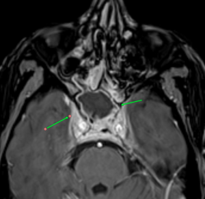
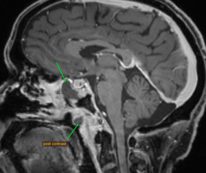
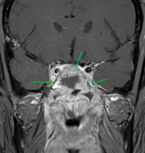
MRI images of mucormycosis with cavernous sinus involvement in a patient with COVID-19.

Imaging findings on CT or MRI that suggest the diagnosis are non-enhancing hypointense mucosa over nasal turbinate and nasal septum (black turbinate sign), concomitant involvement of paranasal sinuses and orbit, bone erosion or destruction, cavernous sinus involvement (figure), and ischemic stroke.

The diagnosis is confirmed by demonstrating fungus with special stains in tissues obtained from nasal scraping or surgery.

Mucormycosis is a medical emergency, and treatment should start with antifungal drugs, surgical intervention, and correction of metabolic abnormalities or risk factors. Amphotericin B is the first-line drug of choice. Liposomal form of Amphotericin B (LAmpB) is preferred in view of a better side effect profile. The efficacy of conventional deoxycholate amphotericin B (dAmpB) is not inferior to the liposomal form but has more side effects. However, the cost of LAmpB is 10 times more. Nephrotoxicity is the most common side effect of dAmpB.

Other drugs used in the treatment are posaconazole and isavuconazole. Oral medication can be used for longer duration. The duration of treatment is not known and has to be decided on a case-to-case basis. Ampho B is recommended for initial 4-6 weeks, which can be followed by posaconazole or isavuconazole for a few more weeks.

Surgical treatment includes debridement or excision of necrotic tissue. Depending on the site of involvement, an ENT surgeon, ophthalmologist, dental surgeon, and neurosurgeon should be part of the team. Surgical intervention at early stage improves the outcome. Functional endoscopic sinus surgery, paranasal sinus debridement, and orbital exenteration are commonly required surgical procedures in ROCM.

The major drawbacks in managing mucormycosis in India are a gap in treatment protocol and non-affordability of LAmpB due to financial constraints.

The mortality rate of mucormycosis in India is in the range of 28–52%, and it increases to 80% in intracranial extension of disease. High mortality is due to delays in seeking medical attention and diagnosing the disease and challenges in managing the advanced stage of infection. There is an urgent need to increase awareness among the general public and doctors for early diagnosis and treatment.

The intriguing question is why there has been such a recent spurt of cases of mucor in India? Though mucormycosis is globally distributed, its estimated prevalence even before the COVID-19 pandemic was 70 times higher in India as compared to global data. The possible reason for the high prevalence is the abundant presence of Mucorales in the community and the hospital environment and the high prevalence of diabetes in the Indian population. The sudden spurt during the second wave might be due to a lowering of immunity by the new strain of COVID-19 making these patients susceptible for fungal infection. •

Chandrashekhar Meshram is chair of the The Tropical and Geographical Neurology Specialty Group of the WFN.

COVID 19 and the AAN-WFN Continuum Program in Mumbai

By Satish Khadilkar and Sarosh Katrak

The COVID pandemic had universal effects on medicine, and Mumbai was no exception. In fact, being a densely populated city with lots of international travelers, the cases appeared early and the magnitude was high, throwing the health system off gear.

While most of the younger neurologists continued to work, they had to be involved with COVID duties as well. The focus shifted to service medicine and service neurology in a major way, and academic activities had to take a back seat.

The AAN-WFN Continuum Program has been operational in Mumbai since 2005, initiated by S. M. Katrak as per the advice given by Ted Munsat. The first event was held at Mumbai on

July 17, 2005, on multiple sclerosis and was attended by postgraduate students from five institutions. Following the success of the program at Mumbai, teachers in other cities of India also conducted Continuums in their cities, and the programs were well regarded by students and practitioners.

Satish Khadilkar had attended the programs since the beginning and participated more actively in the later years, taking up the responsibility as co-convenor of the program with Katrak’s guidance and advice, and as convenor since 2021.

Virtual platforms gradually developed, and the government opened up online consultations in 2020. We decided to shift to an online platform for this CME program. Having worked with physical copies of Continuum and in-person meetings, it was a challenge to acclimatize to this form of learning.

Interestingly, it did not prove too difficult, particularly for younger colleagues. We conducted the first online Continuum program Dec. 27, 2020, on the topic of neurocritical care. It was attended by 48 participants.

In 2021, we have conducted programs on dementia, epilepsy, and demyelinating disorders, which were attended by approximately 50-85 students from various institutions in Mumbai, other places in the state of Maharashtra, and a few from the state of Gujrat. We incorporated the Google forms for virtual feedback. All participants do not complete the forms, and we are exploring ways to facilitate the process.

We have recently adopted a system wherein a mentor helps the presenter to prepare his slides. This approach has much increased the clarity and utility of the presentations, time management, homogeneity of the group, and the individuals find themselves more involved. We intend to persist with this pattern and are in discussion with other centers in India to adopt this method in their centers for these Continuum programs. •

New Paths of Sharing Science for Neurology Education in the Era of the COVID-19 Pandemic

By Abderrahmane CHAHIDI, Zouhayr SOUIRTI, Mohamed CHRAA, Najib KISSANI, Reda OUAZZANI
Moroccan Society of Neurophysiology & Moroccan League Against Epilepsy, Morocco.

Abderrahmane CHAHIDI

Health care innovation, notably in clinical neurology and neurophysiology, has been rapidly growing over the last few decades, both in terms of therapy and diagnosis. Hence, it is necessary to spread and share information along the various possible paths. In this time of confinement, and to keep abreast of current news, mainly in the developing regions of the world, the Moroccan Society of Neurophysiology IFCN Chapter and the Moroccan League Against Epilepsy ILAE Chapter organized a series of Expert Webinars over three months (May, June, and July 2020) and focused on topics of concern to the clinicians in their daily practice, particularly in the field of clinical neurology and neurophysiology (EEG, EMG, EP, IONM, PSG, TMS, Epilepsy, Neuromuscular diseases, Sleep, Neurostimulation, etc.). The webinar language can be English or French, and many renowned experts in the different fields of clinical neurology and neurophysiology kindly accepted our invitation.

Zoom session snapshot from Prof. Kaji’s talk.

This innovative initiative aims to animate the field of clinical neurology/neurophysiology in our context and boost synergies between the players in various medical and surgical disciplines involved in neurology. Highly supported by the scientific community, the Clinical Neurophysiology Expert Webinars discussed the daily challenges faced by practitioners in teaching clinical cases.

Example of a full Q&A session after Prof. Kaji’s talk.

These webinars had the following objectives: ensuring the continuity of the teaching of young people and the continuing education of specialists and practitioners, substituting for the scientific days, meetings, congresses, and training courses usually organized at this time of the year, perpetuating solidarity and collaboration between national and international professionals, and reinforcing the relationships between the SMNPH, IFCN, and WFN. Finally, another objective is to unify the efforts of scientific societies to address the multiple global health challenges caused by the various pandemic threats to humanity, such as the COVID-19 pandemic.

This initiative was inaugurated with an EEG and epilepsy course led by Prof. Ron Lesser focusing on EEG interpretation, including normal EEG interpretation (techniques, artifacts, and normal aspects) and pathological EEG interpretation (epileptic abnormalities and non-epileptic abnormalities), and Prof. Gregory Krauss, who presented a lecture on nonepileptic events often misidentified as seizures (psychogenic seizures and syncope).

The second session of lectures, dedicated to EMG and neuromuscular diseases, took place the next day. Prof. Henry Kaminski from George Washington University talked about the diagnosis and treatment of ocular myasthenia gravis. In addition, we were gratified by the participation of Prof. Ryuji Kaji (First Vice President, WFN, Japan), as our special invited speaker, who provided an update on the electrodiagnosis of ALS and related disorders.

The Moroccan Neurophysiology Society (MNPS) has been excited to receive the feedback from the participants who have enormously appreciated these new ways of scientific meetings and who keep asking for more webinars.

The program, which concluded July 30, included more than 40 presentations that were presented by a variety of expert speakers and moderators. •


In the Midst of the COVID-19 Pandemic

Since my last column, the COVID-19 pandemic has spread through most developed countries courtesy of international travel, tourism, and high-density populations. It is now set to decimate less developed countries in South America, Asia, and Africa, consequent upon insufficient resources and less sophisticated public health services. Those least well-resourced countries face, like in epidemics of old, the twin blights of the epidemic and economic collapse and risk ensuing anarchy. Even some better-off countries have pursued different strategies in balancing the health requirements and when to re-open their economies with differing results now and no doubt differing results in the longer term.

William Carroll, MD

What is different with this pandemic and the plagues and epidemics of the past, and even the more recent SARS, Ebola, and Zika emergencies, is the concatenation of the modern world by the internet. While the world looks for a more unified inter-governmental response, peoples of the world, and in particular scientists, clinicians, and concerned citizens, have joined a multitude of international, regional, and global groups to contribute to the care, surveillance, and data collection of those affected. Through the internet and modern communications, infection rates, mortality rates, and survival rates issue daily from most countries irrespective of denominators. Most of us are involved in one or more such activities while we watch with concern — and at times with incredulity — as nations make individual decisions to minimize the human and economic costs of this pandemic. We all hope to see a successful formula develop.

The WFN also hopes that all of its individual member neurologists have remained safe and in good health. Happily, the London office staff have done so and the same applies to the trustees and officers.

In the meantime, let me outline the WFN activities during the current situation.

Communication

The WFN has implemented a program to improve the communication between the London office and its members. In a time when communication is so important, it has been heartening to see the response of member societies to this initiative. Once complete, and this first part is almost there, the office will commence a rolling dialogue, contacting approximately 10 members each month to ensure the details are current. This will see the details for each of the 122 societies being updated annually. In addition to direct communication by email and phone, the WFN website is now regularly updated with changes being advised by social media. More active use of social media also follows each monthly trustees teleconference on the second Thursday of the month. It is hoped that members will visit the WFN website more frequently and advise the London office of necessary changes to their contact details.

The WFN is participating with the Brain Health Unit of the WHO in its COVID-19 neuro endeavor and with other groups interested in collecting data about potential neurological involvement in the course of COVID-19 infection. The WFN’s Environmental Neurology Specialty Group has advised that it will be keeping a registry of member societies of members who are doing this at a national level. Links to members of the Global Neurology Alliance, who are posting information related to their specialty and COVID-19, are available on the WFN COVID-19 website and undergo regular updating. WFN member societies also have available a bulletin board on the WFN website to post news on the pandemic situation in their countries.

Education

Like most global or large regional organizations, the WFN had been moving to a more electronic base for education. The COVID-19 pandemic has provided an additional spur to this direction. With the cancellation of the African Sub-Saharan Regional Teaching Course for 2020, which had been scheduled for Kampala in September, AFAN and the WFN have proposed an e-learning day. Plans are advancing for this to occur as the WFN/AFAN e-Learning Day focused on “Stroke: A Treatable and Preventable Disease.” It is likely that the EAN and AAN will contribute to this very necessary alternative. More information will be found on the WFN website. Plans for the XXV WCN in October 2021, at the new state-of-the-art La Nuvola Conference Centre in Rome, are advancing with the valuable assistance of the Society of Italian Neurology. The scientific and teaching course programs are now being finalized with additional flexibility to permit virtual programming if required.

Promotion

There are a number of other activities engaging the WFN at present which, while being broadly educational, also promote the visibility and good name of the WFN. World Brain Day is the most imminent of these. As most will know, World Brain Day (WBD) has continued to evolve. It typically begins on July 22 each year, the anniversary of the founding of the WFN, which partners with a member of the Global Neurology Alliance closely representing the WBD topic. WBD runs for two to three months to provide greater dissemination and authority of its message. This year the WFN is excited to partner with the International Parkinson Disease and Movement Disorder Society and believes that the 2020 WBD will achieve similar goals as those that the International Headache Society brought to the 2019 campaign.

A second promotional activity being developed is the Brain Health Initiative. This will focus on the importance of brain health in general and at present will comprise five modules of related topics. The Brain Health Initiative is a matter that dates from the GNA meeting held during the Lisbon EAN meeting in 2018. Several members of the Global Neurology Alliance expressed a desire for the WFN to consider such a campaign. The initiative is planned to run in parallel with the more disease-specific WBD campaign.

Other WFN Activities

There are other activities engaging the WFN at present which I will enumerate. First, the Annual General Meeting (AGM) of the Council of Delegates will be a virtual meeting on a Zoom platform commencing at 12 p.m. on Sept. 9, British Summer Time. Details of this important meeting and its agenda will be disseminated directly to member societies by email and to the membership at large on the website. Second, the election of a new trustee to fill the vacancy created by Prof. Riadh Gouider after completing his term will be undertaken in the week before the AGM, in order not to disadvantage member societies in time zones where the AGM might be occurring at unsociable hours.

While on the topic of WFN trustees, it is with much pleasure that I welcome Prof. Marianne de Visser as a co-opted trustee. Her experience, wisdom, and integrity will complement the work of the WFN. (See box above.)

Third, notification has been sent to member societies in the Asian and Oceanian Association of Neurology requesting expressions of interest in hosting the 2025 (XXVII) WCN. To date four member societies have done so.

Let me also, on behalf of the trustees and all member societies, express my gratitude to the work of the London office during the pandemic. They have been locked down, out of the office, but their output and the quality of work has remained exemplary.

Finally, I wish all members of the WFN, their families, and patients a safe passage through this pandemic.


Long-Haul COVID

By Avindra Nath and B. Jeanne Billioux

By Avindra Nath and B. Jeanne Billioux

It is becoming increasingly apparent that many patients who recovered from the acute phase of the SARS-CoV-2 infection have persistent symptoms. This includes clouding of mentation, sleep disturbances, exercise intolerance, and autonomic symptoms. (See Tables 1 and 2 below) Some also complain of temperature dysregulation and lymphadenopathy. Although there are no peer-reviewed papers at the moment on these patients, many news articles have been written about this phenomenon1-4 and apparently there are Facebook pages where there are several thousand patients with these symptoms. They describe themselves as the “Long-Haul-COVID” or “Long-Tail COVID.” Many of these patients are health care workers who had massive exposure to the virus early in the pandemic and describe having symptoms for 100+ days. Since many of the symptoms are neurological in nature, these patients are seeking us out for help. It is important that we characterize these patients and try to document the objective findings and then determine how best to study their pathophysiology to develop proper guidelines for treatment.

Most of these patients were in excellent health prior to getting infected with SARS-CoV-2. They all had myriad symptoms during the acute phase; however, as the fever and respiratory symptoms improved, they are left with persistent systemic symptoms, some of which are gradually improving, but not all are following that course. While some were admitted to the hospital due to pulmonary symptoms, the majority were isolated at home. Many of these symptoms overlap with those of patients with myalgic encephalomyelitis/chronic fatigue syndrome (ME/CFS). The cause of ME/CFS remains unknown despite decades of research of the syndrome. Many of these patients similarly report a viral infection as a trigger, but since they come to our attention months and years after symptom onset, it is impossible to know what may have triggered the symptoms.

Symptoms of Long-Haul COVID

  • Insomnia or frequent awakenings
  • Inability to concentrate
  • and think clearly
  • Easy fatiguability despite normal lung function
  • Anorexia or increased appetite
  • Temperature dysregulation
  • Lymphadenopathy
  • Dysautonomia

Table 1. Symptoms.

Long-Haul COVID thus represents an excellent opportunity to study the pathophysiology of ME/CFS that may have broader implications. It is unclear at present if these patients may have endocrine abnormalities, but certainly with SARS-CoV-1, hypothalamic/pituitary dysfunction and adrenal insufficiency has been reported5. It would, of course, be equally important to determine if there might be any premorbid conditions or medications that might contribute to these symptoms.

Autonomic symptoms in Long-Haul COVID

  • Tachycardia upon mild exercise or standing
  • Night sweats
  • Gastroparesis
  • Constipation
  • Peripheral vasoconstriction

Table 2. Autonomic symptoms.

Even at this early stage, it might be important to determine what the potential pathophysiological mechanisms might be. So far there is no convincing evidence for widespread infection of the brain with the virus. The virus has been detected in CSF and the brain in very rare cases; however, there is evidence for widespread glial cell activation that may be related to metabolic dysfunction or to the massive immune activation in the periphery. Other possibilities are specific immune responses targeted against specific regions of the brain and autonomic nervous system. Depending on the predominant underlying pathophysiological mechanism at play, targeted treatment might be possible.

Unfortunately, it looks like the pandemic has gotten out of control. It is spreading rapidly across the globe, and even if we have an effective vaccine, we might never have enough dosages to vaccinate the eight billion inhabitants on this planet. And we also have a subset of the population that is opposed to any kind of vaccination. So COVID-19 is here for the long haul, and neurologists are going to play a critical role in the management of these “Long-Haulers.” Several efforts are underway to prospectively follow these patients with persistent symptoms, including studies at the intramural program at the United States National Institutes of Health in Bethesda, Maryland. Hopefully we can get to the bottom of these manifestations soon and find solace for these patients in a timely manner. •

Avindra Nath, MD, is chief of the Section of Infections of the Nervous System and Clinical Director, National Institute of Neurological Disorders and Stroke (NINDS), National Institutes of Health in Bethesda, Maryland.

Jeanne Billioux, MD, is staff clinician and head of the program in International Neuroinfectious Diseases within NINDS. Her research focus is on emerging infectious diseases and conducting research on the neurological consequences of infections in an International setting.

References:

  1. https://www.wsj.com/articles/three-months-in-these-patients-are-still-ravaged-by-covids-fallout-11593612004
  2. https://www.washingtonpost.com/health/could-covid-19-cause-long-term-chronic-fatigue-and-illness-in-some-patients/2020/05/29/bcd5edb2-a02c-11ea-b5c9-570a91917d8d_story.html
  3. https://www.bloomberg.com/news/videos/2020-06-10/the-lingering-symptoms-of-covid-19-video
  4. https://www.washingtonpost.com/health/2020/06/11/coronavirus-chronic/?arc404=true
  5. Leow MK-S, Kwek DS-K, Ng AW-K, Ong K-C, Kaw GJ-L, Lee LS-U. Hypocortisolism in survivors of severe acute respiratory syndrome (SARS). Clin Endocrinol. 2005;63(2):197-202.

COVID-19: A Neurologist’s Perspective

By Avindra Nath and B. Jeanne Billioux

The crisis we are currently facing is unprecedented in every way. Just a few months ago, we were talking about developing targeted gene therapies for a spectrum of diseases, including ultrarare diseases. Only a few weeks later, the health care system finds itself overburdened and undersupplied to the point where we are talking about rationing health care1. Maintenance care has been pushed to telemedicine clinics and elective procedures have ground to a halt. Many patients sick with respiratory symptoms are being sent home to isolate themselves, and some are dying at home. There is an acute shortage of ventilators to the point that in some hospitals one ventilator is being shared by multiple patients2.

Figure 1. Distribution of comorbidities in patients requiring inpatient care due to COVID-19.

Several basic medicines are in limited supply. Although many hospitals and institutions recognized the need to stockpile personal protective equipment, several hospitals have run out of masks and gowns due to a limited supply chain. This crisis has tested community-based ingenuity, and in some hospitals, personal protective equipment is being fashioned by staff and community volunteers out of plastic visors and trash bags. Many doctors on the front line have succumbed to the infection, and many others are quarantined, a sobering reminder of these dire circumstances. The words here just a few months prior would read as a work of fiction, but this is the unfortunate reality of the crisis we face – COVID-19. Nearly every country and every major city in the world has been affected by the infection. On April 12 alone, there were over 10,000 new infections and nearly 1,000 deaths in a single day in New York. What started in Wuhan in November 2019 has become a global pandemic necessitating drastic changes in our way of life.

About COVID-19

COVID-19 is caused by the virus SARS-CoV2, a single-stranded RNA virus. Merely 60 nm in size, the virus that can only be visualized by an electron microscope has caused massive devastation. Although many pandemics have occurred in the past several decades, SARS-CoV2 has an array of features that have made it incredibly effective in spreading through the population. Perhaps the most important among these features is that asymptomatic and pre-symptomatic hosts can spread it. These asymptomatic carriers can infect large populations without knowing that they are infected with the virus. In fact, every time we speak, we release droplets into the air that can carry the virus a few feet.3 This property is the reason that distancing of at least six feet from one another and use of masks even made of cloth by the general public can be effective at lowering the degree of spread. Evidence suggests the virus can be easily inactivated by a wide variety of cleansing and disinfecting agents, including proper use of soap and water4.

Since the virus is spread through the respiratory passages, it manifests predominantly with respiratory symptoms. Most patients develop fever, dry cough, and fatigue/malaise, with many also reporting headache, myalgias, rhinorrhea, and anosmia with ageusia. Gastrointestinal symptoms occur in some patients. The symptoms may last for one to two weeks with nearly full recovery. Some symptoms such as fatigue may take longer to recover. However, nearly 20% to 30% patients may develop much more severe pulmonary symptoms. Toward the end of the first or second week, when other symptoms are improving, these patients develop dyspnea due to massive inflammation in the lungs caused by a viral pneumonia resulting in an acute respiratory distress syndrome. These patients require ventilatory support, and mortality rates are high. However, those patients who manage to survive the ordeal can recover with few residual symptoms, although the long-term consequences of the pulmonary damage are currently not known5. Patients who require hospitalization shed virus for an average of 20 days (range 8 to 37 days) from the time of symptom onset. The possibility that the virus may get reactivated has been raised. The Korean CDC is following 51 such patients who were thought to be cured but became positive again after leaving quarantine. If the virus is capable of reactivation, and whether reactivated virus is capable of infection, remains an open question. However, the findings in the Korean patients are likely related to false negative PCR testing.

Complications and Risk Factors

Multiple systemic complications may occur in patients who have severe respiratory symptoms. This may include myocarditis, which can be fatal in nearly 50% of those who develop it. A coagulopathy may occur in others resulting in both venous and/or arterial occlusions. Renal failure is a late complication of the disease.

Several risk factors have been identified for the severe manifestations of the illness. (See Figure 1). Interestingly, children only develop a mild illness and generally recover fully. Older adults have the highest risk. The complications seem to be more common in males. Hypertension and diabetes are also major risk factors which account for nearly 50% of the comorbidities in hospitalized patients6. The reasons for this are not entirely clear. One hypothesis suggests that since angiotensin converting enzyme 2 (ACE2) is the receptor for SARS-CoV-2, the use of ACE inhibitors to treat hypertension or diabetes can induce the expression of the receptor making the cells more vulnerable to infection with the virus. Clinical studies are underway to test this hypothesis. Current recommendations are to keep patients who are already on ACE inhibitors and ACE receptor blockers on their medications, as the risk of adverse events of discontinuing these medications may outweigh the minimization of risk for COVID-19.

As neurologists, we worry about our patients who have a chronic neurological illness. Can the illness itself or the medications that they are on put our patients at greater risk of severe illness? These questions are particularly important in the context of nursing homes, where neurologic comorbidities are common, and the virus has displayed rapid spread. Most certainly, patients with diseases such as Parkinson’s disease, stroke, myasthenia gravis, or other diseases that can impair mobility may also impair lung function. Patients with immune-mediated disorders such as multiple sclerosis, neuromyelitis optica, and myasthenia gravis who are on immunosuppressant drugs may be at risk for more severe complications of the illness. Various organizations such the National Multiple Sclerosis Society are collecting data on patients who develop COVID-19. These data repositories are going to be helpful in determining what medications pose greater risk of complications from the infection. In the meantime, recommendations and guidelines are emerging from various societies based on our current knowledge for the management of patients with stroke7, multiple sclerosis (nationalmssociety.org), epilepsy (ilae.org) and myasthenia gravis8.

Neurological complications are rare but are being increasingly recognized9. These complications can involve the entire neuro-axis. They may occur during active viral infection and as a post-viral syndrome. (See Table 1). Some patients may present with altered mental status in the absence of respiratory or other typical COVID-19 symptoms as their sole initial presenting feature of SARS-CoV2 infection10. Anosmia is a common symptom of any upper respiratory tract infection. But anosmia with COVID-19 has received special attention. It seems to be one of the most common symptoms and often occurs in the absence of rhinorrhea. This suggests involvement of the olfactory nerve or pathway by the virus. As the majority of patients with anosmia recover their sense of smell and taste after the acute phase of the illness, the nerve endings or the cells surrounding the nerves may be affected, allowing for regeneration to occur. In a case report of a patient with sudden anosmia due to COVID-19, it was found that the olfactory clefts were inflamed, with relative sparing of the olfactory bulb11. In a mouse model of coronavirus infection, the virus can be transmitted via olfactory pathways trans-synaptically to the brain and to the brainstem12. This has raised concern about the potential long term consequences of anosmia in COVID-19. However, the mouse coronavirus uses a different receptor and hence may not replicate the human disease. Nevertheless, it is important to prospectively monitor the patients to make sure they do not develop any long-term sequelae since we know that anosmia is a recognized early symptom of neurodegenerative diseases such as Parkinson’s disease and Alzheimer’s disease.

Strokes with COVID-19

Presentation

Venous sinus thrombosis
Ischemic strokes in multiple arterial distributions
Small blood vessel occlusions
Watershed Infarcts

Pathophysiology

Coagulopathy: elevated D-dimer, PT, aPTT
Antiphospholipid antibodies
Cardioembolic
Hypoperfusion
Risk factors
Myocarditis
Known vascular risk factors
ARDS and multiorgan impairment

Table 2.

Myalgia frequently accompanies the illness. Most viral illnesses can cause body aches and pains. However, in some patients with COVID-19, the muscle aches can be quite severe. Muscle tenderness may last for several days after all other symptoms have resolved. They can involve the back muscles. A case of rhabdomyolysis13 was reported similar to what was also seen with the SARS14, although this patient was also on lopinavir/ritonavir which may have contributed to the myolysis. Since the onset of these symptoms is early in the course of the illness, it is possible that the virus invades the muscle to cause myositis, however, pathological findings have not yet been described. Importantly, these patients need proper hydration to prevent kidney damage. Also, it should be noted that potential medications used in the treatment of COVID-19 (including some protease inhibitors) may cause patients to be predisposed to muscle damage.

Meningitis and encephalitis are rare. Dull headaches are common and typically occur at the onset of the illness and resolve within a few days. They are not accompanied by any signs of meningeal irritation. However, a classical presentation of a viral meningitis has been described with COVID-19 and virus can be detected in the CSF. Encephalitis is harder to diagnose. Most patients who become comatose do so after development of ARDS and multi-organ failure, hence the CNS symptoms are attributed to hypoxia and metabolic abnormalities. Fever itself can cause delirium. However, a few cases of encephalitis where patients developed generalized seizure and coma are now being described. In one such patient from Japan, the patient had mild pleocytosis and detectable virus in the CSF. An MRI showed lesions in the temporal lobe and adjacent ventriculitis15. Few neuropathological findings have been published, but one study found low levels of SARS-CoV-2 RNA in the brain by PCR of 4 different COVID-19 patients at autopsy15a. Another case study found evidence of betacoronaviral infection of the brain with postmortem electron microscopic evaluation15b. From the earlier SARS epidemic in 2003, autopsy findings showed that the virus could be detected in the brain by multiple techniques in all patients evaluated (n=8)16. Spread of SARS-CoV-2 into the brain could involve an array of mechanisms. The virus can spread via the vasculature and enter the brain carried by infected leukocytes. Transneuronal spread has been hypothesized to also occur from the lung via the vagal nerve or from the nasal passages via the olfactory nerve.

Strokes are being increasingly recognized in this population and occur as the presenting symptom of the infection or any time during the illness. (See Table 2.) In a study from China, 5% (n=11) of 211 patients admitted with COVID-19 had acute ischemic strokes, 0.5% (n=1) had cerebral venous thrombosis, and 0.5% (n=1) had cerebral hemorrhage17. While most often these patients have underlying vascular risk factors, there are several patients where nothing other than the SARS-CoV-2 infection can be identified as a cause of the stroke. The virus is known to invade endothelial cells and can also cause a coagulopathy. Elevated D-dimer levels and increased PT and activated PTT have been described. Antiphospholipid antibodies have also been detected18. Some may develop disseminated intravascular coagulation. The virus can also cause a cardiac myositis19 which could also cause a stroke by hypoperfusion or embolism. Some patients may simultaneously develop deep vein thrombosis or vascular occlusions in other organs.

Atypical Acute Respiratory Distress Syndrome is the major cause of death in patients with COVID-19. What is atypical is that these patients have severe hypoxemia even when the lung capacity and mechanics are well preserved20. Even when the pCO2 is rising the patients are not hyperventilating and lose their respiratory drive. They develop what seems like an Ondine’s Curse. However, these patients do not have any other brainstem signs so the pathophysiology of this condition remains unclear at the present time. However, it is critical that these patients be treated with oxygen, and prone positioning also seems to help. Early ventilatory support should also be considered.

Post-viral syndromes occur when the patient is seemingly improving from the viral syndrome at about a week to three weeks after the onset of the viral prodrome. An isolated case of acute necrotizing hemorrhagic encephalopathy has been described21. This patient had bilateral thalamic lesions and other lesions in the temporal lobes which are typical of the syndrome. It is thought to be mediated by cytokine storm. A patient with transverse myelitis with quadriparesis, a sensory level and bowel and bladder involvement has been described22. However, MRI or CSF evaluation was not reported. A single case of Guillain Barre Syndrome (GBS) has been published in a patient from China23. A case series from Italy of five COVID-19 patients who developed GBS described three patients with an axonal form of GBS and two with a demyelinating process24. We recently communicated with a patient who had a sensory variant of GBS. The illness was self-limiting and did not require intervention. Acute disseminated encephalomyelitis has been recently described in an adult patient with SARS-CoV-224a; similarly, several cases have been described with the human coronavirus-OC4325 and with MERS26.

However, multiple challenges in the evaluation of patients with neurological complications exist. It is difficult to get neuroimaging when patients are acutely infected for fear of contamination of the scanners. Performing surgery or autopsies are also challenging due to the production of aerosols and lack of proper safety measures.

Therapeutic Debate

Anti-virals: Even though currently there is no proven antiviral therapy for the human coronaviruses, several drugs are being considered for clinical trials and empirical treatment of patients. (See Table 3.) There are 287 studies on coronavirus registered on www.clinicaltrials.gov. In vitro studies have shown some efficacy with chloroquine and hydroxychloroquine. These drugs cause acidification of the endosome-lysosomes and prevent viral replication. They have an anti-inflammatory effect. However, it requires pretreatment of cells prior to infection and has only a minimal effect post infection. While clinical trial results remain unpublished, these drugs have been utilized in clinic off-label in COVID-19 patients at several institutions. Well-controlled studies are necessary to know whether these drugs are efficacious against the virus. There is now a scarcity of the drug, and some countries have banned its export. Several HIV protease inhibitors have been shown to bind to the SARS-CoV-2 protease but clinical experience in small numbers of humans infected with the virus have failed to show clinical efficacy with lopinavir/ritonavir combination. Many clinical trials are currently underway that include nucleoside analogs such as remdesivir, and convalescent serum or intravenous immunoglobulin. Although the ability of most of these agents to enter the CNS is unknown, animal studies of remdesivir (GS-5734) have shown evidence of CNS penetrance, albeit at lower levels than other tissues27. Interestingly, a few drugs used to treat patients with multiple sclerosis such as teriflunomide and beta-interferons are considered to have anti-viral effects. But their effect on SARS-CoV-2 is still unknown.

Anti-inflammatory drugs: The most common cause of death is the massive immune response in the lungs leading to consolidation of the lungs with inflammatory infiltrates. Several immune suppressive medications are being used empirically. These include corticosteroids and IL-6 blockers. A case for the use of methotrexate has been made due to its broad anti-inflammatory properties and good CNS penetration.

What the Future Holds

As we continue to face the ongoing crisis, early results show reasons for optimism. In several states in the U.S., exponential growth trends have tapered. Distancing and preventive measures seem to be effective in flattening of the curve and helping institutions lower concomitant caseloads. The number of new infections and deaths are not rising as rapidly. Optimistically, we will soon face a new challenge of when and how to reopen our clinics and operating rooms. But what will this clinical environment look like? Social distancing is likely to play a role, and providers may see patients and enter any public spaces with masks on and maintain a distance of six feet from each other. Telemedicine will likely continue to play a much larger role in routine health care. A safe and effective vaccine could solve many of these issues, though development and testing of such a vaccine prior to administration to the general populace will take significant time. Another possibility is host adaptation. Most viruses are cyclical in nature. Mutations may occur that make the virus less virulent. Early signals suggest this might be the case with SARS-CoV-2. A 382 nucleotide deletion in open reading frame 8 has been identified in some circulating strains. A similar deletion also emerged in the SARS virus in 2003 that was associated with poor replication fitness28. However, until then, we will continue to see patients with COVID-19, and as neurologists we need to be vigilant for potential complications that require our attention and intervention. It is our duty to protect and advocate for the most vulnerable. •

Avindra Nath, MD, is chief of the Section of Infections of the Nervous System and Clinical Director, National Institute of Neurological Disorders and Stroke (NINDS), National Institutes of Health in Bethesda, Maryland.

B. Jeanne Billioux, MD, is staff clinician and head of the program in International Neuroinfectious Diseases within NINDS. Her research focus is on emerging infectious diseases and conducting research on the neurological consequences of infections in an International setting.

Correspondence
Avindra Nath, MD; Room 7C-103; Bldg. 10;
10 Center Drive, Bethesda, MD, 20982; U.S.
301-496-1561; e-mail: natha@ninds.nih.gov

References

1. Emanuel EJ, Persad G, Upshur R, et al. Fair Allocation of Scarce Medical Resources in the Time of Covid-19. N Engl J Med 2020.
2. Truog RD, Mitchell C, Daley GQ. The Toughest Triage – Allocating Ventilators in a Pandemic. N Engl J Med 2020.
3. Lewis D. Is the coronavirus airborne? Experts can’t agree. Nature 2020;580:175.
4. Kampf G, Todt D, Pfaender S, Steinmann E. Persistence of coronaviruses on inanimate surfaces and their inactivation with biocidal agents. J Hosp Infect 2020;104:246-251.
5. Guan WJ, Ni ZY, Hu Y, et al. Clinical Characteristics of Coronavirus Disease 2019 in China. N Engl J Med 2020.
6. Zhou F, Yu T, Du R, et al. Clinical course and risk factors for mortality of adult inpatients with COVID-19 in Wuhan, China: a retrospective cohort study. Lancet 2020;395:1054-1062.
7. Leadership AASC. Temporary Emergency Guidance to US Stroke Centers During the COVID-19 Pandemic. Stroke 2020.
8. International MGC-WG, Jacob S, Muppidi S, et al. Guidance for the management of myasthenia gravis (MG) and Lambert-Eaton myasthenic syndrome (LEMS) during the COVID-19 pandemic. J Neurol Sci 2020;412:116803.
9. Mao L, Jin H, Wang M, et al. Neurologic Manifestations of Hospitalized Patients With Coronavirus Disease 2019 in Wuhan, China. JAMA Neurol 2020.
10. Filatov A SP, Hindi F, et al. . Neurological Complications of Coronavirus Disease (COVID-19): Encephalopathy. Cureus 2020;12:e7352. doi:7310.7759/cureus.7352.
11. Eliezer M, Hautefort C, Hamel AL, et al. Sudden and Complete Olfactory Loss Function as a Possible Symptom of COVID-19. JAMA Otolaryngol Head Neck Surg 2020.
12. Dube M, Le Coupanec A, Wong AHM, Rini JM, Desforges M, Talbot PJ. Axonal Transport Enables Neuron-to-Neuron Propagation of Human Coronavirus OC43. J Virol 2018;92.
13. Jin M, Tong Q. Rhabdomyolysis as Potential Late Complication Associated with COVID-19. Emerg Infect Dis 2020; 26.
14. Tsai LK, Hsieh ST, Chang YC. Neurological manifestations in severe acute respiratory syndrome. Acta Neurol Taiwan 2005;14:113-119.
15. Moriguchi T, Harii N, Goto J, et al. A first Case of Meningitis/Encephalitis associated with SARS-Coronavirus-2. Int J Infect Dis 2020.
15a. Menter, JD; Haslbauer, R; Nienhold, S, et. al. Post-morten examination of COVID-19 patients reveals diffuse alveolar damage with severe capillary congestion and variegated findings of lungs and other organs suggesting vascular dysfunction. Histopathology. 2020 May 4.
15b. Paniz-Mondolfi A, Bryce C, Grimes Z, et al. Central Nervous System Involvement by Severe Acute Respiratory Syndrome Coronavirus -2 (SARS-CoV-2). J Med Virol. 2020 Apr 21.
16. Gu J, Gong E, Zhang B, et al. Multiple organ infection and the pathogenesis of SARS. J Exp Med 2005;202:415-424.
17. Li YW, W.; Zhou, Y.; et al., . Acute Cerebrovascular Disease Following COVID-19: A Single Center, Retrospective, Observational Study. Lancet 2020:https://ssrn.com/abstract=3550025.
18. Zhang Y, Xiao M, Zhang S, et al. Coagulopathy and Antiphospholipid Antibodies in Patients with Covid-19. N Engl J Med 2020.
19. Kim IC, Kim JY, Kim HA, Han S. COVID-19-related myocarditis in a 21-year-old female patient. Eur Heart J 2020.
20. Gattinoni L, Coppola S, Cressoni M, Busana M, Rossi S, Chiumello D. Covid-19 Does Not Lead to a “Typical” Acute Respiratory Distress Syndrome. Am J Respir Crit Care Med 2020.
21. Poyiadji N, Shahin G, Noujaim D, Stone M, Patel S, Griffith B. COVID-19-associated Acute Hemorrhagic Necrotizing Encephalopathy: CT and MRI Features. Radiology 2020:201187.
22. Zhou KH, J.; Dai, D.; Feng, Y.; Liu, L.; Nie, S. Acute myelitis after SARS-CoV-2 infection: a case report. https://wwwmedrxivorg/content/101101/2020031620035105v1fullpdf 2020.
23. Zhao H, Shen D, Zhou H, Liu J, Chen S. Guillain-Barre syndrome associated with SARS-CoV-2 infection: causality or coincidence? Lancet Neurol 2020.
24. Toscano G, Palmerini F, Ravaglia S, et al. Guillain-Barre Syndrome Associated with SARS-CoV-2. N Engl J Med 2020.
24a. Zhang T, Rodricks MB, Hirsh E. COVID-19-Associated Acute Disseminated Encephalomyelitis: A Case Report. medRxiv 2020.04.16.20068148; doi: https://doi.org/10.1101/2020.04.16.20068148
25. Yeh EA, Collins A, Cohen ME, Duffner PK, Faden H. Detection of coronavirus in the central nervous system of a child with acute disseminated encephalomyelitis. Pediatrics 2004;113:e73-76.
26. Arabi YM, Harthi A, Hussein J, et al. Severe neurologic syndrome associated with Middle East respiratory syndrome corona virus (MERS-CoV). Infection 2015;43:495-501.
27. Warren TK, Jordan R, Lo MK, et al. Therapeutic efficacy of the small molecule GS-5734 against Ebola virus in rhesus monkeys. Nature 2016;531:381-385.
28. Su YCF, Anderson, D. E., Barnaby, Y. E, et al.,. Discovery of a 382-nt deletion during the early evolution of SARS-CoV-2. Biorxiv 2020;
https://doi.org/10.1101/2020.03.11.987222.

It’s All About COVID-19

William Carroll, MD

This is the most important event in the last century. Arguably, aside from the tragic loss of life, the widespread and virtually simultaneous shutdown of global and regional economies, and the restriction of individual movements and travel with the enforcement of social distancing has no precedent. Wars, nuclear bombs, AIDS, global financial crises, and the threat of global warming have not done so much, so rapidly, to so many. Countries, governments, peoples, and individuals are all learning, some the hard way, that without methods to prevent the mutation of new zoonotic viruses and vaccines to combat them, public health measures are all we have to reduce the toll of human lives and the economic consequences which, in turn, also impact human life and livelihoods. The constraints effected by the implementation of public health restrictions and the unparalleled rise in the use and need for health, hospital, and ICU resources affects all aspects of health care globally and at all levels of socioeconomic standing.

In doing so, it increases the risk, both directly and indirectly, to those vulnerable and those not so vulnerable with varying levels of disability at all ages and to those who are in need of care, particularly neurological care. The dislocation of livelihoods and risk to health is greatest for the vulnerable. This also includes those who are in foreign countries for a variety of economic reasons, but usually to support themselves or their families in their home country as well as those seeking refuge from conflict and strife. I believe the WFN has a role to play at this time. As bad as the rapidity of infection and rise in the death toll have been to date, even worse is to contemplate the ravaging effects upon countries without the resources of Europe, North America, and the advanced nations of Asia. Africa, India, parts of central and east Asia, South America, and a host of smaller less-resourced countries around the world are at risk over the coming year or more.

The WFN Has an Important Role in This Pandemic.

First, we need to support the WHO. Despite being criticized for being slow to call COVID-19 a pandemic and for bias in its assessment of the situation in the Wuhan province of China early on, it is the only global public health organization. In its daily updates during the executive board meeting of the WHO in Geneva in February, which I attended, it was active, alert to the need for resources in Africa and in Iran, and successful in its procurement and deployment. It cannot mandate actions nor enter countries without permission. The WHO continues to operate amid a growing distrust of globalized intervention and previous criticism. Nevertheless, the advice from Geneva and the regional WHO bodies has been consistent and remains appropriate. Countries must institute and relax measures as they see fit for their particular circumstances.

Second, it is important to support national and regional endeavors through each of our national health services to both meet the impact of COVID-19 on health and life, control the rate of spread of the virus, and ensure patients with neurological disorders are not overlooked as well as endeavors to mitigate the economic burden of the pandemic. This is no easy task. Although principles to guide countries and populations are emerging, they will undoubtedly require local and national interpretation to fit the circumstances. It is clear that different countries will have different tolls at different times and some, if not all, will experience similar differences with successive waves of the infection.

Third, the WFN and its members have a dual role neurologically over and above assisting health services to meet the pandemic. First, as I have already mentioned, the importance of maintaining the high standard of neurological care for those with neurological illnesses and the mitigation wherever possible of the effects of the pandemic on them, whether it be on support services, infusion centers, clinics, or diagnostic services. It is also important to be alert to the possibility of neurological injury caused by COVID-19 and to papers and publications claiming the same prematurely and creating unnecessary alarm. In this time of fear and heightened sensitivity, it behoves us all to analyze carefully reports purporting to identify neurological injury as part of the pandemic. While the SARS-CoV-2 virus has not been proven to be neurotropic, there have been several letters and reports, some in reputable journals, of small series of SARS-CoV-2 patients with severe infections having neurological signs and deficits. To my knowledge, many of these reports to date have not involved trained neurologists nor have they adequately assessed the effects of comorbidities or been followed up. Furthermore, there have been none claiming neurological disease from those with milder infections. At this time, there have been at least two reports totaling a handful of cases with Guillain Barre Syndrome in association with COVID-19 infection (including one who tested negative for COVID-19 after the onset of neuropathy) and two of acute necrotizing encephalopathy, neither of which could be described as a significant risk for COVID-19 patients. It is up to us to see clearly in this “fog of war.”

The WFN has adopted the policy on its website to post material only from peer-reviewed journals and if it has not been peer-reviewed optimally to warn readers to assess the material even more carefully. The WFN is alert to the need for all neurologists to be vigilant and to look for evidence of neurological injury during this pandemic. All of us are aware of the Von Economo-Cruchet disease of encephalitis lethargica described in 1917 and merging then among the 1918-1919 “Spanish flu” pandemic. The WFN has supported its Environmental Neurology Specialty Group chaired by Prof. Gustavo Roman to establish a registry of WFN member societies who are collecting country-wide or center-based data and is aware of moves to establish a “COVID-19 Neuro Research Coalition.” On a more individual note, the WFN now has a bulletin board on its COVID-19 page for member societies to post brief reports (c-19@wfneurology.org) on the situation in general or on specific aspects in their country or region to apprise the federation and the membership. It goes without saying that such sharing has a number of advantages, not the least in maintaining the feeling of community while we are all separated.

Effects of COVID-19 on the WFN

Briefly, I should address the WFN timetable in this new world. First, you will have seen the cancellation of many important meetings already and the possibly optimistic postponement of others. The Annual General meeting of the WFN Council of Delegates remains scheduled for 9 a.m. to Noon, Sept. 9,

during the ECTRIMS/ACTRIMS meeting in Washington. But it may well be changed to a virtual meeting. The WFN was already moving to enhance its electronic activities for a number of reasons before the pandemic so it will have two new video productions available shortly on its website. The first is about the WFN now and the second about the 2021 World Congress of Neurology in Rome. The World Brain Day program on Parkinson’s disease and shared with the International Parkinson Disease and Movement Disorders Society is proceeding well, and a new initiative on brain health will run in parallel. I will have more to say on both in my next column.

Finally, on behalf of the Trustees and staff of the WFN, I wish you and your members and their families all safe passage through this crisis. •

William Carroll
WFN President

Editor’s Update for the Journal of the Neurological Sciences Regarding COVID-19

By John D. England, MD

John D. England, MD

John D. England, MD

The COVID-19 pandemic has created an unprecedented medical, social, and economic crisis. Although the respiratory system is the major site of infection with SARS-CoV-2, many other complications are now recognized. Although not initially suspected, neurological features of COVID-19 do occur. In fact, for patients who recover from the respiratory disease, neurological complications may be one of the severe lasting features of COVID-19. New information on COVID-19 becomes available very quickly, often on a daily basis.

To facilitate rapid dissemination of new results and observations on COVID-19, the Journal of the Neurological Sciences and its companion journal, eNeurologicalSci have agreed to review rapidly all manuscripts relevant to COVID-19 and to publish expeditiously all accepted papers. Additionally, all such articles are freely available

upon publication. To facilitate ease of access to COVID-19 articles and guidelines, Elsevier and the World Federation of Neurology have both developed new platforms specific to COVID-19. These sites are refreshed regularly as new research, guidelines, and commentary become available.

If you are interested in learning what articles and resources are available, please access the following sites: COVID-19 Health care Hub (covid-19.elsevier.health) and World Federation of Neurology. •

John D. England, MD, is editor-in-chief of the Journal of the Neurological Sciences, the official journal of the WFN.