Essential Brain Anatomy & Neuropathology (EBA&N)

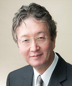
Nobutaka Arai

The Tokyo Metropolitan Institute of Medical Science (TMiMS) began its operations in April 2011 as a new institute integrating the formerly independently operated three institutes; the Tokyo Metropolitan Institute for Neuroscience, Tokyo Metropolitan Institute of Psychiatry, and Tokyo Metropolitan Institute of Medical Science. The Laboratory of Neuropathology (LONP) in TMiMS manages the neuropathological assets of both the former institutes and the brand new one.

LONP has created a digital archive of normal and pathological images of the human central nervous system, and operates Essential Brain Anatomy & Neuropathology (EBA&N) that can be viewed at: https://pathologycenter.jp/english/en_index.html

This system was awarded the Tokyo Metropolitan Association of Medical Welfare and Health Excellence Award in 2014 and the Tokyo Metropolitan Government Staff Award (Governor’s Award) in 2016. In 2017, this website was certified as an intellectual property  of TMiMS.

EBA&N consists of various whole slide images (Cellular Neuropathology, Disease Cytopathology, Normal Histopathology, Muscle Diseases), in addition to brain cutting videos and many photographs. It is very easy to understand because the commentary is written directly on the images and videos, not as footnotes.

We are pleased to issue an individual account to those who wish to view it, so please apply by using the application form in the sample room on the EBA&N site. Registration is completely free. No special browsing software is required. We welcome anyone interested in neuroanatomy and neuropathology. •

Sylvius and “The Quack”

Masterpieces at a Leyden Home

By Peter J. Koehler

When reading the name Sylvius, most neurologists will think of neuroanatomic structures like the aqueduct and the lateral fissure of the hemispheres. Descendant from a Protestant family from northern France (the city of Cambrai that at the time was situated in the southern Netherlands), Franciscus dele Boë (1614-1672), also known as Sylvius, was born in the German town of Hanau, east of Frankfurt-am-Main.

Sylvius by Frans van Mieris I (1665, oil on
panel, 18,8×14,2 cm, New York, private
collection; see also The Leiden Collection
FM-104).

He studied medicine in Sedan and Leyden, made a peregrination to southern German universities and defended a thesis at the university of Basel, Switzerland (1637). He graduated a second time the following year at Leyden university.

He became famous for his anatomy lessons, at the time including physiology. He demonstrated the circulation of the blood as published by William Harvey (1578-1657) a few decades previously (De motu cordis, 1628).

“Many students, and certainly not the worst ones, attended his courses, so that it seemed as if only he could understand and explain anatomy.” Among his students was Danish Thomas Bartholinus (1616-1680), son of the famous anatomist Caspar (1585-1629). Thomas’s notes of Sylvius’s course of the early 1640s was included in his father’s Institutiones anatomicae (1641 edition). Caspar Bartholinus wrote “we cannot pass by the very accurate anatomist D. Franciscus Sylvius; from his noble mind and ingenuity we take the admirable new structure of the brain.”

Following a period of practicing general medical in Amsterdam (1641-1658), where he lived at the Keizersgracht [canal], and during which he enjoyed chemistry and anatomy in his leisure time, he returned to Leyden to become professor of medicine. He gave bedside teaching in the nearby Caeciliahospital and studied the pathological changes at autopsy.

Fissure of Sylvius

Fissura Sylvii.

He attracted many foreign students, including Danish Niels Stensen (“Steno”; 1638-1688), who wrote that Sylvius’s method of brain dissection was a combination of the ancient Galenic approach and the more recent method of Costanzo Varolio (1543-1575).

The latter cut the brain from its base to above, thereby improving the visualization of structures at the base, including the cranial nerves and the pons (Varolius). Another pupil, Gabriel Ypelaer, defended a thesis under Sylvius’s supervision, in which he presented a description of the lateral fissure (1660). Sylvius published it three years later in his own Disputationem medicarum decas.

In 1774, the Swiss Albrecht von Haller (1708-1777) emphasized that this was probably the first description of the fissure. Sylvius’s name became also associated with the aqueduct, although many refer to the homonymous teacher of Andreas Vesalius (1514-1564), Paris anatomist Jacobus Sylvius (1478-1555).

Probably the structure had already been observed by others before Jacobus Sylvius, although there is discussion on whether they really saw the small canal. The Leyden Sylvius, however, described the “canalis vel aquae-ductus” between the converging “roots of the spinal cord” and the corpora quadrigemina below “our bridge” (Disputationem (1663; chapter 21).

With respect to physiology, Sylvius rejected the classical qualities and humors, retaining the concept of animal spirits that were used in the brain and sent to the muscles through the nerves. He adhered to a kind of chemical humoral pathology that became known as iatrochemistry, in which effervescense, a vehement reaction between acid and alkaline secretions occur, for instance, in the right ventricle of the heart or the duodenum.

Collecting Masterpieces

Sylvius and his wife / “The music lesson”
by Frans van Mieris I (1672, oil on
panel, 41×31 cm, courtesy Staatliche
Kunstsammlungen Dresden – Gemäldegalerie
Alte Meister, Dresden, inv. no. 1743).

Sylvius’s first wife, Anna de Ligne, whom he married in 1649, died the year before he moved back to Leyden. Their two children also died at a young age. Arriving in Leyden, he bought a nice house at Rapenburg (on a canal) no. 31, not far from the university building. He had it converted (for 10.500 guilders) into a distinguished home with dining room and salon.

Other houses at the Rapenburg were owned by upper-class Leiden citizens, including city official, merchants, and like Sylvius, university professors. An inventory of his house in 1673, half a year after his death, shows that he had been the owner not only of a considerable library, comprising hundreds of medical, chemical, and religious books, but also one of the largest painting collections of the city.

It seems that at the time he was a patron for contemporary painters, including Experiens Sillemans (1611-1652/3; six paintings) and Simon Luttichuys (1610-1661; six) from Amsterdam, and Gerrit Dou (11) and Frans van Mieris I (7) from Leiden. Among the latter painter’s works, Sylvius owned “The music lesson” with portraits of Sylvius and his second wife Magdalena L. Schletzer, whom he had married in 1666. During the plague of 1669, Sylvius became severely ill, but survived. However, his young wife (21 years old) died, even before Van Mieris finished the painting. It was hanging in Sylvius’s front room. For Van Mieris, Sylvius was “zynen Mecenas” [his Maecenas]. Dutch writer and painter Arnold Houbraken (1660-1719) wrote: “Professor Silvius, welke laatste dikwerf verzogt, dat al wat hy maakte voor hem mogt wezen” [Professor Sylvius, the latter who often asked that all what he made might be for him] (Houbraken, 1721, p. 3).

Next to the painting with his second wife, Van Mieris painted another portrait of Sylvius (1665, see above, he inscribed the age of the person 52, which should have been 50 or 51).

Furthermore, Sylvius owned works by Roelant Savery (1576-1639), Paulus Potter (1625-1654), and Philips Wouwerman (1619-1668), in particular animal scenes. Imagine living in such a kind of art gallery! A reconstruction of his home showed that he had 22 paintings in his dining room (including the five senses by Jan Miense Molenaer), 34 in his large salon, 26 in a side chamber, 16 in the upper back chamber, and 42 (including Van Dyck’s portrait of King Charles of England) in the master bedroom that was arranged as a kind of Kunstkammer.

Apparently, he did not collect everything from his surroundings, as is demonstrated by the fact that he did not have any of the moralizing genre pieces by Jan Steen (1626-1679) or landscapes of Jan van Goyen (1596-1656).

The Quack and (Brain)Stones

Sylvius’s house in Leyden at Rapenburg 31.

Since Jheronimus Bosch’s painting “Cutting the stone,” similar scenes have been depicted in paintings and engravings during the 16th and 17th century (see World Neurology 2017, January, pp. 6-7). Adriaen Brouwer’s (c.1605-1638) “Quacksalver,” now possessed by the Kunsthalle in Karlsruhe), was hanging in Sylvius’s entry room. Although it is not a representation of the actual procedure of “cutting the stone,” stones are depicted hanging on pieces of red string on the quack’s banner, in a way that can observed in several other paintings. It would indicate examples of stones that the quack had removed in the past from the heads of other patients. Even a knife has been attached to the banner.

The place of action is a market square, where a quack, who has relatively short legs, with long hair, a beard, and moustache, is standing on a partly sawed off or buried barrel. He is wearing a red hat with feathers, a yellow coat with blue stripes, and brown trousers. These clothes remind of the Italian comedy (commedia dell’arte), which was sometimes applied by painters. A dagger is attached to his belt. He seems to be looking at the viewer.

In his left hand, he has a small pot and with the index finger of the right hand he probably takes some ointment. Around the table in front of him, we see adult people, probably peasants listening and looking to the objects on the table. There are also some children. The one with the red beret with white feather and pink coat may be doing some mischief. The woman in brown blouse and white bonnet behind the table, laughing to the quack possibly took the plug from the bottle with the blue fluid, or is she emptying a small pot? To the right-hand side, a woman in pink coat with white bonnet and apron, is sitting on a low bench. A man in front of her seems to be explaining what is happening; he is pointing to the quack.

Two other persons, one with a red cap and the other a black beret, behind that man are discussing the scene. A man, leaning on the windowsill of the opened window above, and a woman behind him, are also listening. A number of objects are displayed on the table, including a small bottle with a plug, a spatula – it may be pointing in an intended direction –  and a bowl with a triturator. A big knife is hanging from the edge of the table.

At the background to the left, we see a farmer’s house and a church (and some persons, one of whom is obeying nature’s call near the tree) and to the right façades of six houses in a row. Deception of the credulous peasants seems to be the main subject of this scene.

If the viewer would still have doubts, the painter added, on the upper right corner of the banner, some letters: “JAN CERNAKEL.” This is a reference to Bruegel’s “Witch of Mallegem,” where it is spelled with a “k” instead of “c.” The German world “Karnickel” has several meanings, including rascal or bellwether.

The Pineal Gland and Brain Stones

“Der Quacksalber” [Quacksalver] by Adriaen Brouwer that once belonged to Sylvius.
(c. 1630, oil on panel, 45,2×61,8 cm, courtesy Staatliche Kunsthalle, Karsruhe, inv. no.1897).

Although the artists of the paintings and engravings that directly or indirectly refer to “cutting the stone” had moralistic or satirical intentions, rather than showing actual practices, it is funny to see that Sylvius at least owned one such painting. This is particularly interesting as he lived in a time, in which the pineal gland, by some, including René Descartes (1596-1650), Henricus Regius (1598-1679), and Louis de la Forge (1632-1666), was believed to be the organ in which the soul is localized.

The finding of calcifications, stones, in this organ led to several speculations. Some reasoned that these findings could never be compatible with the idea of localization of the soul in the pineal gland. Others, who had found such stones in persons with behavioral disorders (including a woman who committed infanticide), believed rather that it proved that the cause was in that organ. By its calcification, the physiological flow of humors in that area was believed to be prohibited (see World Neurology 2017, January, pp. 6-7).

In these discussions, the name of Sylvius (and English physician/anatomist Thomas Wharton [1614-1673]) was often found as opponents to the idea of attributing such a function to the pineal gland. The town physician of Nijmegen, subsequently professor of medicine and anatomy in Utrecht (1649), IJsbrand van Diemerbroeck (1609-1674), published a new edition of his Anatome Corporis Humane (1679; first edition 1672), in which he gave a description of the anatomy and position of the pineal gland, mentioning “arena & calculi” [sand and calcification] that occupied half the volume of the gland, with a reference to Sylvius, who indeed referred to the pineal gland in his Disputationes Medicarem IV (De Spirituum Animalium in Cerebro, Cerebelloque Confectione, per Nervous Distributione, atque Usu Vario). He was much in doubt about its function.

“Know Thyself”

This ancient Greek aphorism is one of the Delphic maxims depicted on a column of the Temple of Apollo. Smith (1999) believed that Sylvius’s painting collection “emphasized the importance of self-knowledge and the effort to achieve moderation in all things, like the numerous fruit and flower still lifes in his collection, which alluded to the ephemeral nature of human existence and the vanity of the things of the world, the message of neostoicism.”

Brouwer’s “Quacksalver” would also fit in this idea of admonition to temperance and several of the themes, also used in the literary arts of the period, including self-knowledge and “insight in the deceptive essence of the world.” It would be recognizable in Sylvius’s inaugural address, when he became professor in Leyden (1658). The painting in Sylvius’s collection gives evidence of his sense of humor as well as the perception of Van Mieris’s and Brouwer’s work among the public. •


FURTHER READING:


Baumann ED (1949). François dele Boe Sylvius. Leiden, Brill


Beukers H (2000). The Sylvian Fissure. In: Koehler PJ, Bruyn GW, Pearce JMS (eds). Neurological Eponyms. New York, Oxford University Press, pp. 51-5.


Houbraken A (1721/1976). De groote schouburgh der Nederlantsche konstschilders en schilderessen. Part III Amsterdam, Israël (see also http://www.dbnl.org/tekst/houb005groo01_01/colofon.php accessed October 1st, 2020).


Smith PH (1999). Science and Taste: Painting, Passions, and the New Philosophy in Seventeenth-Century Leiden. Isis 90 :421-461.

St. Olavs Hospital, Norway

By Dr. Abdoul Bachir Djibo Hamani

With Prof. Christian Samsonsen, head of
the department of neurology at St. Olav’s
Hospital

I was honored to be accepted to do a department visit at the prestigious Norwegian University of Science and Technology (NTNU) at the St.Olavs Hospital in the Department of Neurology in Trondheim in Norway.

For this I would like to express my sincere thanks to:

  • The World Federation of Neurology (WFN) for this wonderful opportunity to acquire this new experience in the care of people suffering from neurological disease, especially to Jade Levy, who was in charge of the visit
  • The Norwegian Neurological Association’s president, Prof. Anne Hege Aamodt
  • All the staff of the Department of Neurology
  • Prof. Christian Samsonsen, who was my training supervisor. Prof. Samsonsen had confidence in me and supported and encouraged me throughout the duration of my training.
  • Dr. Elisabeth Kvisvik and Dr. Thanh Pierre Doan for having accepted me into this company and who have facilitated my relations with other colleagues in the departments of neurology, neurophysiology, and stroke.
  • All my supervisors and colleagues who are too numerous to share our experiences. I spent an excellent stay in your company.
  • The entire university hospital administration.

Planned from March 3-27, 2020, my program of visit of the neurology department at St. Olav’s Hospital actually lasted from March 3 to July 28, 2020, because of the coronavirus pandemic, which made my return trip to Africa impossible.

During this stay, I had the opportunity to make daily visits to patients in the neurology department and also to participate in the outpatient consultation where I had the chance to strengthen my skills in the diagnosis and management of neurological pathologies, more specifically epilepsy. I also experienced a number of neurodegenerative and autoimmune pathologies, such as multiple sclerosis, amyotrophic lateral sclerosis, and severe myasthenia gravis.

I also took part in botulinum toxin injections, upper occipital nerve block, and the performance and interpretation of electroencephalograms. I visited some fantastic places in the city and learned to speak Norwegian (Takk=thank you).

Since my return, I have performed several blocks of the upper occipital nerve, and I have been able to set up an electroencephalogram reading club for a better improvement of our electroencephalogram laboratory.

Our neurology department today has adopted several therapeutic protocols resulting from my stay in Norway.

I am sure that this experience will serve all of Africa in general and my country in particular. Thank you again. •


This Doctor Can: A Journey From a Bedwin Boy in Eastern Sudan to a Professor of Neurology

By Prof. Osheik Ab’Asha Seidi, MB BS, MRCP (U.K.), ABiM, CCST (U.K.), FRCP

Osheik Ab’Asha Seidi

I was born in a village called Aroma, named by the British for the nice smell of its naturally growing wild roses, in the northeast African country Sudan. My parents could barely read or write, as they did not complete primary school.

My family was large and wanted all their kids to go to school. From the outset, I was required to do many jobs as well as attend school. This included early morning shopping from the local market for fresh vegetables and meat, to the afternoon and evening milking and feeding of our goats and cows.

The educational system in my country of origin, Sudan, was strong and free. So, despite my family’s limited financial abilities, I was blessed by being able to make my way through primary, intermediate, and high schools successfully, eventually ending up in the Faculty of Medicine at the University of Khartoum.

The university was originally opened in 1924 under the name “Lord Kitchener’s School of Medicine” and was linked to the University of London until 1956 when Sudan became independent from British colonization. I graduated with distinctions but soon realized that I would not be able to support my family as the local economy was in complete disarray. I left for Yemen while most of my colleagues expatriated to the Gulf countries.

I managed to make some money in Yemen, reasonably helped my family, got married, and passed part 1 of the MRCP (U.K.). In the early 1990s, I moved to Saudi Arabia as a GP in a remote village that had no electricity, TV transmission, or paved roads. Soon, I was selected after a tough competitive test to join the Arab Board of Internal Medicine residency program in Abha city, in the Asir Region of southwestern Saudi Arabia.

That was a 5-year residency program. Luckily, in my second year of the rotation, I managed to pass the written and clinical parts of the MRCP (U.K.) and soon after obtained my ABiM (Arab Board of Internal Medicine) degree. I was then promoted to consultant physician at age 35, an unusual thing in that setting.

My passion for neurology led me to resign my job in Saudi Arabia and move to the U.K. in 1998, with my supportive wife and three young kids. I started as a senior house officer, but in a few months, I was promoted to clinical lecturer / registrar at Sheffield University Hospitals NHS Foundation Trust.

Still, my ambition was to excel in medical research as well as clinical skills, so I kept an eye on suitable opportunities, until I succeeded in getting a job as a clinical research associate at Guy’s, King’s College, and St. Thomas’ School of Medicine in 2000. There, I discovered new abilities in myself as a researcher and combined basic laboratory research with participation in international randomized clinical trials on many drugs for multiple sclerosis.

I learned a lot from my seniors at the department of neuroimmunology and was particularly influenced by Prof. Richard Hughes and his impressive range of scientific styles and leadership abilities. I worked with many people of extremely high caliber with comprehensive knowledge in medicine and many other sciences Sharing the teamwork of my research committees, I published widely and participated in international neurology conferences as a speaker. The U.K. gave me what I was looking for – opportunities to further discover my personal strengths.

During that time, I became aware of many misconceptions about Black people, particularly those coming from Africa and more so from Sudan when the war in Darfur was at its peak. I had many personal discussions with my colleagues and really felt that I must do something to clear the mist.

I moved to northeast England as a training specialist registrar in neurology, and in Newcastle upon Tyne, I met many Africans, including Dr.  Peter Adegbie, an inspiring and kind Christian priest originally from Nigeria. He studied theology and had similar feelings as mine regarding the need to clear the misconceptions about Black Africans. Together, with many other African friends in the northeast of England, we participated in a major project called African Voices, led by Dr. Peter Adgebie and funded by the European Union Fund.

We prepared and carried out a comprehensive program to shine a light on African cultures and peaceful diversity. Training was for all ages, but mainly targeted young people in the U.K. We also prepared a curriculum for children ages 5-9 years with a simple approach to let them have true ideas about Africans. Through the stories and experiences of members of the African diaspora communities in Bristol and Newcastle, this resource aimed to challenge young peoples’ perceptions of African countries and provide opportunities to question the way in which we see our world.

The program was extraordinarily successful, and the teaching resource is widely used.

Over the past 10 years, I have moved back to Sudan and joined the Faculty of Medicine at the University of Khartoum. I progressed to professor of internal medicine and neurology in 2014, and was a founding member for the African Academy of Neurology (AFAN) and acted as the vice president for East Africa on the first steering committee (2015–2017). With the solid support of many colleagues in Sudan, we managed to establish a PACES center in Sudan in 2012, and I have supervised all the MRCP (U.K.) examinations in Sudan from 2010 until now.

I also actively participated in starting the Medical Training Initiative (MTI) and hosted the first trainee from Sudan when I was a consultant in Sunderland, U.K., in 2007. I still help in the selection process of suitable candidates from Sudan with leadership of my colleague, Prof. Alaadin Hassan Ahmed, currently dean of the Faculty of Medicine at the University of Khartoum.

I hope my story will inspire many young Black people and other ambitious people of all colors and walks of life.•


This article is reprinted with permission from the Royal College of Physicians. https://www.rcplondon.ac.uk/news/doctor-can-journey-bedwin-boy-eastern-sudan-professor-neurology

First WFN/AFAN e-Learning Day

By Riadh Gouider

The World Federation of Neurology (WFN) and the African Academy of Neurology presented their first virtual one-day teaching course—the first WFN/AFAN e-Learning Day—which took place from 8:45 a.m. to 6 p.m. (BST), Saturday, Oct. 10, 2020.

Collaborators

This educational event was held with the support of the European Academy of Neurology (EAN), the American Academy of Neurology (AAN), and the World Stroke Organization (WSO).

Organizing Committee

The WFN/AFAN Day was organized by Dr. Riadh Gouider, Dr. Wolfgang Grisold and Dr. Walter Struhal, under the direction of the e-Learning Committee on behalf of the WFN and the Board of Directors of the African Academy of Neurology (AFAN).

Local Organizers

The e-Learning Day was locally organized by the Tunisian Society of Neurology (ATN) and the Tunisian College of Neurology and Neurosurgery (CNN).

Professional Congress Organizer (PCO)

The Professional Congress Organizer company is: Meet-in-TV, Millesima Events.

Main Topic

The topic was “STROKE: a Treatable and Preventable Disease.”

Opening Ceremony

The opening of the e-Learning Day was jointly directed by Prof. William Carroll, WFN president, and Prof. Foad Abdallah, AFAN president.

Quizzes

There were five Quiz sessions, animated by Prof. Riadh Gouider and Dr. Saloua Mrabet. 151 attendees participated in the quizzes. The quizzes were operated by the Voting Platform “Mentimeter.” The Quiz Winner was Dr. Ahshish Govind from South Africa.

Quality Assurance Survey

The Quality Assurance Survey link was sent out to all the participants by the PCO. We received more than 200 answers to the survey made about the opinion of the participants about this event. Much appreciation of the quality of the educational content and technical platform was shared by more than 90% of the participants. The Certificate of Attendance will be managed and sent out by Kimberly Coard Karlshoej from the WFN Office.

Second WFN/AFAN e-Learning Day

After the success of the First e-Learning Day, we propose to have a second e-Learning Day on Epilepsy. •


Scientific Program Report

The program was treated in four Scientific Sessions, with a parallel session in French.

  • Session I: Stroke Burden and Challenges in Africa
  • Session II: Stroke Risk Factors and Subtypes
  • Session III: Stroke Imaging
  • Session VI: Stroke Management (with parallel session in French)

Conferences: 10 conferences were pre-recorded in advance and broadcast Oct. 10 with two conferences by session.

Invited Speakers: 10 speakers from all over the world presented lectures and provided deep insight into the latest developments in the field of stroke and vascular diseases.

Prof. Foad Abdallah (Egypt, AFAN)

Prof. Rufus Akinyemi  (Nigeria, AFAN)

Dr. Nobuaki Yamamoto (Japan, WFN)

Dr. Naeem Brey (South Africa, AFAN)

Prof. Bruce Campbell (Australia, WSO)

Prof. Kurt Niederkorn (Austria, WFN)

Prof. Pierre Fayad (Nebraska, AAN)

Prof. Valeria Caso (Italy, EAN)

Prof. Faouzi Bel Hassen (Maroc, AFAN) – French Session

Prof. Yacoube Mapoure (Cameroun, AFAN) – French Session

Moderators: 10 moderators animated the discussions and the interaction between the speakers and the audience during sessions.

Prof. William Carroll (Australia)

Prof. Augustina Charway Felli (Ghana)

Prof. Lawrence Wong Ka Sing (Hong Kong)

Dr. Marie0me Soda Diop (Senegal): She couldn’t connect to the live streaming. Dr. Imen Kacem moderated the conference instead.

Prof. Marc Fisher (Boston)

Dr. Sarah El-Sadig Misbah (Sudan)

Dr. Julien Razafimahefa (Madagascar)

Prof. Athanase Millogo (Burkina Faso)

Prof. Chokri Mhiri (Tunisia) : French Session

Dr. Imen Kacem (Tunisia)


Participation Report

The WFN/AFAN Day brought together neurologists, residents in neurology and health care professionals from Africa and around the globe.

Registration: There were 1,118 registrations from 90 countries worldwide, 944 registrations from 37 African countries. As shown in the statistics below, registrants were from:

Tunisia: 180 registrants

Ghana: 161 registrants

South Africa: 101 registrants

Nigeria: 85 registrants

Egypt: 83 registrants

994 physicians registered for the e-Learning Day, with 63% of them neurologists and residents in neurology.

Attendance Report

Among those who registered, 576 participants from 60 countries followed the event, with 513 participants from 31 African countries.

The participation rate reached 51.1 % of the registration.

About 520 physicians participated in the e-Learning Day with more than 350 neurologists and residents in neurology.

WFN Committees and Specialty Groups

e-Communications Committee and the Aphasia, Dementia, and Cognitive Disorders Specialty Group

By Wolfgang Grisold

This column focusing on WFN committees and specialty groups has a dual purpose:

  • to inform and raise awareness on the important contribution of these groups for the functioning of the WFN
  • to raise interest among the readers for the work of the WFN, and perhaps find and recruit persons to work closer with the WFN, in the interest of neurology.

The work of the WFN depends on the contribution of many members worldwide. Several committees support the trustees in their work (https://wfneurology.org/committees) and also develop ideas and consult. A good example is the e-Communications Committee, chaired by Walter Struhal, which identifies and helps to integrate digital technologies into the WFN. The work of this committee is summarized by Dr. Struhal.

The WFN also has several Speciality Groups, which were previously known under ARGs (Applied Research Committee https://wfneurology.org/wfn-specialty-groups). Here, the work of the Specialty Group is introduced by Suvarna Alladi, a neurology professor from India.

The WFN e-Communications Committee has been created to facilitate the trend of remote learning, and e-learning’s purpose is enhanced by the present COVID crisis. The new and advanced digital techniques are now rapidly evolving and will also serve to improve teaching in remote areas of the world.

WFN e-Communications Committee

Walter Struhal, MD

The origins of this committee stem from the Website Committee. Dr. Struhal got involved in this committee in 2010 as one of the founders of the worldwide young neurologists’ group—International Working Group for Young Neurologists and Trainees. Since 2014, he is in charge of the website and initiated social media channels and the WFN online footage with the close help of Chiu Man, who for more than a decade acts as WFN`s webmaster. He is assisted by Surat Tanprawate, Tissa Wijeratne and Wolfgang Grisold.

This was the early core of the current online presentation of WFN as well the roots of the present e-Communication Committee. The website was completely renewed and redesigned into a “responsive” design that allowed reader to view the same content with equal quality on mobile devices as on computer screens.

While social media increased over the years to a tremendous audience with more than 11,000 friends on Facebook, reaching with our posts from the last 30 days alone >17,000 followers, in addition to >2,700 followers on LinkedIn and >4,800 followers on Twitter.

Social media became one of the strategic core activities within WFN’s online footage and one way of reaching out to neurologists worldwide.

Today, this committee headed by Dr. Struhal consists of members worldwide (below). And the committee is actively supported by Simona Milenkova, a social media expert working for Kenes.

The main objective is continuously informing our audience on important developments in neurology on the global scale, on news from WFN, and recently in employing our growing online presence in e-Learning. One of the first meetings was the joint WFN/AFAN e-learning Day (https://wfneurology.org/2020-09-18-wfn-afan), which was well received worldwide. The large amount of information reaching this committee provides a large and challenging workload.

Aphasia, Dementia, and Cognitive Disorders Specialty Group (ADCD SG)

The WFN Aphasia, Dementia and Cognitive Disorders Specialty Group (ADCD SG) is an international community of cognitive neurologists and allied specialists dedicated to promoting research and improving clinical practice in aphasia, dementia, and other cognitive disorders globally. The group has been actively pursuing this mission since 1966 through its biennial meetings and has grown to having members from Europe, the U.K., North and South America, Australia. West, South and East Asia join its community over the years

The mission of the ADCD SG aims to stimulate scientific discussion in the field of aphasia, dementia, and cognitive disorders and to translate research findings into better assessment, management, and treatment of patients through teaching courses, biennial meetings, and participation in the World Congress of Neurology as well as other meetings. It is multidisciplinary, welcomes members of different specialties, across cultures, and seeks to collaborate with other organizations, within and outside the WFN.

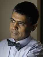
Suvarna Alladi

The chair of the ADCD SG is Suvarna Alladi, professor of neurology of the National Institute of Mental Health and Neurosciences in Bangalore, India. Her clinical and research group focuses on providing multidisciplinary care for persons with dementia, cross-cultural issues in cognitive neurology, developing cognitive tests in different Indian languages and literacy levels and risk factors. Organizing community support for dementia, she co-founded ARDSI (www.ardsihyd.org) and strengthened policy for dementia (www.stride-dementia.org).

The Executive Committee is composed of distinguished experts from across continents: Prof. Morris Freedman, Canada; Stefano Cappa, U.S./Italy; Lorraine Obler, U.S.; Manabu Ikeda, Japan; Eneida Mioshi, U.K.; Peter Nestor, Australia, Matt Lambon, U.K.; Thomas Bak, U.K.; and Facundo Manes, Argentina.

The biennial meetings and teaching courses are the most impactful of the group’s activities. The first biennial meeting of the Specialty Group (formerly Applied Research Group) was held in 1966. The biennial meetings have traveled from venues in Europe to South America, Cambridge U.K., Edinburgh, and then eastward to Istanbul, Hyderabad India, and Hong Kong. The 50th anniversary meeting returned in 2016 to Lake Como, followed by Portugal and the next meeting is planned to take place in Nara, Japan.

The meetings have a tradition of putting the emphasis on quality rather than quantity and to create a forum for discussion and a genuine exchange of ideas. The symposia are based on a wide range of topics, in the traditional clinical areas in aphasia and cognitive disorders, along with newer areas in cognitive science, biomarkers, and technology.

During the COVID-19 pandemic, an online sharing of knowledge and continued interaction is planned. The expert group has developed a rich clinical and research resource across multiple disciplines of cognitive neurology, cognitive psychology, psycholinguistics, speech, and language pathology among others. The SG also has a repository of cognitive-assessment tools in multiple languages, including English, Spanish, Italian, Indian languages, Chinese, and Japanese, among others.

The global expertise of the group has focused on developing joint recommendations for adaptation of diagnoses, assessments, and treatments of aphasia and cognitive disorders across the world. Through its Forum for Young Researchers (FYRE), the SG encourages and nurtures young talents globally. •


WFN e-Communications Committee
Chiu Man: Webmaster (WFN U.K.)
Daniel Gams Massi: Pan-Africa (Cameroon)
Laura Druce: WFN office (WFN U.K.)
Maria Benabdeljlil: Pan-Arab (Morocco)
Riadh Gouider: Pan-Africa (Tunisia)
Surat Tanprawate: Asia-Oceania (Thailand)
Tissa Wijeratne: Asia-Oceania (Australia)
Wolfgang Grisold: WFN Secretary General (WFN, U.K.)


Dr. Juan Rafael Santoni Mendoza (1935-2020)

By Prof. Anna Robles and Prof. Marco T. Medina

Dr. Juan Rafael Santoni Mendoza

Undeniably a father of Dominican neurology,  Dr. Juan Rafael Santoni Mendoza was honest, teacher by vocation, tirelessly hardworking, researcher. He was devoted to his followers, but first of all to his patients. He gave us everything we expected from a great teacher and a great man.

Prof. Santoni Mendoza, affectionately known as “Fao,” passed away Sept. 29, 2020, after a short but aggressive systemic gastrointestinal pathology. Until then, he had been active fulfilling his professional activities as well as his academic and family responsibilities.

He was born in Santo Domingo, Dominican Republic, on March 7, 1935. He went on to marry  Monica Howard Williams de Santoni and was a father to four children: Marcos, Maria Luisa, Alexius Eduardo, and Carlos Juan. He was a neurologist and former president of the SDNN (Dominican Society of Neurology and Neurophysiology).

Dr. Santoni studied Medicine at the Universidad Central de Madrid in Spain, nowadays known as “La Complutense de Madrid,” received a degree in medicine and surgery in 1959. He studied under the distinguished and illustrious Prof. José Casas, Cátedra de Patología General de la Facultad de Medicina de Madrid in 1953.

His primary school education was at Colegio Santo Tomás in Santo Domingo and was followed by the two first years of high school at Hackley School in Tarrytown, New York, where his undisputed leadership led him to be elected class president. He returned to the Dominican Republic so as to finish high school at the Escuela Normal de Varones of Santo Domingo.

In 1959-1961, he did his internal medicine training internship at St. Vincent’s Hospital Dublin, Ireland. He chose to specialize in neurology and neurophysiology at the Institute of Neurology, Queen Square London (1961-1963), received his degree in clinical neurology and electroencephalography with Prof. William Cobb (who frequently invited H. Gastaut from Marseille). He attended as invited neurologist and neurophysiologist (1963-1965) at the Fundación Jiménez Díaz and Gran Hospital de la Beneficencia in Madrid, neurosurgeon Professor Sixto Obrador Alcalde.

Dr. Santoni performed an intense pedagogical work: professor of neuroanatomy and neurology at the Universidad Nacional Pedro Henríquez Ureña (UNPHU) until 1986; professor of neurology for the Universidad Autónoma de Santo Domingo (UASD), for more than three decades until 2008.

He belongs to the group of neurologists who founded and integrated our society in 1968, a distinguished member for over 50 years. He was a pioneer of neurophysiology and epileptology formation for our country. He was a founding member of the Sociedad Dominicana de Cefaleas (SODOC) in 1999.

Dr. Santoni worked as a neurologist at different hospitals in Santo Domingo: Hospital Moscoso Puello, head of the neurological department (1966-1976); Instituto Nacional de la Diabetes (INDEN), consulting neurologist (1979-1996); Hospital Robert Reid Cabral for Children, honorary assesor and consultant neurologist since 1970; Centro de Rehabilitación foundation 55 years (1965), appointed in 1974 head of the EEG department.

He published approximately 120 papers covering the most diverse range of topics on epilepsy and neurophysiology. He wrote dozens of scientific articles and chapters for neurology books with his elder son

Dr. Carlos Santoni Williams as collaborator.

Prof. Santoni has been member of prestigious academies and neurological societies, national as well as international, was a national delegate to the World Federation of Neurology representing our country with dignity for many years; and was life founding member of the Dominican Society of EEG and Clinical Neurophysiology.

He was founding member (1968) of the Dominican Yachting Federation and president from 1972 to 1977. He was a tango dancer and student of Prof. Doña Milkeya Melo.

Prof. Juan Santoni represents six decades of exquisite world-class neurological practice. He was diligent, exacting, and persistent; born with the researcher’s inquisitive curiosity. He released  scientific papers at exemplary rhythm. And on he goes, remaining light, fresh, efficient. As if the passing by of years renewed his energies, to carry on the walk along his brilliant road of life.

It was a privilege to consult him and imitate his example. Fao Santoni was a human miracle made neurologist, and today, with honors, we say good-bye.

Undoubtedly with the absence of Dr. Santoni, the Dominican Republic and Latin America have lost a great man and professor of neurology, but the world of neurosciences has also been affected.


Prof. Ana Robles, FAAN, is former WFN regional director of Latin America. Prof. Marco T. Medina, FAAN, FEAN, Chevalier is WFN regional director for Latin America and PAFNS president.

Simplifying the Diagnostic Criteria for ALS

New Criteria are Aimed to Facilitate Enrollment of Patients into Clinical Trials

By Matthew Kiernan, for the ALS Consensus Committee

The World Federation of Neurology recently joined forces with the International Federation of Clinical Neurophysiology and patient support groups, including the Motor Neurone Disease Association (UK) and the American ALS Association, to convene a consensus meeting to address diagnostic criteria for amyotrophic lateral sclerosis (ALS; motor neurone disease).

Matthew Kiernan, Chair of the ALS/MND Specialty Group, World Federation of Neurology. ALS Consensus Committee: Ammar Al-Chalabi (London, UK), Mark Baker (Newcastle upon Tyne, UK), David Burke (Sydney, Australia), Li-Ying Cui (Beijing, China), Mamede de Carvalho (Lisbon, Portugal), Andrew Eisen (Vancouver, Canada), Julian Grosskreutz (Jena, Germany), Orla Hardiman (Dublin, Ireland), Robert Henderson (Brisbane, Australia), Ryuji Kaji (Tokushima, Japan), Matthew Kiernan (Sydney, Australia), Jose Manuel Matamala (Santiago, Chile), Hiroshi Mitsumoto (New York, USA), Waulter Paulus (Gottingen, Germany), Jeremy Shefner (Phoenix,USA), Neil Simon (Sydney, Australia), Michael Swash (London, UK), Kevin Talbot (Oxford, UK), Martin Turner (Oxford, UK), Leonard van den Berg (Utrecht, The Netherlands), Renato Verdugo (Santiago, Chile), Steve Vucic (Sydney, Australia) Photo Legend: ALS Consensus Committee, Gold Coast, Australia.

The broad aim of the consensus meeting was to improve the process of diagnosis of ALS in the early stages of the disease, when clinical symptoms are minimal, thereby giving therapies the best chance of success. Previous ALS criteria, dating back to the original El Escorial and later Airlie House and Awaji criteria, used degrees of diagnostic certainty from possible to definite ALS. While such an approach makes sense, it also carries a degree of uncertainty for patients, their families, and clinicians. From a practical level, patients with a label of possible ALS may be denied entry to clinical trials, even though such patients would likely benefit from instigation of trial therapies compared with those recruited later in their disease.

Given that uncertainty around a diagnosis of ALS constitutes a potential barrier to patient enrollment in clinical trials, the consensus meeting held on the Gold Coast in Australia began by considering a broad range of data across different phenotypes, clinical presentations, and outcome measures, in addition to discussions that evaluated diagnostic technologies.

Prior to defining consensus criteria, a collective understanding of ALS was established based around key tenets: that ALS represented a progressive disorder of the motor system, that it involves dysfunction of upper and lower motor neurone compartments of the nervous system, and that there is typically a focal onset. It was accepted that while upper motor neuron signs were not always clinically evident, involvement of the lower motor neuron was more often apparent through clinical examination. In terms of diagnostic technologies, supportive evidence of lower motor neuron dysfunction has tended to be derived from electromyography and neuromuscular ultrasound, particularly the detection of fasciculations in multiple muscles. Supportive evidence of upper motor neuron dysfunction was more limited, relying on transcranial magnetic stimulation studies of the central motor nervous system, MRI, and neurofilament levels, although it was accepted that a diagnosis of ALS does not require these investigations. And finally, while ALS may include cognitive, behavioral, and psychiatric abnormalities, these features were not essential for a diagnosis.

Accepting these broad concepts of disease, combined in an integrated fashion, a consensus was reached, whereby ALS was defined by the presence of:

  • Progressive motor impairment, documented by history or repeated clinical assessment, preceded by normal motor function.
  • Upper and lower motor neurone dysfunction in at least one body region (in the same body region if only one body region was involved), or lower motor neuron dysfunction in at least two body regions.
  • Investigation findings that excluded alternative disease processes.

In adopting these simplified criteria for ALS, the previous diagnostic categories of possible, probable, and definite were abandoned. Such assessment of a likelihood of disease served to generate misinterpretation by patients, who may inadvertently have considered that such terms represented the likelihood that ALS was causing their symptoms.

In reality, the consensus group acknowledged that nearly all patients diagnosed as possible ALS continue to progress and ultimately die from ALS. Moving forward from these new consensus criteria, it is anticipated that ALS patients will be fast-tracked into clinical trials. With the advent of these new criteria, the diagnosis of ALS can be made early, and definitively. •

Reference:

Shefner JM, Al-Chalabi A, Baker MR, et al. A proposal for new diagnostic criteria for ALS. Clin Neurophysiol 2020;131:1975-1978.


Becoming Acquainted With the Pandemic

By the time this edition of World Neurology is available, it will have been more than eight months since the world had to learn to deal with COVID-19. In that time, our lives and practices have changed dramatically. Social distancing, travel restrictions, and educational activities have forced us to adapt to what is effectively a new world. In some countries, the pandemic is flaring again, resulting in death, disability, and the consumption and diversion of resources. In others, populations are variably safer but still constrained.

William Carroll, MD

It is nevertheless heartening to see and experience the intent of many of our colleagues at local, national, and international levels collaborating to record quality data so as to better understand the short-, medium-, and long-term effects of COVID-19. Groups such as the European Academy of Neurology’s “EAN NEuro-covid ReGistrY (ENERGY),” the World Health Organization’s Neuro-COVID Global Forum, and the COVID-19 Neuro Research Coalition are all actively involved in developing platforms and directions to facilitate the capture of neurological involvement in the pandemic. In many countries, clinicians, public health workers, and hospital facilities are struggling to care for those affected. Within this activity, we are also witnessing a disturbing trend of disbelief and even outright opposition toward the need for change in social behavior. There is a niggling sense that beneath the surface, there is a latent feeling of every person for themselves.

It is essential in such environments that professional organizations, such as ours, remain united in our efforts to care for those with neurological diseases, record accurately those with COVID-19-related neurological illness, support colleagues in the battle with COVID-19, and maintain the training of our next generation of neurologists.

As I have mentioned before, the WFN was already moving to a more electronic-based medium for its educational and communication activities prior to COVID-19. There are now several changes underway within the WFN that will likely drive new and innovative programs. These may result in transformation of WFN operations and potentially persist after the pandemic has subsided.

In communication, the London office has completed the first step to improve its ability to reach all member societies and their individual neurologists. Almost all 122 member societies have contributed to the updating of their contact details, for which the trustees and office staff are most grateful. The second step will be to ensure that material disseminated to member societies is timely, meaningful, and reciprocal. In an era where physical meetings may never be the same again, this is an important and essential development. Examples of how this development has already proven beneficial are the recent Council of Delegates Annual General Meeting, the trustee election, and the promotion of three successful educational programs. In all of these, up-to-date information was communicated to all member societies and through many of them to their memberships.

For the first time in the history of the WFN, a virtual Annual General Meeting was held on Sept. 9. This was notable also for the large number of member society delegates, given that in a non-World Congress of Neurology year, as 2020 is, attendance numbers are typically on the low side. Business was conducted in the usual format and essential motions proposed and carried. Dr. Morris Freedman was declared the new elected trustee following a smooth electronic electoral process contested by six high quality candidates. The clarity, rapidity, and accuracy of the election were greatly appreciated by the office staff, who are usually tasked with such matters manually. While this format lacked the usual intense discussions and face-to-face canvasing, it allowed many more societies to participate without having to travel or to arrange proxies.

Improved communication with member societies has also aided the promotion and the production of educational activities. Two WFN specialty groups and a regional organization have illustrated this aspect effectively. First, the Neuromuscular Disorders Specialty Group, chaired by Prof. John England, partnered with the Spanish Neurological Society to hold a virtual International Congress on Neuromuscular Diseases (ICNMD) Sept. 11-14. Some 800 registrants from almost 50 countries attended the congress and will be looking forward to the next congress in July 2021 in Valencia. Second, the Tropical and Geographical Neurology Specialty Group has just begun the second series of its highly popular and successful, mainly fortnightly, Neuroinfection sessions. Chaired by Prof. Chandrashekhar Meshram and supported by the Indian Academy of Neurology, the topics have updated many common and not-so-common neurological infections. This series will continue through to Dec. 12. The most recent WFN-related educational event was the AFAN/WFN E-Learning Day held on Oct. 10 and run out of Tunis. Eight lectures in four sessions dealing with stroke, with particular emphasis on Africa, were delivered by experts from the African, the European, and the American Academies of Neurology and from the World Stroke Organization. It was a great example of collaborative effort. Altogether there were approximately 1,100 registrants from 80 countries. Thanks go to all involved in the execution of this event and especially to Prof. Riadh Gouider, who was tireless in his efforts to ensure a rewarding e-learning experience. These meetings were all promoted by the WFN through the usual media sources and by direct communication with member societies.

World Brain Day 2020 also exemplified the value of improved communication in promotional terms. The WFN was greatly benefited by the partner organization, the International Parkinson and Movement Disorder Society (IPMDS), both around the July 22 commencement and at the final press conference held during the IPMDS virtual Congress in September. This year, the promotions and activities related to World Brain Day reached more than 50 million people globally, just outdistancing the numbers from the 2019 WBD. Other key social media parameters generated exceptional numbers for WBD. The WBD Committee, chaired by Prof. Tissa Wijeratne, was extremely successful in generating local and regional activities on and around July 22. Many of these are shown on the WFN website: www.wfneurology.org/wbd.

The WBD Committee is already planning the 2021 program. The two promotional videos prepared earlier this year dealing with WFN activities and the upcoming 2021 World Congress of Neurology were shown during the inaugural AFAN/WFN E-Learning Day lunch and coffee breaks and will no doubt be employed through to the World Congress of Neurology. The Brain Health Initiative is close to presenting the first of a series of five videos aimed at the general population. We hope that it and its successors will promote improved awareness of the importance of brain health and the provision of quality neurological care.

While we all miss the pleasure of meeting and communicating face-to-face with our colleagues, the resumption of that personal interaction with the developments described above will significantly enhance the cohesion and success of the WFN. Meanwhile, I wish everyone a safe coexistence with the pandemic. •

Prof. William M. Carroll
President, World Federation of Neurology

Online Education for Virtually Every Neurologist

By Steven L. Lewis, MD, Editor,
and Walter Struhal, MD, Co-Editor

Steven L. Lewis, MD, Walter Struhal, MD

We would like to welcome all neurologists worldwide to the October/November 2020 issue of World Neurology. This issue begins with the report from Matthew Kiernan for the ALS Consensus Committee, operating with the support of the WFN, about the development and publication of the new ALS diagnostic criteria, which are aimed to diminish ambiguity of patients’ diagnoses while also facilitating enrollment of patients into clinical trials. In this issue’s President’s Column, WFN President William Carroll reports on the current global efforts to understand, collaborate, and report on the neurologic aspects of COVID-19 and also updates us on the recent Annual General Meeting of the Council of Delegates, the success of the recent World Brain Day 2020, and recent and current successful efforts related to virtual education in the current era from the WFN, the national societies, and WFN specialty groups. In his recurring column on the WFN Committees and Specialty Groups, WFN Secretary-General Wolfgang Grisold updates us on the activities of the WFN Congress Committee and the WFN Specialty Group on Rare Neurologic Diseases.

In this issue’s history section, Aernoud T.L. Fiolet and Ingrid M. Daey Ouwens describe the history of malaria fever therapy as a treatment for general paralysis of the insane from neurosyphilis, which they provide as a cautionary tale in the current pandemic era with regard to potential pitfalls in urgently developed treatments. Also in this issue, Gustavo Roman, who was recently appointed as editor-in-chief of Cerebral Circulation, issues a call for papers for this new publication.

This issue is distinguished by having a number of articles highlighting a sampling of some of our member societies’ successes related to World Brain Day 2020, including the report from India from Chandrashkhar Meshram, Pramod Kumar Pal, Gagandeep Singh, Nirmal Surya, and Vinay Goyal; a report from Pakistan by Abdul Malik; a report from Man Mohan Mehndiratta and colleagues from New Delhi on his suggestions for a successful virtual advocacy effort; and finally, a report by Marcelle Smith and Patty Francis on the current status of Parkinson’s Disease in Africa, particularly from their South African perspective.

Much of the remainder of this issue is devoted to the important new world of international virtual conferences related to the WFN, in addition to the recent very successful AFAN/WFN E-Learning Day, which was held on Oct. 10 and so skillfully led by Riadh Gouider, WFN Trustee, and discussed in the President’s column. This issue features reports from Abderrahmane Chahidi, Zouhayr Souirti, Mohamed Chraa, Najib Kissani, and Reda Ouazzani from the Moroccan Society of Neurophysiology and Moroccan League Against Epilepsy on their highly attended and successful International Webinars of Experts in Clinical Neurophysiology, and the report by Chandrashkhar Meshram on the WFN Neuroinfection Series (cosponsored by the Forum for Indian Neurology Education [FINE]) that is now on its second set of highly attended and remarkable lectures and case presentations on neuroinfectious disease. These internationally presented and globally attended lecture series represent highly effective and (and in these cases, free) educational efforts; in fact, the creation of these remarkable online (live and archived) educational opportunities are the true silver linings of the current pandemic and we encourage neurologists worldwide to attend and learn from as many of these virtual lecture series as possible.

On a similar note, John England, Wolfgang Grisold, and Juan J. Vilchez report on the recent International Congress on Neuromuscular Diseases (ICNMD), which was both highly successful and well attended in its virtual format for 2020.

Finally, the issue concludes with a heartfelt report from Guillermo Delgado-García, who details his visit to “The Neuro” (the Montreal Neurological Institute) under the auspices of the WFN and the Canadian Neurological Society.

We look forward to receiving reports from the field as you care for your neurological patients throughout the world during this pandemic, and wish you all the best of health and safety. •