By Peter J. Koehler
In the first chapter of Peau de chagrin (1831), one of the novels in the Comédie Humaine series by French writer Honoré de Balzac, it is written “Là dormait un enfant en cire, sauvé du cabinet de Ruysch, et cette ravissante créature lui rappelait les joies de son jeune âge.” [There, a child in wax slept, saved from the cabinet of Ruysch, and that splendid creature reminded him of the joys of his youth; p.36].
Apparently, 100 years after the death of Dutch physician and anatomist Frederik Ruysch (1638-1731), his work was still known in non-medical circles in Paris. Moreover, almost 1,000 of his anatomical preparations are still displayed at the Kunstcamera Museum in St. Petersburg.
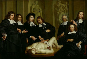
Figure 1. Anatomical lecture of Frederik Ruysch by Adriaen Backer (1670; courtesy Amsterdam Museum).
Ruysch’s Life
Following his examination to become an apothecary, Frederik Ruysch studied medicine in Leiden. (See Figure 1.) At age 28, he became praelector anatomiae of the Amsterdam surgery guild, succeeding Johannes Deijman, whose name was immortalized in Rembrandt’s “Anatomical Lesson of Dr. Deijman” (1656, Figure 2). Ruysch kept the position for over 60 years. He developed a successful method to embalm corpses, probably based on alcohol. Child bodies seemed like sleeping children. (See Figure 3.) His collection (“cabinet”) of anatomical (and other) preparations became a real touristic Amsterdam attraction.
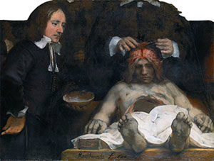
Figure 2. Rembrandt’s Anatomical Lesson of Dr. Deijman (1656); Deijman is working on the brain of the cadaver; his head and several other persons on the original painting were lost by a fire (1723). Courtesy Hermitage, Amsterdam.
In 1717, he sold the collection to tsar Peter the Great of Russia for the amount of 30.000 guilders. He preferred to teach with his preparations rather than with recently deceased corpses. As an empirical physician, his device was “come and see.” Next to anatomy and teaching, Ruysch examined midwives and was appointed professor of botany. In this position, he taught at the city’s botanical garden, which had been founded in 1638.
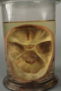
Figure 3. Picture of a child’s head taken from above. (Courtesy, Kunstkamera, St. Petersburg, Frederik Ruysch’s Collection 4070 – 176).
Ruysch as (Neuro)Anatomist
Today, Ruysch’s Opera Omnia are available online, but unfortunately only in Latin and Dutch. (See Figure 4.) These contain the descriptions of the anatomical preparations including the figures. Furthermore, we find his correspondence with other scholars on anatomical subjects. These include the German anatomist Abraham Vater (1684-1751), the physician Michael Ettmüller (1644-1683), and Andreas Goelicke (1670-1744), who spent some time in Leiden and Amsterdam. Ruysch improved his method to prepare anatomical specimens in the 1690s by adding vascular injections with fluid wax. Preparations could now be conserved in liquor balsamicus for longer periods. Moreover, this technique also allowed him to study the blood vessels. Herewith, he was able to refute the theory of the Italian physician Marcello Malpighi (1628-1694) who believed the cerebral cortex consisted of glands. After all, by his method, Ruysch was able to show that the cortex consisted largely of small blood vessels. However, his findings were not accepted by everyone. He wrote:
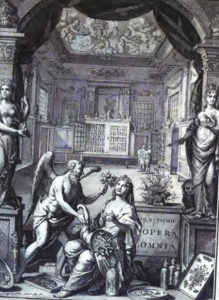
Figure 4. Frontispice of Ruysch’s Opera Omnia (1721)
Marcellus Malpighius, in the treatise on the bark of the brain, writes that this consists of an abundance of very small glands that spring in the twisting wrinkles; and apparently pressed against each other, constitute the external surface of the brain. After I filled the blood vessels with such a great accuracy that the smallest branches were filled and appeared like down, we may clearly see that the bark-like substance of the brain consists merely of joined blood vessels (Ruysch, 1744, p.350)
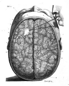
Figure 5. Brain preparation (air is blown between the pia mater and arachnoid)
One of the figures in the Opera Omnia (Tablet 10, Figure 5) “shows the head of a young man that has been kept for several years already as if it was alive” and shows a “small tube put between the arachnoid and pia mater so that these will yield by wind blown between.” Another figure shows the lifted arachnoid. Another interesting figure depicted in his Opera Omnia is that of “the brain taken from the skull of an approximately 10-year-old boy, to which is added the course of the arteries spread out through the pia mater, as well as the emerging nerves from the medulla oblongata.” (See Figure 6.) Ruysch and his contemporaries distinguished 10 cranial nerves. “Our” 7th and 8th were considered one pair. Only at the end of the 18th century, German physician Thomas von Sömmering (1755-1830) described 12 cranial nerves.
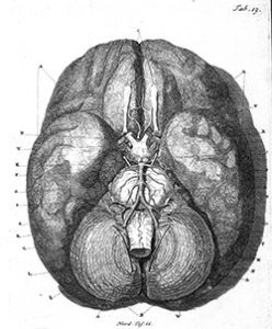
Figure 6. Brain preparation with arteries and 10 cranial nerves.
Ruysch as Physician
In this period, many physicians published case reports, and so did Ruysch. Usually, disorders of the whole body were discussed a capite ad calcem [from head to feet]. In his Hundred anatomical and surgical comments, several are of neurological interest. Case 34 describes a “child with a spine split in two” as well as a “lumbar growth” (Spina bifida; Figure 7). The child also suffered from hydrocephalus.
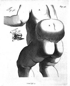
Figure 7. Spina bifida and split spine.
“When we have examined this growth meticulously, we will see clearly that this is a dropsy of a part of the spinal cord and that this is the same ailment as which is called hydrocephalus in the head of an infant.” Ruysch was aware of the grave prognosis. “With respect to the treatment of this ailment, it can hardly or never be dispelled. As none of the children that I have seen, survived.”
Referring to his Amsterdam colleague Nicolaas Tulp (1593-1674; of Rembrandt’s “Anatomical lesson of Dr. Tulp”) he warned not to open the cyst “as we experienced, when it is opened or burst, that death was precipitated.”
“A Headache that Stroke Root”
Another case describes the 18-year-old daughter of a merchant who suffered from headaches that did not react to the usual treatments. “Laxatives, bloodletting alternating from the feet and other locations, Spanish flies [used in the past for blistering, nowadays as aphrodiciacum], sniffing, cupping, etc. At last, she permitted an incision crosswise on the skull … as proposed by us, upon which a considerable amount of blood was lost, but with the slightest relief.” Ruysch now intended to perforate the skull with a small drill,” but beforehand tried a seton on the skin of the neck.
“Soon after the start of this treatment, the pains disappeared.” After a few days, she removed the seton, after which the headache recurred, but after the installment of a new seton, “the pains that tormented her stopped immediately and being more prudent than the first time, she kept it until nature expelled the seton.” Following a third seton, the headache did not recur at all, and she “started to live cheerfully and merrily and she remained in this condition up to the present.”
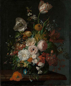
One of Rachel Ruysch’s flower still lifes (courtesy Rijksmuseum, Amsterdam).
Apparently, the case report had impressed Gerard van Swieten (1700-1772), who published it in his own Commentaries on the Aphorisms of Boerhaave (his Leiden teacher). This practice was not unusual in those days.
In 1715, Ruysch became fellow of the prestigious Royal Society and continued to teach anatomy up to the age of 85. He received letters, of which he was only able to answer a quarter. He received famous foreign physicians, including his former pupil, the Danish-born French anatomist Jakob Winslow (1669-1750), who had become a member of the Académie de Sciences in Paris. He referred to Ruysch as “plus grand pionnier parmi les anatomistes de ce siècle” [the greatest pioneer among the anatomists of the century].
Another visitor was the French surgeon Guillaume Desnoues (1650-1735) well-known by his wax anatomy model) who, in the guest book of Ruysch’s cabinet, wrote, “qu’il a surpassé les autres anatomistes dans les injections des vesseaux” [that he had surpassed other anatomists by injections into blood vessels].
Ruysch died at age 93 in 1731. His daughter Rachel became a well-known painter of flower still lifes, many of which are displayed at the Rijksmuseum in Amsterdam (See Figure 8.) •
Literature
Koehler PJ, Boes CJ. A history of non-drug treatment in headache, particularly migraine. Brain 2010;133:2489-500.
Kooijmans L. De doodskunstenaar. De anatomische lessen van Frederik Ruysch. Houten, NTVG/Bohn Stafleu Van Loghum, 2004.
Ruysch W. Alle de ontleed- genees- en heelkundige werken. Amsterdam, Janssoons van Waesberge, 1744.