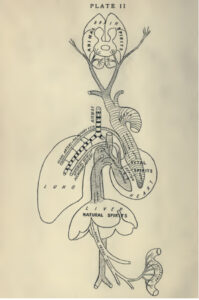
Figure 1. Human physiology according to Galen (129-c.199).
A look back at the relationship between electricity and the brain on the 100th anniversary of the first human EEG recording.
by Peter J. Koehler
This year marks the centennial of the first registration of a human electroencephalogram (EEG). Why is it that just a few hundred years ago physicians were still thinking in terms of animal spirits (spiritus animalis) flowing through cerebral ventricles and hollow nerves, when today we can create brain-computer interfaces to help disabled persons control their wheelchairs?1
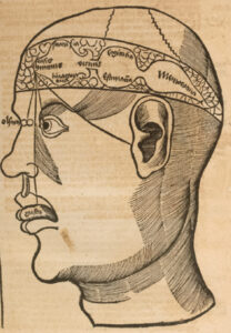
Figure 2. Ventricular or cell doctrine from the 1508 edition of Gregor Reisch’s Margarita philosophica.
“Neurophysiology” Before the 18th Century
The doctrine of pneuma (spiritus in Latin, a very fine and volatile material principle of life) states that natural spirits (spiritus naturalis) arise in the liver, according to the physiology of Galen (129-c.199). These are converted into vital spirits (spiritus vitalis) after passing through tiny pores between the right and left parts of the heart. After passing through the rete mirabele — a vascular plexus of blood vessels at the base of the brain surrounding the pituitary gland that is present in mammals, but not in humans — they become animal spirits (spiritus animalis),2 flowing through the brain cavities and nerves. (See Figure 1.) Greco-Roman medicine, with the even older humoral pathophysiology in addition to this pneuma doctrine, was influential for many centuries.
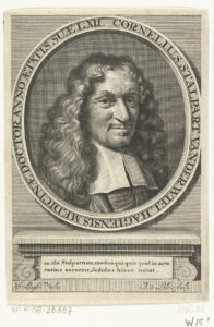
Figure 3. Cornelis Stalpart van der Wiel contested the localization of the soul in the pineal gland.
In the Middle Ages, the material part of the soul was still localized in the brain cavities — a system also known as ventricular or cell doctrine. It was assumed that in the first cell — our first and second ventricles — the information from the senses comes together
(Figure 2; sensorium commune). This is also where fantasy and imagination reside. In the second cell (today’s third ventricle), thinking and judgment were localized, and in the third cell memories were stored. The spiritus animalis could reach the muscles through nerves, which were assumed to be hollow.
In Traité de l’Homme, written in the 1630s but published posthumously in 1664, René Descartes (1596-1650) described his reasons for localizing the material part of the soul in the pineal gland (glandula pinealis) rather than in the ventricles. In the following 150 years, a debate arose as to whether this gland was the seat of the soul, particularly because of the discovery of stones (calcifications) often found in this gland at autopsy. Some denied it for this reason, such as the Dutch physician Cornelis Stalpart van der Wiel (1620-1702, Figure 3).3
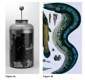
Figure 4a. Leyden jar (left) at the Museum Boerhaave, Leiden, and Figure 4b. Electric eel from Albertus Seba’s Thesaurus: Naaukeurige beschryving van het schatryke kabinet der voornaamste seldzaamheden der natuur [Accurate description of the treasury of the principal rarities of nature] (Locupletissimi rerum naturalium thesauri accurata descriptio (4 Vols. 1734-65)).
Others reasoned instead that people with such stones exhibited behavioral disorders. For example, a case involved a young woman who was convicted of infanticide of her newborn in the 17th century. She was decapitated and at autopsy a pineal gland stone was found that would have explained her behavior.
In the 18th century, electric fish, specifically the electric eel, found in Dutch colonies in the West Indies — present-day Surinam and British Guiana — played a role in the realization that electricity is connected to nerve function. These fish can build up a potential of 700 to 800 volts, much more than the electric ray (torpedo), which had already been described in Ancient Greece.
Dutch colonists, who had felt the effects of a Leyden jar with Pieter van Musschenbroek (1692-1761) from 1745 onward, recognized this feeling when they encountered the electric eel in the West Indies. Experiments were conducted, and correspondence took place with the Hollandsche Maatschappij der Wetenschappen in Haarlem (Netherlands), which had been founded in 1752. The final proof of the electrical properties occurred when sparks were drawn at demonstrations before the Royal Society in London in the 1770s. Thus arose the concept of animate electricity from Luigi Galvani (1737-1798) which, incidentally, was opposed by Alessandro Volta (1745-1827).4,5
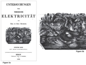
Figure 5a. (left) Title page of Dubois Reymond’s book (second volume), on which we see a drawing of a horse falling paralyzed into a stream by the discharge of an electric eel. Figure 5b. Apparently the drawing was made by him, as we read (to the left below): “EdBRdel” (E dBR delineavit).
Neurophysiology in the 19th Century
Finally, it was Emile Dubois Reymond (1818-1896) who was able to demonstrate the action potential (he called it “negative variation”) in a peripheral nerve with a sensitive galvanometer (1848). With this, he provided irrefutable proof that nerves were actually “electric,” which greatly influenced thinking about the etiology and treatment of nervous diseases. Shortly thereafter, the inventor of the ophthalmoscope (1851), Hermann von Helmholtz (1821-1894), was able to determine the velocity of the signal that propagates along the sciatic nerve of a frog.
Meanwhile, various physicians had begun to apply electrotherapy. In fact, this had been possible since the aforementioned invention of the Leyden jar, which allowed electricity to be brought outside the walls of the laboratory. With electromagnetic equipment and increased interest in the electrical properties of the nervous system, the use of electrotherapy gained interest.
This interest in electricity gradually shifted to the central nervous system, especially when the physician-electrotherapist Eduard Hitzig (1838-1907) got the idea of stimulating the brain of laboratory animals after a chance observation.6,7 With his sensitive stimulation method, he and Gustav T. Fritsch (1838-1927) were the first to be able to elicit muscle contractions in the limbs of a dog (1870; Figure 6). Due to the Franco-German war, however, their publication was hardly noticed. Three years later, it could be confirmed by David Ferrier (1843-1928), who did more detailed neurophysiological research in several species of animals.
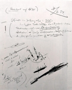
Figure 7. Berger did not publish his first human registration. This is the protocol of his registration on June 28, 1924, with a drawing showing the area of the skull defect and position of the electrodes.
Electrodiagnostics
Shortly thereafter, Richard Caton (1842-1926) in Liverpool was able to observe “feeble currents of varying direction” with electrodes on the cortex of a rabbit using a sensitive coil galvanometer combined with optical magnification.9 Several other researchers engaged in similar studies, including Adolf Beck (1863-1942) in Kraków. In 1890, Beck observed in laboratory animals that spontaneous fluctuations ceased after sensory stimulation. He also observed desynchronization of cortical activity upon sensory stimulation.
In 1901, Leiden physiologist Willem Einthoven (1860-1927) invented the string galvanometer, which shortly thereafter enabled him to record the ECG. He received the Nobel Prize for this in 1924. Hans Berger (1873-1941), who was interested in psycho-physical correlation through a telepathy (he did not use that term) incident, used this instrument — and later the Edelmann string galvanometer — for the registration of brain waves in humans. This year marks the centennial of his first registration from the human cortex in someone with a skull defect. (See Figure 7.)
It was not until five years later, in 1929, that he began to publish papers about it.10 In fact, recognition came only after confirmation by Edgar D. Adrian (1889-1977) and Bryan Matthews (1906-1986), who presented their work to the British Physiological Society in May 1934. In an article in Brain in the same year, they proposed the eponym Berger rhythm in reference to the alpha rhythm, which they believed could be found occipitally. Berger’s role during the Nazi period — some sources originally claimed he was forced by the Nazis to retire and commit suicide because he disagreed with the regime — turned out to be less courageous than previously suggested.11
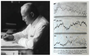
Figure 8. Hans Berger and the EEG of his 16-year-old daughter Ilse at rest (top), during a math problem (middle) and after she had solved it (bottom). The recordings were published in Berger’s 12th report of 1937.
The discovery of EEG had a huge impact, both on science and the general public. The latter has been fascinatingly described by medical historian Cornelius Borck in his book Hirnströme. Eine Kulturgeschichte der Elektroenzephalographie.13 His book was later translated into English.14
Brain Organ
Some years ago, I received a box with historical material from the son of Dutch clinical neurophysiologist Willem Storm van Leeuwen (1912-2005). The box contained recordings from the 1950s carrying the name “Brain organ.” After digitization, I was able to hear the sonified EEG signal, a registration made by Jan Willem Storm van Leeuwen in the 1950s. The sound changed tone with the opening and closing of the eyes. He published a paper on it in 1958.15
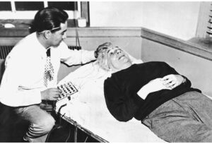
Figure 9. Albert Einstein’s EEG being registered and compared with other scientists at Massachusetts General Hospital in 1950.
As Storm Van Leeuwen had trained in Bristol with William Grey Walter (1910-1977) and in Cambridge with Adrian in the late 1940s, it is likely that he got the idea from the latter, who had written that by listening to the nerve discharge during an experiment, he had been able to save a lot of time and photographic material. Moreover, Adrian sometimes found it an advantage to be able to both hear and see the rhythm as he took the EEG from himself.16 Listening provided a useful complement to visual analysis, as several EEG phenomena could be more easily identified with hearing.
Cybernetic theory-inspired physicist Edmond Dewan (b. 1931) — a friend of the pioneer in that field, the mathematician and philosopher Norbert Wiener (1894-1964) — designed a “brainwave control system” in the 1960s that allowed him to turn off a bedside lamp without using motor skills.
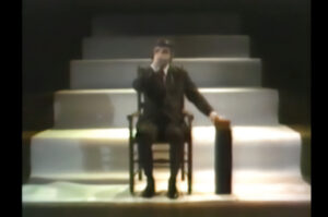
Figure 10. Alvin Lucier during a performance of Music for Solo Performer, which can be found on YouTube (Alvin Lucier – “Music For Solo Performer” (1965) – YouTube).
As an amateur organist, Dewan befriended experimental composer Alvin Lucier (1931-2021), who was inspired to create the first composition based on brainwaves. Music for Solo Performer was first performed in 1965. Sitting on a chair with his eyes closed, Lucier’s EEG was transmitted to numerous speakers scattered around the auditorium. Because the amplified alpha rhythm was below human audible range, the speakers were placed directly opposite various percussion instruments, which were then activated by vibration. As Lucier tried to refrain from mental activity, percussion sounds gradually emerged in the room (Figure 10). These then suddenly stopped again when he opened his eyes, engaged in mental exercises, or when his attention was drawn to sounds from the audience.
Brain-computer music interfaces became very popular. In 1995, when I attended a meeting of the European Club for the History of Neurology in Oslo, Norway, the opening ceremony took place in the Town Hall. The Norwegian composer Arne Nordheim (1931-2010), a pioneer of electronic music in Norway, produced music from signals routed to a computer from the EEG recording of a mother and her epileptic child. Today, such an experiment would probably raise eyebrows and ethical questions, but at the time, it was reported in Norwegian newspapers.
The sonification of brain waves took a new turn toward the late 1960s, when cybernetic theories and scientific breakthroughs gave rise to the field of biofeedback, in which biological processes were measured and fed back to the same individual to gain control over those processes. The sonification of EEG provided an interaction between neurophysiology and experimental music. Although initially applied by neurophysiologists as an adjunct visual EEG analysis, experimental composers turned to brainwave sonification to explore the sonic boundaries between mind and machine.17
Brain-Computer Interfaces
During the past decades, the EEG signal became important as a diagnostic tool for communication with patients suffering from disorders of consciousness.18 In the field of rehabilitation of paralyzed patients, intracortical brain-computer interfaces are being used to restore movement and communication by decoding movement signals from the brain. Brain-computer interfaces enabled functional restoration of movement and communication, including robotic arm control, reanimation of paralyzed limbs through electrical stimulation, cursor control, translating attempted handwriting movements into text, and decoding speech.19
EEG became a promising tool for bridging the gap between mind and machine, enabling the integration of mental and computer processes into one comprehensive system. Berger probably would have been thrilled.
Acknowledgement
I am grateful to Ragnier Stien for making available the program of the performance in Oslo. •
Literature
Vansteensel MJ, Pels EGM, Bleichner MG, Branco MP, Denison T, Freudenburg ZV, Gosselaar P, Leinders S, Ottens TH, Van Den Boom MA, Van Rijen PC, Aarnoutse EJ, Ramsey NF. Fully Implanted Brain-Computer Interface in a Locked-In Patient with ALS. N Engl J Med. 2016 Nov 24;375(21):2060-2066.
McHenry LC. Garrison’s History of Neurology. Springfield, Illinois, Thomas, 1969, p. 13.
Koehler PJ. Brain stones. World Neurology 2017 (January): 6-7.
Koehler PJ, Piccolini M, Finger S. The “Eels” of South America: Mid-18th-Century Dutch Contributions to the Theory of Animal Electricity. Journal of the History of Biology 2009;42(4):715-763.
Koehler PJ. Medical observations by European physicians in the colonies. Philippe Fermin’s observations in 18th century Surinam. World Neurology July 2016.
Hagner M. The electrical excitability of the brain: toward the emergence of an experiment. J Hist Neurosci. 2012 Jul;21(3):237-49.
Koehler PJ. Eduard Hitzig’s experiences in the Franco-Prussian War (1870-1871): the case of Joseph Masseau. J Hist Neurosci. 2012 Jul;21(3):250-62.
Fritsch G, Hitzig E. Über die elektrische Erregbarkeit des Grosshirns. Archiv für Anatomie, Physiologie und Wissenschaftliche Medicin 1870;300-32.
Caton R. The electric currents of the brain. British Medical Journal 1875;2:278.
Berger H. Über das Elektroenzephalogramm des Menschen. Arch. Psychiat Nervenkrankh 1929;87:527-70.
Zeidman LA, Stone J, Kondziella D. New revelations about Hans Berger, father of the electroencephalogram (EEG), and his ties to the Third Reich. J Child Neurol. 2014 Jul;29(7):1002-10.
Berger H. Über das Elektroenzephalogramm des Menschen XII. Arch. Psychiat Nervenkrankh 1937;106:165-87.
Borck C. Hirnströme: Eine Kulturgeschichte der Elektroenzephalographie. Göttingen, Wallstein, 2005.
Borck C. Brainwaves: A Cultural History of Electroencephalography. London/New York, Routledge, 2018.
Storm van Leeuwen W, Bekkering DH. Some results obtained with the EEG-spectrograph. EEG Clin. Neurophysiol 1958; 10:563–70.
Adrian ED, Matthews BHC. The Berger rhythm: potential changes from the occipital lobes in man. Brain 1934; 57: 355–85.
Lutters B, Koehler PJ. Brainwaves in concert: the 20th century sonification of the electroencephalogram. Brain. 2016 Oct;139(Pt 10):2809-2814.
Galiotta V, Quattrociocchi I, D’Ippolito M, Schettini F, Aricò P, Sdoia S, Formisano R, Cincotti F, Mattia D, Riccio A. EEG-based Brain-Computer Interfaces for people with Disorders of Consciousness: Features and applications. A systematic review. Front Hum Neurosci. 2022 Dec 5;16:1040816.
Deo DR, Willett FR, Avansino DT, Hochberg LR, Henderson JM, Shenoy KV. Brain control of bimanual movement enabled by recurrent neural networks. Sci Rep. 2024 Jan 18;14(1):1598.