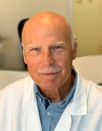
A View From the Top

Wolfgang Grisold

Wolfgang Grisold

This is the second issue of World Neurology this year, and I will take the opportunity to update you on the activities of the WFN. I will comment on the global situation, and then describe the internal developments of the WFN also in regard to future aspects, the persistence of COVID, the important advent of the “Intersectoral Global Action Plan” (IGAP) and World Brain Day 2022, “Brain Health for All.”

Global Situation

Due to ongoing wars, conflicts, and crises worldwide, I would like to emphasize the WFN‘s statements on armed conflict and wars and also encourage donation to professional organizations.

The global situation on wars, conflicts, refugees, displaced persons, and the effects on neurology and neurological patients is severe, and the WFN is deeply concerned. The WFN condemns any conflicts of war. It cannot be stressed enough that our role is to advocate for neurology, patients and caregivers who are endangered in these situations, and the reduction of access to care and treatment. Any armed conflict will also cause new casualties and victims, many of them with neurological sequelae and also subsequent mental conditions.

The role of the WFN as a scientific society, composed of 123 member societies, is building bridges between societies, members, medical disciplines, patients, and caregivers.

The WFN encourages the support to migrants, refugees, displaced and stateless persons, and victims of conflict worldwide. As a charity, we are primarily concerned with people with neurological disorders, their access to care, and the provision of essential drugs. We have indicated organizations that are experienced in global crises, and encourage donations for the purpose to support neurological patients.

Internal Developments

The year 2022 marked the time of a new administration. The strategy is to build on established structures and evolution as well as the integration of new developments.

The WFN is working on the improvement of communication with its member societies, further developing educational tools, such as the e-learning hub and educational days, among others. The experience with the previous WFN e-learning days has been successful, and the format and time of these events has been well accepted. The possibility of short-term educational interventions has been demonstrated in a recent SNO-WFN webinar on scientific advances on neuro-oncology, which was well-attended. We want to thank our partners including AFAN, EAN, AAN, IHS-GPAC, and SNO for their support. We believe these newly added educational concepts of virtual interventions will be important.

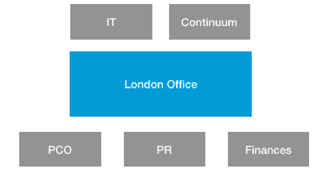
Basic administrative structure of the WFN in London. The office presently has four staff members who administer and coordinate the WFN affairs. External IT aspects and the management of Continuum are conducted by local and experienced collaborators (IT=information technology; PCO=professional conference organizer; PR=public relations)

The WFN has several committees, which act on behalf of the trustees. For the new administrative period, we have decided to adapt some of the committees and also add several subcommittees for specific purposes such as gender and diversity, young neurologists, teaching centers as well as a patient platform, among others. Based on the virtual WFN regional meeting in January-February 2022, we have selected chairs and filled the positions with suggestions from the regions. We have also increased the ratio of females from 10% to 40%, and we are committed to continue to adapt gender balance.

At this point, I also want to thank the outgoing chairs and members of the committees for their devotion and dedication to the WFN. Rotation of positions is necessary in such a large organization, and even if the terms end, we hope that all previous members will continue working with us supporting the WFN or in other future functions. The list of the new committees will be available on the website, and we will continue to introduce committees in World Neurology.

Other current tasks include preparation for the next Council of Delegates (COD) meeting this October 2022 in Amsterdam, where a new Secretary General and and one trustee will be elected. The WFN met with the AAN in a leadership meeting, and this is also planned with the EAN and with AOAN.

The preparation of the WCN 2023 is proceeding, and the WCN program committees have started their work. A preliminary program will appear soon. The preliminary work for WCN 2025 in Korea is also ongoing.

Define the Future

The WFN has reached a critical size and has globally a wide span of activities, which make a permanent and reliable organizing, planning and administrative structure necessary. This includes long-term planning and decisions, and availability with regard to resources both personal and financial.

The WFN, being a U.K. charity, guarantees a strict and well-assessed structure, which is taken care of by the trustees with the support of the WFN staff in London and is regularly audited each year. These administrative tasks, projects, and communicative tasks, are carried out by the office staff and externally with the help of Chiu, Helen Gallagher, our professional conference organizer (PCO), Freedman (our financial consultant), and Yakkety Yak, our public relations consultant, which make the WFN a complex structure.

Congress organization is done by a PCO, and the organization of WBD and Brain Health by a PR expert company.

We appreciate that the interest and trust in the WFN is high and increasing from the standpoint of cooperations. Any project taken up by the WFN needs administration and monitoring, and increasingly, project management tools will have to be used to define capacities to the extent that projects can be supported.

For education, the projects will be based on a needs assessment, which will be the basis of future development of all educational activities. We are also supportive of the concept of CME and Continuous Professional Development, which is lifelong learning for neurologists. There are several worldwide concepts such as the AAN’s Continuum, and the European EACCME model, which offer a widespread and detailed choice of CME educational methods and models. We are happy to say that the WFN congresses (World Congress of Neurology) are always accredited with up to 40 credits from EACCME , and we have always passed the strict and thorough definitions for these meetings. They are valid for AMA and the Canadian Royal College of Physicians.

Education in neurology is needed at all levels and is a continuous process. In addition to practical and scientific content, the importance of advocacy as well as leadership will have to be implemented in the strategy of our educational programs. We are committed to our concepts of department visits and training centers, and will report on these developments in the next issue.

COVID Is Continuing

I want to remind our readers that the COVID pandemic is not over yet, and travel and communication is still at risk. This also relates to the planning of the World Health Assembly (WHA) meeting in Geneva as well as to the COD meeting in Amsterdam.

The WFN has updated the COVID website and the Specialty Group on Tropical Neurology is providing monthly updates. Also Elsevier will provide a collection of COVID papers published in ENS (and in the future, JNS) and can be found on the COVID site: https://www.sciencedirect.com/journal/eneurologicalsci/special-issue/1006JKSX5HX.

The WFN also actively works with the WHO in several working groups on the neurological effects of COVID in both acute and late effects.

The pandemic had and still has catastrophic effects on patients and caregivers, not only as limited access to care and reduced capacities, but also medical and bureaucratic hurdles. It has to be assumed that the indirect damage to acute and chronic neurological patients is high and will take considerable time to return to normal.

As neurologists, we have to take care of the so-called soft facts such as communication, personal interaction, quality of life, and that the needs of patients and caregivers are addressed.

Intersectoral Global Action Plan (IGAP)

Much energy and effort is being invested in the WHO’s International Global Action Plan (IGAP). I want to thank my predecessor William Carroll and Alla Guekht and Kimberly Karlshoej for their continuous efforts. This is a good example of a project that will have a worldwide impact, but is also a good example of worldwide cooperation.

IGAP is about to be accepted at the WHA in Geneva in May 2022. The WFN has been working with other societies such as the ILAE, the World Stroke Organization, Movement Disorders Society, and the International Headache Society on this development.

It is based on a long and fruitful cooperation with the WHO, which has several landmarks and previous books, including two editions of the Atlas and also the development of ICD 11. This IGAP will elevate the importance of neurology worldwide and will enable countries to use this WHO initiative for the establishment or development of neurology.

A brain health unit has been created by the WHO, which indicates the importance of brain health worldwide. (https://www.who.int/health-topics/brain-health#tab=tab_1).

Once the IGAP is accepted, the implementation of the IGAP will need the full attention for new projects with the WHO and with individual societies to implement this exciting program.

World Brain Day Topic

This year‘s World Brain Day (WBD) will be dedicated to “Brain Health for All.”

It is chaired and organized by our Public Awareness Committee by Tissa Wijeratne and David Dodick, with professional assistance from Yakkety Yak, which has taken care of the last WBDs. The committee consists of representatives from the WFN regions, and it is hoped that this WBD will underline the importance of brain health globally. We hope to align with the WHO in this important activity. The intent is not only to reach as many regions as possible, but also customize our WBD tools for individual use and we hope that many, if not all, WFN member societies will be able to celebrate with the WBD in their regions and use this topic to promote neurology at all levels. The topic of this year’s WBD also aligns with the upcoming IGAP, which will be approved by the WHA in May.

The view of the WFN is that brain health has a wide span from intrauterine life toward childhood, adulthood, and into aging in regard to neurological function, dysfunction, rehabilitation, and palliative care.

The selection of the topic “Brain Health for All” is based on the WFN’s 2021 brain health campaign (https://wfneurology.org/brain-health-initiative) and the cooperation with the WHO. Also regional societies including the EAN are committed to brain health, such as the European Brain Health Summit meeting in May 2022 (https://www.ean.org/ean/advocacy/brain-health). There will also be a brain health session at the EAN congress in Vienna.

This was a short update on the current proceedings of the WFN, including several cooperations and developments. Please follow us on the website and social media.

If you have comments or questions, please contact us at info@wfneurology.org. •

Prof. Jun Kimura (1935-2022) Father of Electrodiagnostic Neurology

By Ryuji Kaji

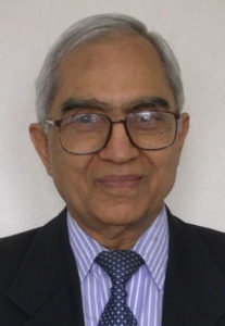
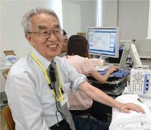
Dr. Jun Kimura was born on Feb. 25, 1935, in Kyoto, Japan, and was brought up in Takayama, a scenic rural place near Kyoto. He once graduated from School of Technology in 1957, but re-entered Medical School of Kyoto University, and obtained MD (1961). He was granted a Fulbright Scholarship (1962-1967), and began his career at the University of Iowa School of Medicine as a medical resident and fellow (1962-1968), associate professor (1972-1977), and professor (1977-1988).

I first met him in my junior neurology residency in 1981, when he was invited to give a lecture on blink reflex and facial palsy at the time of the World Congress of Neurology in Kyoto. I was fascinated by his unique way of presentation, both informative and amusing. After his talk, I tried to talk to him in English, but was surprised to hear his Japanese reply. Having had a few minutes of conversation, I became aware of his kindness, generosity, and humility. He published a famous textbook, “Electrodiagnosis in Diseases of Nerve and Muscle: Principles and Practice,” in 1983, which at once became the standard for neurologists, and determined my career as an electromyographer.

After completing my MD, PhD course in Kyoto, I was fortunate to have a position of clinical fellow at the Hospital of the University of Pennsylvania in 1985, and Dr. Austin Sumner became my first mentor of EMG. Dr. John England, currently the editor of Journal of the Neurological Sciences, was my colleague at Dr. Sumner’s EMG laboratory. We used to bet a box lunch on the diagnosis. At that time (1985 – 1986), Dr. Kimura served as president of American Association of Electrodiagnostic Medicine. In 1987, I received an international call from the dean of my alma mater, the head of the search committee, who asked me to persuade Dr. Kimura to go back to Kyoto University as professor and chair of the department of neurology. First, he declined the offer, since he already had a solid background, reputation, and family in the United States. I had to visit his home in Iowa to ask him to change his mind. After a while, he agreed to go back to Kyoto on the condition that I follow him to help adjust to the Japanese style of life. For possible projects, we discussed starting botulinum toxin injections for neurological disorders with the skill of EMG for the first time in Japan, and Kyoto became a center for botulinum toxin therapy.

His wife, Mrs. Junko Kimura, was kind enough to go back with him, which made my task much easier. In fact, she was helping edit the journal Muscle and Nerve in Kyoto, while Dr. Kimura was the chief editor. Dr. Kimura thus became my second mentor of EMG, and I learned much from him not only on science, but also on career. He was soon elected as the president of the International Federation of Clinical Neurophysiology (IFCN) in 1990.

Despite his busy professional activities, he encouraged us to run research activities in Kyoto: discovery of the use of IVIg for multifocal motor neuropathy (1989, 1991), clinical trials of Japanese botulinum toxins including type F, exploring the pathophysiology of dystonia, and clinical development of ultra-high dose methylcobalamin for ALS, which will be officially approved soon in Japan.

He was humble enough not to put his contribution too much on every work, and was kindly looking after every young fellow, including myself. His mottos included “Care for the patients” and “To see central (nervous system) from peripheral.” He attracted dozens of medical students to become neurologists. In 1991, we were fortunate enough to have Dr. Hiroshi Shibasaki as the professor of clinical neurophysiology, while Dr. Kimura served as the neurology chair, and both groups collaborated successfully, making Kyoto the mecca of clinical neurophysiology then.

During those productive years, we decided to convene the International Congress of EMG and Clinical Neurophysiology (10th ICEMGCN), the official congress of IFCN, 1995, in Kyoto. Because Dr. Kimura had few connections with the industry then, we tried our best to raise funds for the congress. Fortunately, the meeting was very successful with a record number of participants, and the surplus funds were donated to the national funds for neuropsychiatric diseases.

After he was elected as the first vice president of the WFN in 1998, he asked the national fund to further donate the money to help publish World Neurology. On his retirement in 1998 in Kyoto, he returned to Iowa, but was often seen in Kyoto with his wife and disciples in later years.

When he was inaugurated as president of the WFN in 2002, he made a number of reforms to the federation, such as constitution and bylaws, finance, and membership of the WFN. During and after his presidency, he was keen about education of young neurologists in Asian, Arab, and Latin American countries, and traveled virtually all around the globe many times. Wherever he went for EMG hands-on, he packed the room with young neurologists. His educational activities went on tirelessly for the rest of his life. When he was asked to visit some developing countries, he was kind enough to travel in economy class using his mileage of the airline company.

I was appointed as the chair of the neurology department in Tokushima in 2000, and thanks to his promotion, I took an executive position (member-at-large) of IFCN during 2001-2006. At the end of my term, Dr. Kimura, Dr. Andrew Eisen (treasurer of IFCN), and I discussed the need for new electrodiagnostic criteria for ALS, which make it possible to start any test drug for ALS as early as possible in clinical trials, since we knew that the number of spinal motoneurons are decreased below 30% of normal at the time of diagnosis using El Escorial criteria. Thus, we held a consensus meeting in Awaji Island near Tokushima to set the currently known “Awaji Criteria.” A pivotal phase 3 trial of ultra-high dose methylcobalamin (JETALS), funded by the Japanese government, used these criteria for entry, and was successful in detecting ALS at the early stage.

During 2007-2013, I served as trustee of the WFN for two terms. For the second term, I was appointed as the chair of the Asia Initiative by Dr. Vladimir Hachinski, then-president of WFN, to revitalize neurology in Asia, which is the most densely populated region in the world. The mission was tough, but Dr. Kimura’s seeds in young Asian neurologists became the fruitful core of many Asian neurological societies.

At the WCN 2016, the second congress in Kyoto, I was elected as first vice president of the WFN and worked mainly on organizing the congress. At the end of my term in 2021, Dr. Kimura was helping me to run for the president with all his might. After the results were announced, he seemed to be extremely exhausted. He passed away unexpectedly in his sleep on March 3, 2022. My last e-mail to him was to celebrate his 87th birthday on Feb. 25. He replied with a message that he was writing the new version of his textbook and looking forward to coming back to Kyoto in April.

He is survived by his wife Junko and three sons: Ken, Ray, and Joe and his wife Lori, and his grandchildren.

His last but the most important motto was “use EMG like a hammer,” and I believe he is still enjoying teaching EMG in heaven. •

Ryuji Kaji, former vice president of WFN, is a Distinguished Professor of Neurology at Tokushima University. He is also the director of Utano National Hospital in Kyoto.


From the editors

Steven L. Lewis, MD, Walter Struhal, MD

We’d like to welcome all readers to the March-April 2022 issue of World Neurology.

The issue begins with the obituary of Dr. Jun Kimura (1935-2022), former WFN president and renowned electrodiagnostic neurologist, written by his colleague and former mentee, Ryuji Kaji.

In the President’s Column, WFN President Dr. Wolfgang Grisold discusses a number of items, including implications of the current global situation, internal developments and plans at the WFN, an update on COVID with regard to the WFN, the International Global Action Plan (IGAP) of the WHO, and World Brain Day 2022, devoted to Brain Health for All.

Dr. Chandrashekhar Meshram announces the important news that the Padma Shri Award, the highest civilian honor of India and conferred by the president of India in New Delhi, has been awarded to Dr. Bhimsen Singhal.

Dr. Meshram also summarizes Dr. Singhal’s remarkable accomplishments.

Drs. Marina Alpaidze, Tsotne Samadashvili, and Alex Razumovsky discuss the successful teaching course sponsored by the Neurosonology Specialty Group of the WFN held in Tbilisi, Georgia, in late 2021, which discussed the use of transcranial Doppler ultrasonography as an essential daily modality in the critical-care setting.

In this issue’s History Column, Dr. Peter Koehler discusses the early endeavors to build a CT scan, with particular reference to the Ukraine pioneers who were integral in this development.

This issue also includes a number of important announcements, including World Brain Day 2022 devoted to Brain Health for All, an announcement for the WFN’s call for applications for 2022 Grants, and an announcement of the upcoming 17th International Congress of Neuromuscular Disease, which will take place in Brussels in early July 2022.

This issue also updates us about a recent important position statement from the American Epilepsy Society about the serious risks associated with use of valproate by women of childbearing potential.

Finally, this issue also includes an obituary of Dr. Paul Kleihues, a world-renowned neuropathologist in the field of brain tumor research who was integral in the WHO classification of human tumors.

We thank all readers for their interest in World Neurology and invite you to submit ideas for contributions. Please send your ideas to stevenlewi@gmail.com. •


World Brain Day 2022; Brain Health For All

By Prof. Tissa Wijeratne and Prof. David Dodick

World Brain Day (WBD) 2022 is dedicated to the theme “Brain Health for All”  as our brains continue to be challenged by pandemics, wars, climate change, massive disparities in health equity and myriad preventable diseases.

The brain consists of about 100 billion neurons and up to five times as many glial cells. Each neuron connects to about 10,000 other neurons by way of trillions of synapses. The length of these connections end-to-end would wrap around the earth four times. Connections between these neurons help us read, write, watch, learn, plan, think, feel, move, love, and solve problems daily.

The 2022 WBD “Brain Health for All” campaign will focus on five key messages globally:

  1. Awareness: Brain health is vital for mental, social, and physical well-being.
  2. Prevention: Many brain diseases are preventable.
  3. Advocacy: Global efforts are required for optimal brain health.
  4. Education: Education is key to brain health for all.
  5. Access: Equitable access to resources, treatment, and rehabilitation are essential for brain health.

We invite neurologists, neuroscientists, health professionals, trainees, technologists, advocacy organizations, patient support groups, professional societies, and other activists to become part of the 2022 World Brain Day campaign. You can participate by posting our banner advertisement on your webpage and sharing our posts, messages, and videos with your friends and colleagues. You can partner with local neurology societies and organizations to promote these activities. Please encourage people with any neurological disorder and their caregivers to contribute by sharing their story via a short video message (less than 90 seconds) and emailing the video to wbd2022@wfneurology.org. You can visit our website and use the freely available educational resources to lead a successful World Brain Day campaign at your institution or organization this year.

On behalf of the WFN, we, the co-chairs of the WFN’s WBD campaign, look forward to virtually visiting your association, society, and regional group to work with you to extend the reach and impact of WBD 2022 to every household in your region and country.

Let us work together to optimize brain health for a better world and brighter future. •

Prof. Tissa Wijeratne and Prof. David Dodick are co-chairs of World Brain Day, World Federation of Neurology.


Padma Shri for Dr. Bhimsen Singhal

By Chandrashekhar Meshram

Chandrashekhar Meshram

The Padma Awards are the highest civilian honors of India announced annually on the eve of Republic Day. They are conferred by the president of India at an investiture ceremony in New Delhi. The award recognizes public service and significant achievements across a variety of disciplines such as art, engineering, medicine, social work, literature, education, sports, and others.

This year, Dr. Bhimsen Singhal has been selected for the Padma Shri award. Dr. Singhal is one of the most respected and eminent neurologists who has contributed significantly to the growth of neurology in India over the last 60 years. Born in Mount Abu in 1933, he received his training in neurology under Prof. Noshir Wadia at Grant Medical College in Mumbai. He is currently director of neurosciences at the Bombay Hospital Institute of Medical Sciences and previously served as professor and head of the department of neurology at Grant Medical College and Sir. J. J. Group of Hospitals in Mumbai. He has trained over 200 neurologists who provide care in urban and rural areas across India.

Dr. Bhimsen Singha

Dr. Singhal is renowned for his clinical skills and compassionate care. He has conducted clinical research, authoring several book chapters and scientific articles. He is recognized for his special expertise in multiple sclerosis, myasthenia gravis, and Parkinson’s disease. He described the entity of megalencephalic leukodystrophy, especially prevalent in the Agarwal community and with international collaboration, identified its gene defect.  He has been associated with the WHO Working Groups for Parkinson’s Disease and Multiple Sclerosis. Dr. Singhal has served as regional director of the World Federation of Neurology (WFN) and has been on its various committees. He continues to be active, seeing patients daily, attending conferences, and spending much of his time on philanthropic activities.

Dr. Singhal has received several prestigious national awards, such as the Dr. BC Roy National Award, the RD Birla National Award, the Priyadarshini Academy Award, the Wockhardt Award for Medical Excellence, and the Dhanvantari Award. He has been honored with named lectureships at the WFN and the Indian Academy of Neurology conferences. The Padma Shri award is particularly special. It recognizes his clinical and academic contributions as well as public service through his non-profit national foundations, highlighted in this article.

In 1998, Dr. Singhal established the Neurology Foundation with its mission of “knowledge and care.” Assistance is provided to patients with neurological disorders through its various programs like Sahay (financial assistance for medical treatment), Upchar (rehabilitation therapies for children and adults from low socio-economic areas), Aanchal (community outreach) and Parvarish (support for professional organizations such as the Multiple Sclerosis Society of India and the Indian Epilepsy Association).  Since 1998, its highly popular biennial Neurology Update conferences have been attracting neurologists and physicians from all over India. World-renowned neurologists are specially invited to these meetings to interact and update the knowledge of the audience.

President of India conferring Padma Shri on Dr. B.S. Singhal.

In 2001, Dr. Singhal also founded the Parkinson’s Disease and Movement Disorder Society (PDMDS)—a nonprofit charitable organization supporting and rehabilitating persons with Parkinson’s disease and their caregivers. Over the years, the PDMDS has impacted the lives of thousands of persons with Parkinson’s disease. They regularly attend free sessions in 65 “support centers” spread across India. The majority of them continue to receive support throughout their lives. This PDMDS model has been acclaimed internationally as an effective model of care for developing countries.

In all these activities, Dr. Singhal has been well-supported by his family, colleagues, and grateful patients. His wife, Dr. Asha Singhal, an obstetrician and gynecologist, has devoted her life to the care of underprivileged people. His son, Dr. Aneesh Singhal, is a stroke neurologist at the Massachusetts General Hospital in Boston, and his daughter Dr. Seema Singhal is a hemato-oncologist (multiple myeloma) at Northwestern University Hospital in Chicago.

Dr. Singhal richly deserves this Padma Shri award. It will undoubtedly inspire future generations of neurologists and promote the growth of neurology in India and other parts of the world. •

Chandrashekhar Meshram is a co-opted trustee of the WFN.

Early Endeavors to Build a CT Scan: The Ukraine Pioneers

by Peter J. Koehler

Some years ago, I presented a lecture on the history of CT and MR at the History of Neurology course of the American Academy of Neurology (2018). Soon after the invention of X-rays by Wilhelm Conrad Röntgen (1845-1923) in 1895, two steps needed to be made to improve brain imaging, notably the introduction of contrast enhancement and the production of 3D images.

The first step was taken by the use of air contrast in the ventricles of the brain, inserted directly (1918) or by lumbar puncture (1919). Arterial contrast followed in the 1927. The technique was called arterial encephalography as it was the brain rather than the arteries, in which physicians were interested at the time.

The second problem was, at least temporarily, solved by the introduction of planigraphy, better known as tomography, which was introduced in the early 1930s1. For many decades, a combination of air contrast X-rays and analogous tomography with small amounts of air was applied (PEG or pneumoencephalography). The procedure was quite uncomfortable for the patients. After the introduction of the air directly in the ventricle or by lumbar punction, the patients were rotated in a somersault chair to bring the air into the place of interest in the head. I remember having seen such a patient returning to the ward suffering from severe headache and vomiting. A short film clip of the procedure can be found here: ((46) The Scanner Story (Part 1 of 2 of documentary covering early CT development) – YouTube and go to the section 3.04-4.00 minutes).

Mathematics: The Inverse Radon Transform

Johann Radon (ca. 1920; Wikimedia Commons, public domain).Johann Radon (ca. 1920; Wikimedia Commons, public domain).

In the late 1950s and early 1960s, the first attempts to look for a more patient-friendly method were conducted. Interestingly, the mathematical method for the reconstruction of an object from multiple projections had already been discovered by the mathematician Johann Radon (1887-1956) in 1917. Born in Tetschen, Bohemia (now Děčín in the Czech Republic), he studied at the University of Vienna. Due to his near-sightedness, he was exempt from the draft. He was active as a professor of mathematics at several universities in central Europe. His 1917 publication did not have immediate practical applications and was not used by subsequent CT pioneers.

Pioneers in Kiev: Rediscovering the Inverse Radon Transform

What is less well-known is that in the late 1950s, Ukraine pioneers were working on a reconstruction project. Doing research at the Igor Sikorsky Kyiv Polytechnic Institute (KPI) in Kiev and named after the Kiev born aircraft designer Igor Sikorsky (1889-1972), two men tried to make the experimental arrangement for reconstruction. Semyon Isaakovich Tetelbaum (1910-1958) studied at the KPI, after which he became electrical design engineer working on projects involving radio and television. In 1940, he became professor at the KPI. Boris Isakovich Korenblum (1923-2011) was born in Odessa, a city in the south of Ukraine, on the Black Sea, and changed careers from the violin to one in mathematics after winning a mathematics competition. He studied mathematics in Kiev. Being of Jewish descent, he escaped from being killed by the Nazis in Babi Jar. Losing his job during Stalin’s anti-Semitic campaign of 1952-1953, he got a position at the Construction Engineering Institute in Kiev, where he stayed until his emigration to Israel and later to the United States.

In 1956, Tetelbaum published “About the Problem of Improvement of Images Obtained With the Help of Optical and Analog Instruments” in Bulletin of the Kiev Polytechnic Institute2. The subsequent year another article by him followed in the same periodical, notably “About a Method of Obtaining Volumetric Images by Means of X-ray Radiation.3” (See Figure 1.) In 1958, Korenblum, Tetelbaum, and Tyutin published an article titled “About a Scheme of Tomography” in the Proceedings of Higher Educational Institutions – Radiophysics4.

Semyon I. Tetelbaum (left) and Boris I. Korenblum.

The articles remained unnoticed in the Western world until Harrison H. Barrett (Department of Radiology and Optical Sciences Center), William G. Hawkins (Department of Mathematics), and Michael L.G. Joy (Institute of Biomedical Engineering) of the University of Arizona published a “Historical Note on Computed Tomography” in 1983. The authors wrote that Tetelbaum “formulated the tomography problem in terms of line integrals and found the exact solution, which is now referred to as the inverse Radon transform.” Korenblum, Tetelbaum, and Tyutin “gave a detailed account of the theory, including a method of fan-beam correction, equivalent to reordering the data, and a discussion of a practical way of handling the singularity in the integrand of the inverse Radon transform.” Furthermore, in their paper, the Ukraine scientists “presented a block diagram of television-based analog computing system for implementing the reconstruction” and estimated the system should be capable of “reconstructing a 100 x 100 image in five minutes.” (See Figure 2.)

One of the authors of the historical note (Hawkins) programmed the reconstruction algorithm and concluded that it performed satisfactorily, although “because it was not computationally efficient, only a 32 x 32 image was reconstructed.”5 As the papers were published in the Russian language, it was difficult to access the contents. More recently, two of the three articles have been translated by Alex Gustschin, chair of biomedical physics in the Department of Physics and Munich School of Bioengineering, Technical University of Munich.6,7 He also provided comments and biographical data on Tetelbaum and Korenblum, but was unable to find information about Tyutin.

So why didn’t Korenblum and his colleagues build the machine in 1958? In 1983, Barrett and colleagues wrote that they “continue to search unsuccessfully for later references to the reconstructed images.” Gustschin suggested that the unexpected death at age 48 of Tetelbaum in 1958 may have caused the end of the project.

Second Rediscovery of the Radon Transform

Figure 1. From Tetelbaum’s 1957 publication.3

Unaware of the publications of Radon or the Ukraine scientists, William Henry Oldendorf (1925-1992), a neurologist working at UCLA, built a prototype to transmit a beam of X-rays through the head and reconstruct its image in 1959-1960. Although the method was patented, he was unable to get it funded.

Figure 2. Functional block diagram of the television-based computing device from Korenblum et al. 1958.4

Working in Cape Town, South Africa, approximately in the same period, and later moving to Tufts University, Allan McLeod Cormack (1924-1998) formulated mathematics of reconstruction for simple attenuating objects with certain symmetry and applied a reconstruction technique (1957) with some success. He constructed an experimental scanner (1963) and only in 1970 he became aware of the solution found by Johann Radon in 1917. The story of Godfrey Newbold Hounsfield (1919-2004) is probably well-known. Working at EMI in London since 1949, he led a design team for building the first all-transistor computer (1958) to be constructed in Britain. Then he did early tests with a laboratory CT using a gamma ray source and single detector.

Figure 3. First clinical CT-scan (October 1971).

In October 1971, the first clinical scan was made. High-resolution images of a 41-year-old woman with a frontal tumor were produced in cooperation with neuroradiologist James Ambrose (1923-2006) at Atkinson Morley Hospital. (See Figure 3.) Cormack and Hounsfield received the Nobel Award for Medicine or Physiology in 1979. A 15-minute documentary on Hounsfield and Ambrose can be found at (46) The Scanner Story (Part 1 of 2 of documentary covering early CT development) – YouTube. •


References

  1. Lutters B, Koehler PJ. Cerebral pneumography and the 20th century localization of brain tumours. Brain. 2018 Mar 1;141(3):927-933.
  2. Tetelbaum SI. About the problem of improvement of images obtained with the help of optical and analog instruments” Bulletin of the Kiev Polytechnic Institute 1956;21:222.
  3. Tetelbaum SI. About a method of obtaining volumetric images by means of X-ray radiation. Bulletin of the Kiev Polytechnic Institute 1957:22:154-60.
  4. Korenblum BI, Tetelbaum SI, Tyutin AA. About a scheme of tomography Proceedings of Higher Educational Institutions – Radiophysics 1958;1:151-7.
  5. Barrett HH, Hawkins WG, Joy ML. Historical note on computed tomography. Radiology. 1983 Apr;147(1):172
  6. Gustschin A. Translation of Tetelbaum SI: About a Method of Obtaining Volumetric Images by Means of X-ray Radiation. arXiv:2001.03806.
  7. Gustschin A. Translation of Korenblum et al. About a scheme of tomography. arXiv:2004.03750.
  8. Webb S. From the Watching of, 1990. Shadows: The Origins of Radiological Tomography Bristol & New York. Hilger


Announcing ICNMD 2022

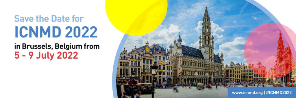
The 17th International Congress of Neuromuscular Disease will take place July 5-July 9 in Brussels.

On Tuesday, we will have eight teaching courses and two hands-on courses (four hours each with a break in between).

The TC02 (TC02: Diagnosis and Treatment of GBS, CIDP and Autoimmune Nodopathies) & TC06 (Diagnosis of Inherited Neuropathies) are endorsed by the ICNMD/PNS Society

From Wednesday, the program includes a daily morning plenary followed by concurrent scientific sessions (22), workshops (12) and overarching sessions (11). Included are nine sessions for oral presentations and a special session for Regional Aspects of Neuromuscular Diseases – a joint WFN-ICNMD session.

Here are the titles of the four plenary sessions: 

Wednesday, July 6 | Trial Readiness and Advances in Therapy in Hereditary and Inflammatory Myopathies

Thursday, July 7 | The ALS-FTD Overlap Syndrome

Friday, July 8 | Guillain-Barré Syndrome: State of the Art and Challenges (Endorsed by ICNMD/PNS Society)

Saturday, July 9 | New Developments in Diagnosis and Treatment of Myasthenia Gravis and Congenital Myasthenic Syndrome

Here is the link to the preliminary program: https://icnmd.org/program2022/

For more information, contact Barbara Bricalli, program manager of ICNMD 2022 via www.icnmd.org. •


Neurosonology Specialty Group WFN Teaching Course

Meeting discussed use of transcranial Doppler ultrasonography as an essential daily modality in the critical-care setting.

By Marina Alpaidze, MD, PhD, Tsotne Samadashvili, MD, PhD, and Alex Razumovsky. PhD, FAHA, NVS

Conference room at Caucasus Medical Center.

The Neurosonology Specialty Group of the WFN is dedicated to the promotion of science and research as well of education and training in the field of ultrasonic techniques and its clinical utilization. Therefore, international cooperation and the dissemination of scientific information within the field of neurosonology is part of the WFN Neurosonology Specialty Group activities.

The WFN Neurosonology Specialty Group course considering clinical applications of transcranial Doppler (TCD) utilization in neurocritical care and neurosurgery was held Nov. 5, 2021, at the Caucasus Medical Centre in Tbilisi, Georgia. The meeting was conducted under the auspices of the NSG WFN, Georgian School of Anesthesia and Intensive Therapy, and Georgian Chapter of NSG WFN, and the Georgian Society of Cerebral Hemodynamics and Neurosonology.

Razumovsky (top left), L.Tsikarishvili. (bottom left), T. Samadashvili (top right), M. Alpaidze (bottom right).

Among the faculty were Drs. T. Samadashvili, MD, PhD, president of the Georgian School of Anesthesia and Intensive Therapy (NGO) and chair of the anesthesiology department at Caucasus Medical Centre; Lado Tsikarishvili, MD, PhD, chair of the neurosurgery department at Caucasus Medical Centre; M. Alpaidze, MD, PhD, professor in the department of radiology at the Tbilisi State Medical University; and Alexander Razumovsky, PhD, FAHA, NVS, secretary of the WFN Neurosonology Specialty Group.

(From left to the right) L. Tsikarishvili (neurosurgeon), T. Samadashvili (anesthesiologist), M. Alpaidze (radiologist), and A. Razumovsky (neurophysiologist).

This course was designed for different medical specialists, including neurologists, neurocritical care physicians, and neurosurgeons.  Presentations were related to the clinical yield of TCD for neurocritical care applications, specifically for patients after subarachnoid hemorrhage and traumatic brain injury. Different aspects of neurosurgical, critical care, and neurosonology were discussed in detail. Among them were management strategies of cerebral vasospasm after SAH and TBI. Some new trends in the clinical utilization of neurosonology applications were debated. Due to the COVID-19 regulations in Georgia, the meeting offered simultaneous live broadcast for a wide-ranging audience. •

Marina Alpaidze, MD, PhD, is professor in the radiology department at Tbilisi State Medical University. Tsotne Samadashvili, MD, PhD, is chair of the anesthesiology department at Caucasus Medical Center. Alex Razumovsky. PhD, FAHA, NVS, is secretary of the Neurosonology Specialty Group of the WFN.


American Epilepsy Society Announces Position Statement on Valproate Use in Women of Childbearing Potential

R. Edward Hogan, MD, David G. Vossler, MD

The American Epilepsy Society (AES) president, R. Edward Hogan, MD, recently shared important information about the serious risks associated with use of valproate (valproic acid, divalproex) by women of childbearing potential, as highlighted in the updated AES Position Statement on the Use of Valproate by Women of Childbearing Potential.

Valproate is prescribed across a diverse range of specialties and is approved by the U.S. Food and Drug Administration (FDA) for treatment of epilepsy and bipolar disorder and for migraine headache prophylaxis.

The updated AES position statement highlights specific warnings related to significant risk of fetal teratogenicity and risk of decreased IQ and other neurodevelopmental disorders, including autism, in children exposed to valproate in utero, and a contraindication to its use for migraine prevention in pregnant women and women of childbearing potential not using effective contraception.

The statement cites evidence that increased awareness of these important considerations for women of childbearing potential correlates to reduced fetal exposure and risks. Based on this evidence and the epilepsy care experience, AES encourages provider conversations with patients to mitigate these risks, reduce in utero valproate exposure, and promote safe and effective treatment for women of childbearing potential.

AES Council on Clinical Activities Chair David G. Vossler, MD, cited this AES position statement as an important extension of the ongoing work of the Council’s Treatments Committee to monitor and inform AES members about important Drug News and FDA Alerts related to care of people with epilepsy. •


Dr. Paul Kleihues (May 21,1936-March 17, 2022)

Dr. Paul Kleihues passed away March 17, 2022, at the age of 85 years.

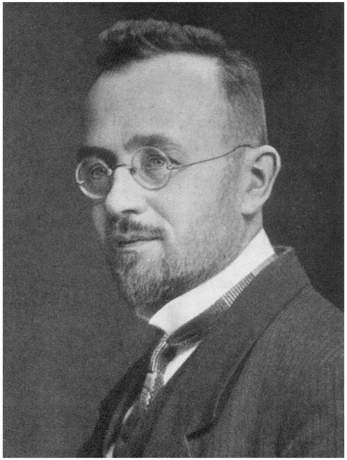
Dr. Kleihues was a world-renowned neuropathologist in the field of brain tumor research. Starting his scientific and professional career at neuroscience and neuropathology institutions in Germany, he later served as director of the Institute of Neuropathology at the university hospital in Zurich, and subsequently as head of the International Agency for Research on Cancer (IARC), World Health Organization (WHO). He took responsibility for the WHO classification of human tumors, and was particularly interested in the integration of clinics, radiology, histology, and molecular genetics as fundamental basis for comprehensive and precise tumor typing. His personal professional and scientific activities, in tight cooperation with Dr. Hiroko Ohgaki, left a sustainable legacy in brain tumor classification.

Within the international neuropathology community, Dr. Kleihues was a highly appreciated and benevolent mentor, colleague, and friend.

The IARC commemorates Dr. Kleihues and his achievements in a respectful and compassionate obituary: https://www.iarc.who.int/news-events/dr-paul-kleihues-21-may-1936-17-march-2022/ •

—Dr. Johannes Hainfellner

Division of Neuropathology and Neurochemistry (Obersteiner Institute) Department of Neurology Medical University of Vienna, Austria