WFN Reinforces Its Commitment to Advancing Global Neurology Education

Report of a press conference held at the World Congress of Neurology, Montreal, October 2023

On behalf of the WFN trustees, assisted by the WFN press support organization, Yakkety Yak.*

Key teaching initiatives bridge gaps in neurological care to promote brain health worldwide.

Neurological disorders are currently the second highest cause of death and the leading cause of disability worldwide. The new Global Burden of Disease (GBD) study shows that the number of people living with brain disease is expected to double by 2050.

One of the central challenges of addressing the growing burden of neurological diseases is the lack of available neurology care in under-resourced parts of the world. The World Federation of Neurology (WFN) is committed to finding new and innovative ways to educate and train an increasingly critical global neurology workforce to close this gap.

The World Health Organization (WHO) recommends one to five neurologists per every 100,000 people, but many low- and middle-income countries fall well below this number. In fact, the contrast in available neurologists between low-income and high-income countries is so stark that some of the lowest-income nations have 70 times fewer neurologists than countries like the U.S. and Canada.

“It’s impossible to develop appropriate care without education. It’s impossible to promote research without education,” said Dr. Alla Guekht, elected trustee of the World Federation of Neurology.

WFN will continue working with its regional neurological associations and member states to increase the availability of neurological care worldwide.

WFN Training Centers

WFN furthers its mission of fostering quality neurology and brain health worldwide by promoting global neurological education and training, with an emphasis on under-resourced parts of the world. WFN celebrates 10 years of WFN Training Centers, which facilitate training and knowledge exchange as well as visiting fellowships in global regions that have severe shortages of neurologists. WFN currently has training centers in Africa (Dakar, Senegal; Cairo, Egypt; Rabat, Morocco; and Cape Town, South Africa) and Mexico.

“The idea is not to go there and teach, but to empower regions to train their own neurologists,” said Dr. Wolfgang Grisold, president of the World Federation of Neurology. “This has been extremely productive, and while it does not fill the gap that is needed for neurologists in these and other regions, it does create important incentives.”

Future education efforts need to extend beyond neurology specialists to include training in neurological disorders and brain health at all levels of the health care system.

“Increasing the number of neurologists alone is not enough,” said Dr. Augustina Charway-Felli, president of the African Academy of Neurology. “We need to increase neurological awareness across health care providers of all levels — primary health care providers, general practitioners, specialists that are not neurologists and allied health care professionals.”

Global Partnerships and Visiting Neurologist Programs

WFN facilitates programs where residents or young neurologists visit hospitals in other countries for four to six weeks to gain exposure to different health systems and bring back their knowledge and experience to their home countries.

These initiatives are enabled by partnerships between the WFN and national neurological societies. They aim to enhance the exposure of young neurologists to their respective national neurological frameworks within a global context. This offers them the opportunity to connect with new peers and promote future collaborations among countries, universities, hospitals, and departments.

“We are very grateful for the many countries that receive either residents or young neurologists for short department visits,” said Dr. Grisold. “This has been very effective because people get exposed to a different health system for four to six weeks and come back with new ideas they can implement in their own departments and health care systems.”

Increasing Virtual Training

The COVID-19 pandemic required many health organizations to adapt to virtual modes of communication to expand access to training and education previously limited to in-person gatherings. WFN plans to continue utilizing virtual platforms to make critical neurology training and education available on a global scale.

“Increased virtual meetings empower us to be more relevant and efficient in all the different parts of the world,” said Dr. Guy Rouleau, president of the XXVI World Congress of Neurology. “Virtual education reduces costs for everyone —  both participants and the ones who generate the teaching — and we are also better able to tailor education and training to local needs.”

Ongoing education efforts must not only be more widely accessible to the global neurology community but also tailored to the specific needs of each country’s health systems and population.

WHO’s Intersectoral Global Action Plan (IGAP)

WHO’s Intersectoral Global Action Plan (IGAP) on epilepsy and other neurological disorders is a comprehensive framework aimed at reducing the global burden of neurological diseases and bolstering health care systems worldwide with the necessary resources and expertise. One of its goals is to increase training in neurological issues for the primary health care workforce and caregivers.

Dr. Tarun Dua, head of the World Health Organization’s Brain Health Unit, emphasized the need for a multifaceted approach to this complex issue beyond simply increasing the number of neurologists.

“The way we are building up our neurological workforce with the current education programs — even if we double the capacity, we are not going to bridge that gap in the nine years needed to meet the goals of the action plan,” said Dr. Dua. “We need to have a paradigm shift in our thinking.”

This paradigm shift must include not just new and expanded ways of training neurologists, but also reframing how we think of brain health as a human rights and global public health issue.

Spreading Education Through the World Congress of Neurology

The 26th World Congress of Neurology (WCN 2023) was held Oct. 15-19 in Montreal, Canada. WCN 2023 was the first in-person conference since the COVID-19 pandemic and brought together over 2,300 face-to-face and 1,300 virtual participants from 132 countries, including neurologists and advocates from the six global regions represented by our 124 WFN Member Societies. WFN also welcomed a new member society from the Republic of Chad.

“The resounding success of this year’s World Congress of Neurology is a testament to the unwavering dedication of the global neurology community,” said Dr. Grisold. “We’ve not only rejuvenated our connections but also extended our reach, embracing a new member society from the Republic of Chad. Together, we’ve paved the way for a brighter future in neurology.”

At WCN 2023, Dr. Steven L. Lewis, WFN secretary general and Congress committee chair, announced the locations for the next two biennial conferences:

  • 27th World Congress of Neurology, 2025, Seoul, South Korea
  • 28th World Congress of Neurology, 2027, Cape Town, South Africa

From 2024 onward, WFN will establish educational interim meetings called “WFN Digital Neurological Updates (WNU),” devoted to updates in neurology. These virtual meetings will be held in years between congresses and are intended to fill the gap between the bi-annual conferences.

“We will continue to find fresh ways to present the newest discoveries and the latest clinical information to attendees from around the globe,” said Dr. Lewis. “Our goal is to create the most accessible platform so that neurologists and other health care providers can bring this information back to their regions, to their hospitals and to their individual patients, to benefit from the latest developments in the field of neurology and to provide the best neurologic care to their patients wherever they may be.”

Looking toward 2024 and beyond, WFN is excited to find new and even more effective ways to support training for the global neurology community, foster regional exchange of knowledge and education, and prioritize brain health as a human right and a global public health issue. •


* This article was composed by our press consulting agency Yakkety Yak, Inc., on behalf of the WFN. It is based on interviews and communications and contains no industry influence or bias.

From the Editors

We’d like to welcome all readers to the December issue of World Neurology, the last issue of 2023.

The issue begins with the President’s Column, where WFN President Dr. Grisold provides an overview and recap of the many activities of the World Congress of Neurology (WCN), which recently occurred in Montreal, including the Council of Delegates meeting. Dr. Grisold also updates us on other ongoing initiatives and global activities of the WFN. In an accompanying article, the many WFN activities that were highlighted at the WCN press conference are summarized. Later in the issue, a photo montage from the WCN shows a sampling of the activities from this remarkable and highly attended event.

Three articles in this issue discuss essential medicines for neurological disorders (issues that the WFN is highly involved in), including a global overview of this important issue by Drs P. Brhlikova, R. Walker, N. Fothergill–Misbah, and AM Pollock, a report by Dr. Grisold and Dr. Pochigaeva on the World Health Organization (WHO) meeting on improving access to neurological medicines, and an update on the recently approved disease-modifying therapies (DMTs) for multiple sclerosis (MS) on the WHO essential medicines list, by Drs. B. Yamout, T. Kalincik, J. Laurson-Doube, B. Hemmer, and S. Viswanatha.

In another article, Dr. Prince Kazadi, Director of Aslek Epilepsy in the Democratic Republic of Congo (DRC) along with his collaborators, Drs. M. Lubendo, D. Tshiyuk, and N. Kimponto, discuss the activities of their organization with regard to research, staff training, community education, and patient care for patients with epilepsy.

In this month’s history column, Dr. Peter J. Koehler describes the history and theories of speculations about the relation between the function of the brain and its movements. In a second historically based article in the issue, Drs. Domenico Inzitari and Vladimir Hachinski provide their analysis of the possible role of Tourette’s syndrome in a famous figure in Spanish history.

Several regional meetings are also reported in this issue, including the report by Dr. Alex Razumovsky from the WFN’s Neurosonology Specialty Group and his colleagues Dr. K. Potapova, L. Sokolova, D. Andreichenko, and S. Dudukina, who report on workshops in Ukraine that discussed clinical use of transcranial Doppler ultrasonography for patients with various disorders, including its application for wartime traumatic brain injury. Drs. Amir Molaie and David Liebeskind report on the 2023 International Symposium on Thrombolysis/Thrombectomy and Acute Stroke Therapy and the Symposium on Collaterals on the Brain that occurred at the University of California, Los Angeles, and was attended by global experts.

Drs. Satish Khadilkar and Gagandeep Singh announce the recent publication of the second edition of the Indian Academy of Neurology (IAN) Textbook of Neurology, a highly comprehensive and up-to-date two-volume textbook relevant to neurologists worldwide.

Finally, this issue features a heartfelt obituary for Prof. Hany Aref, a remarkable neurologic leader and a wonderful man. Although he is so greatly missed by those many of us who were fortunate enough to know him and to learn from him, his legacy as a leader in neurology and in stroke care in Egypt and worldwide will be long-lasting.

In closing, in this last issue of 2023, we want to wish everyone a wonderful new year and look forward to sharing additional information with you about the WFN and neurology and neurologists around the world in our future issues of World Neurology. •

IAN Textbook of Neurology

The book encompasses both the theoretical and practical aspects of neurology.

by Satish V. Khadilkar and Gagandeep Singh

The “IAN Textbook of Neurology,” edited by Satish V. Khadilkar and Gagandeep Singh, is a comprehensive two-volume sourcebook for students and postgraduates in neurology and practicing neurologists. The book has been compiled by an experienced team of authors from various parts of India and international contributors. The book is well structured into various sections, including history and examination methods, neuroanatomy and neurophysiology, the clinical and diagnostic approaches to various nervous system diseases, and therapeutic aspects.

This book is unique in terms of encompassing both the theoretical and practical aspects of neurology. The detailed description of neurological examination methods along with applied aspects of it will be extremely useful to students. The inclusion of case examples in appropriate places makes it an interesting read. The online version also provides students with multiple-choice questions for self-assessment and illustrative case videos for better understanding.

The second edition adds one more distinctive feature of providing a regional context in various neurological disorders where such peculiarities exist. This is a thoughtful attempt to provide insights into unique demographics, epidemiology, and disease behaviors. This should also motivate the researchers in the country. This area should be worked upon further in the next edition to combine the global and Indian perspectives. The data from a country with such a large population will add value to the world literature. In recent times, the fields of neurogenetics and immunology have been rapidly evolving. Although this book provides primers of both these fields, the coming editions will certainly see more incorporation of these sciences and their impact on the neurology practice.

The book features a foreword by
Dr. Wolfgang Grisold, president of the World Federation of Neurology, and is available on various platforms globally. •

History and the Tourette Syndrome: The Case of Conde Duque de Olivares

The neuropsychiatric syndrome that may have affected the Conde Duque de Olivares, who in the early seventeenth century had almost absolute power in deciding internal or foreign affairs of the Spanish Kingdom.

Gaspar de Guzman, Count-Duke Olivares *oil on canvas *67 x 54,5 cm *circa 1638

BY DOMENICO INZITARI, MD, AND VLADIMIR HACHINSKI, MD, DSC

Early in the seventeenth century, France and Spain vied for predominance, led by Cardinal Richelieu (Armand Jean du Plessis de Richelieu,1585-1642) and the Conde Duque de Olivares (Gaspar de Guzmán y Pimentel, 1587-1645) respectively. Gregorio Marañon y Posadillo (1887-1960), founder of Spanish endocrinology, documented in a now classic biography manic depressive swings in the mood of Olivares. Temper and behavior disorders were also reported by other contemporary or modern biographists. We decoded, translated, and examined a secret report of a Medici family’s (Grand Dukes of Tuscany, Italy, at that time) representative in the Spanish Court, extant as original manuscript in the State Archives of the city of Florence, Italy. There, it affirmed that he had an unhealthy mind, and reported about continuous and large movements affecting his body.

We suggest that the likeliest explanation of these abnormal movements in someone with episodic mood swings is Tourette Syndrome, a recognized association.

Ancient and modern history provide examples of individuals capable of changing the destinies of the world, particularly if they gained absolute power in ruling a nation. When a ruler becomes an “Enlightened Prince,” outlined by Machiavelli (Niccolò di Bernardo dei Machiavelli, 1469-1512) it can be a favorable situation. However, when a ruler’s political decisions are influenced by an abnormal personality or a definite neurological disorder, the results might be disastrous.

In this article, we discuss the neuropsychiatric syndrome that may have affected the Conde Duque de Olivares, who in the early seventeenth century had almost absolute power in deciding internal or foreign affairs of the Spanish Kingdom, and was possibly affected by the Tourette Syndrome. The presence of personality traits linked with this neurological disorder may have had a role in Spain’s struggle with France for the domination of Europe.

In addition to the Monanni’s Dispatch, we drew the information for describing the syndrome presented by Olivares from two modern Olivares’ biographies, the one of Dr. Gregorio Marañon1, a Spanish endocrinologist, and the other of the eminent historian John Huxtable Elliot (1930-2022)2. Another source was a book, in which the same Elliot compared Cardinal Richelieu with Olivares3.

Historical Background

In 1621, King Philip III of Spain died, and Philip IV, a boy of 16 left the effective power of kingship in the hands of his former gentleman of the chamber, the Conde Duque de Olivares. Court favorite and prime minister since 1623, Olivares governed Spain with almost absolute authority for nearly 20 years. He embarked on a policy of uniting what he considered Spain’s anachronistic division of kingdoms, which ultimately led to revolts in the 1640s. He involved Spain in the Thirty Years’ War to establish Spanish-Austrian Habsburg domination of Europe.5 This plan was in opposition to those of the French plenipotentiary during the reign of Louis XIII, Cardinal Richelieu, who was also strongly determined to reaffirm the role of France as a great international power. The struggle was conducted by these two men on both diplomatic and military grounds. After alternating vicissitudes, it ended with the success of France and Cardinal Richelieu, and the defeat of Spain and Olivares.

The Disorders of Mood

Monanni4 reported about Olivares: “It is said he has an unhealthy mind, as since he was young, he had clear episodes of insanity, for which he had to be cured.”

Marañon1 described Olivares as the typical cyclothymic personality. Elliot3 confirms Marañon observations: “Moments of euphoria were succeeded by dark periods of discouragement, and both moods may indeed have run to the extremes.” Adverse events such as political failures or attacks by his opponents made him feel defeated and depressed. However, once these feelings abated, he found courage, vigor, and willingness to plan new enterprises and machinations (Contarini, quoted by Marañon).

Depressive phases were characterized by frustration, feelings of sin, fugues. On the other hand, he displayed an inclination for grandiose designs; especially in the first years of power, there was a phase of continuous hypomanic excitement.1

The Movement Disorder

Despite the detailed description of the physical and psychopathological traits of Olivares, Marañon’s biography did not mention any motor abnormalities. In contrast, Monanni’s Dispatch4 includes a brief description of involuntary movements “continuous and large, affecting his body, that is, his hands, head, and legs, while dealing with the most important affairs with his ministers.”

The Temper and Behavioral Disorders

In a background of long-lasting mood oscillations, character instability, and irritability are also reported: Monanni4 describes his character as often mutable, “so that it is impossible to be in sympathy with him.” Based on other contemporary accounts, Elliot writes2,3 that “Olivares suffered from acute hypersensitivity … was prone to sudden, terrifying bursts of temper … he lost his temper with people, with events, with fortune, with himself … he indulged in long tirades, and banged the table in outbursts of fury, although he could be affable, smooth, and indeed ingratiating in private conversations.” According to Marañon1, Olivares inherited from his father the irascible character, excessive meticulousness, and prolixity in details, traits that are typical of an obsessive personality. Marañon also notes that character abnormalities were reported in several other members of the family.

Interpretation of the Neuropsychiatric Syndrome

The available description of abnormal movements is too short for any definition based exclusively on the features of the motor disorder. So, the clinical interpretation can be made considering the whole neuropsychiatric picture. Movements were large and continuous, and supposedly activated by emotional stress (“when dealing with the most important affairs with his ministers”). The possible diagnoses include chorea, torsion dystonia, tics, and other complex involuntary movements that are commonly considered distinct from tics such as stereotypes, hyperekplexia, akathitic movements, and that can occur in the restless legs syndrome.6

Huntington’s disease should be excluded, owing to the absence of any ancestor affected, and of mental deterioration till the late years of Olivares’ life, although death occurred when he was only 58. Hereditary nonprogressive chorea of early onset, although rare, should also be considered. Another possibility is torsion dystonia: We can only postulate that torsion dystonia with the continual hyperkinesia usually twisting in pattern, and the sustained abnormal postures should have been so functionally disabling, to be not easily missed by the Olivares’ biographists.

We suggest that the association of involuntary movements with the typical personality and character traits is the strongest support of the hypothesis of Tourette Syndrome.

Compared with the general population, individuals with Tourette Syndrome appear to have an increased frequency of many different behavioral symptoms, including impulsivity and emotional lability6, anger, and short temper.7 Obsessional compulsive symptoms in particular have received close attention6. It has long been established that there are links between obsessive-compulsive disorder and depressive illness. Depression and anxiety scores are higher among Tourette Syndrome patients compared to normal controls.8 Among patients with Tourette Syndrome, Comings & Comings7 have found an increased prevalence of phobias and panic attacks, obsessions and compulsions, depression, and mania. These authors conclude that all of these psychopathological abnormalities may be linked to a common genetic disorder causing disinhibition of the limbic system.

Conclusions

Although the role of Olivares in relation to the decline of Spain in the seventeenth century is controversial, many clues from contemporary observers and modern historians suggest that his psychological traits undoubtedly influenced his political decisions.

The presence of involuntary movements associated with psychopathologic features commonly observed in Tourette Syndrome make this diagnosis plausible.

Acknowledgments

We thank Dr. Francesco De Feo, Egle Casale Inzitari and Marco Inzitari for their assistance in decoding and translating the Monanni’s Dispatch. •


Domenico Inzitari is with the Italian National Research Council, Neuroscience Institute, Sesto Fiorentino, Florence, Italy. Vladimir Hachinski is with the Department of Clinical Neurological Sciences, Robarts Research Institute, University of Western Ontario, London, Canada, and is a past president of the WFN.

This article represents the opinions and interpretations of the authors and does not necessarily reflect the opinion of the WFN.

References

1. Marañon G. El Conde-Duque de Olivares. 3rd Edition, Madrid, 1952.

2. Elliot JH. The Count Duke of Olivares. Yale University Press, New Haven and London, 2nd Printing, 1988.

3. Elliot JH. Richielieu and Olivares. Cambridge University Press, Cambridge, 3rd Printing, 1987.

4. Monanni B. Monanni’s Dispatch. Mediceo, filza 4963, 14 Nov 1637, State Archives, Florence.

5. The New Encyclopaedia Britannica. Olivares Conde-Duque de. Micropaedia 1984; vol. VII, p.520.

6. The Tourette Syndrome Classification Study Group. Definition and Classification of Tic Disorders. Arch Neurol 1993; 50: 1013-1016.

7. Comings D, Comings B. A controlled study of Tourette Syndrome I-VII. Am J Hum Gen 1987; 41: 701-866.

8. Robertson MM, Shelley C, Baker J, Flynn D. The psychopathology of Gilles de la Tourette’s Syndrome. A Controlled study. Br J Psychiatry 1993; 162: 114-117.

Prof. Hany Mohammed Aref (1961-2023)

Prof. Hany Mohammed Aref

By Riadh Guider and Maged Abdel Naseer

The medical and neurology communities are deeply saddened by the passing of Prof. Hany Aref, on July 26, 2023, at the age of 62 years. He made an indelible impact on neurology and stroke at the national, regional, and international levels.

He was deeply committed to serving neurology in Egypt as head of the neurology department at Ain Shams University in Cairo. He focused on brain health care, clinical education, and research to improve the treatment for patients with neurological diseases. Prof. Aref was the general secretary of the Egyptian Society of Neurology, Psychiatry, and Neurosurgery.

He was the founder of the recently inaugurated stroke unit at Ain Shams University on Sept. 5, 2023. This Center of Excellence for Comprehensive Stroke Services was Prof. Aref’s life dream. It took four years to build, and he raised 100 million Egyptian pounds for its construction. A mammoth and lasting achievement.

His commitment and efforts extended regionally through his significant role in Arab organizations. He was the treasurer of Pan Arab Union of Neurological Societies (PAUNS). He was also a member of the scientific committee of Middle East North Africa Committee for Research and Treatment in Multiple Sclerosis (MENACTRIMS).

Internationally, he served as WFN delegate of the Egyptian Society of Neurology, Psychiatry & Neurosurgery, and lately was a member of the WFN Nominating Committee.

Dedicated stroke bed at the stroke unit at Ain Shams University.

He was an exceptionally valued member of other international neurological organizations; a Fellow of American Academy of Neurology (FAAN), member of European Academy of Neurology, and member of editorial board of the European Journal of Neurology.

In the stroke field, he served as vice president of the Middle East and North Africa Stroke Organization (MENASO). He significantly contributed to the World Stroke Organization (WSO) as a member of the Future Leader Taskforce. He was also a fellow of the European Stroke Organization (FESO).

Although it is tragic to think of Prof. Aref passing so early, he left behind a lifetime of extraordinary memories. He was a great teacher and mentor to many young neurologists and researchers. 

He was a kind person and a great colleague. It was a great pleasure and honor to work alongside him.

Our deepest and heartfelt condolences to his wife Prof. Nevine Al Nahas, his two sons, friends, and colleagues. The global neurological community will fondly remember him. •

WFN Neurosonology Specialty Group: Teaching Workshops

Workshops discussed clinical use of transcranial Doppler ultrasonography for patients with different diseases, including its application for wartime traumatic brain injury.

By Kateryna Potapova, Larisa Sokolova, Dmitro Andreichenko, Svetlana Dudukina, and Alex Razumovsky

The Neurosonology Specialty Group (NSG) of the WFN is dedicated to the promotion of science and research as well of education and training in the field of ultrasonic techniques and its clinical utilization. Therefore, international cooperation and the dissemination of scientific information within the field of neurosonology is part of the NSG activities.

Since the start of the Russia-Ukraine conflict, it has become imperative and clear that Ukraine needs highly skilled and advanced medical training given the magnitude of civilian deaths and injuries.

Monitoring of the brain after acute injury is central to the practice of neurocritical care for patients with a wide range of disorders, including subarachnoid hemorrhage, traumatic brain injury (TBI), ischemic and hemorrhagic strokes, and infectious disease of the central nervous system as well as encephalopathy of various etiologies. The WFN NSG endorsed a transcranial Doppler (TCD) workshop program where there was a description of fundamentals of TCD and advantages relevant to the clinical utilization of TCD in the neurocritical care environment with emphasis on diagnosis and monitoring for cerebral vasospasm and intracranial hypertension in patients after wartime TBI.

In mid-September 2023, NGO Razom Co-Pilot Project assisted in organizing medical training in Kyiv at the O.O. Bogomolets National Medical University. Specialists from the U.S. conducted a three-day workshop there on TCD diagnostics for doctors from all over Ukraine. In Kyiv, there were 72 attendees from six regions of Ukraine and Kyiv. Training was led by Dr. Alex Razumovsky (Specialty Care Consultant, President of TCD Global, Inc. and NSG WFN Advisory Board member) and Dr. Kenneth Green (Retired U.S. Navy commander, managing director of CounteRisk Technologies, Inc. and co-chair for Military Medicine and Veterans Affairs Subcommittee at the Society for Brain Mapping and Therapeutics).

The high professionalism of the instructors provoked great interest among attendees; many practical questions were asked, to which the lecturers gave complete and comprehensive answers. Everyone was given the opportunity to conduct TCD studies independently, under the guidance of faculty. Most of the participants successfully answered test questions after the workshop and received continuing medical education certificates. In addition, as part of these efforts, a modern TCD instrument was donated by DWL company (Compumedics Germany GmbH/DWL USA, Inc) to the University’s Neurology Department in order to conduct detailed examination of cerebral vessels, which can detect strokes due to the thrombosis, stenosis, vasospasm and a host of other causes.

Further learning on TCD clinical utilization took place at I. I. Mechnikov Dnipropetrovsk Region Clinical Hospital in Dnipro where 15 neurologists, anesthesiologists, and ultrasound specialists also practiced their new diagnostic skills and already started performing TCD examinations for their patients. Today, Mechnikov Hospital in Dnipro is receiving a large number of military personnel with war-time TBI. Conducted workshop and TCD clinical utilization provided opportunity to doctors of the Mechnikov Hospital to improve diagnosis, prevent complications, and plan treatment management for patients in neurointensive care, endovascular center, and the neurosurgery department.

All participants expressed great gratitude to the faculty and organizers of the workshop during such a difficult period and expressed hope for further cooperation. •

Epilepsy Care in the Democratic Republic of Congo

by Dr. Prince Kazadi,  Dr. Moise Lubendo, Dr. Daniel Tshiyuk, and Dr. Nazaire Kimponto

The ASLEK Epilepsy Association in the Democratic Republic of Congo (DRC) is the unique national non-profit organization in the DRC, working in the field of research, staff training, community education, and patient care in neurology in general and epileptology  specifically since 2008.

ASLEK take cares of 2,500 patients suffering with epilepsy in the DRC, of whom 1,002 patients have at least one psychiatric entity (anxiety, depression, etc.)

The DRC currently has about 105 million inhabitants. With a minimum prevalence of epilepsy of 1%, or 1,050,000 patients, suffering with epilepsy whose total number of neurologists is 10.

Our areas of action:

  • Epilepsy education
  • Epilepsy management
  • Epilepsy research
  • Medical training
  • Social support

Epilepsy Education

Aslek Epilepsy in DRC organizes awareness campaigns in the community, schools, churches, and public squares to combat the stigma and social discrimination suffered by people suffering from epilepsy in the DRC.

Management of Epilepsy

ASLEK had the privilege of being among the associations supported in Africa by ROW Foundation; we received after six months, a supply of drugs, allowing us to cover the care of 74 patients eligible for levetiracetam and 26 patients eligible for lamotrigine and other patients receive medicines from members’ contributions.

We work with TeleEEG UK, a non-profit organization helping resource-limited countries without many neurologists like the DRC, with EEG interpretations and we also work in collaboration with the Federal University of Rio de Janeiro in Brazil, which supports us with interpretations of EEGs  from rural areas of the DRC.

We are working on the implementation of IGAP in the DRC.

Research on Epilepsy in the DRC

ASLEK works in epilepsy research in the DRC in collaboration with University College London (UCL) for studying the impact of inbreeding on the risk of genetic disorders in three local communities in the DRC.

This perspective is the most unique and has shown a desire to study genetic susceptibility that can potentially explain the high incidence and prevalence of epilepsy in communities with high consanguineous marriage rates. These factors have not yet been explored in resource-limited country contexts due to the very high cost of genetic sequencing, as well as the absence of a laboratory, and much more emphasis has been placed on parasitic diseases associated with seizures.

ASLEK is also involved in research on the study of early detection of cognitive degeneration in collaboration with Neurosteer.

Medical Training

The Democratic Republic of Congo currently has only 10 neurologists for 105 million inhabitants and all of them are in the country’s large cities, and there are no neurologists in rural areas.

This lack of neurologists in the Democratic Republic of Congo remains a major challenge to overcome because it is the basis of erroneous diagnoses and inadequate care.

ASLEK Epilepsy in DRC is the only non-governmental organization training doctors and nurses in rural areas of the DRC on epilepsy, with a view to improving the management of epilepsy through the use of a smartphone which generates a summary to share with neurologists around the world for remote therapeutic guidance (telemedicine) with some challenges related to the internet connection for sharing data from rural areas of the DRC.

There is great interest in training primary doctors in neurology in the DRC through the granting of comprehensive training scholarships.

Social Plans

Epilepsy is considered in several regions of the DRC as a contagious disease. This makes this pathology more stigmatized in the DRC, thus making the lives of people suffering with it very difficult and even unlivable, leading some patients to commit suicide and adopt a life of isolation as a means of defense.

ASLEK Epilepsy in DRC thus fights against the stigmatization of epilepsy in schools, while using students as a bridge to convey the message to parents, churches and public awareness campaigns.

We train and inform the Congolese community about epilepsy by promoting sketches and videos on local and national television channels, referring to the slogan of our association which says epilepsy is not contagious.

In addition to awareness campaigns against stigma and social discrimination, ASLEK, within the limits of its means, also provides material support in kind and in cash to children suffering from epilepsy, most of whom come from very poor families and live with less than one American dollar per day.

Challenges

Challenges we face include the following:

  • Medical staff
  • The remarkable deficit of personnel specialized in neurology.
  • Lack of infrastructure, including lack of clinics specialized in epilepsy ; added to this, is a problem with medical equipment, a notable deficiency in EEG, CT, and MRI for the causal diagnosis of certain forms of epilepsy, and the absence of biochemical laboratories drug deficiencies and the high costs of drugs available on the DRC market such as sodium valproate, levetiracetam, topiramate, etc.
  • Many patients do not accept the diagnosis for fear of stigmatization, social discrimination and rejection, barrier to marriage among young women, lack of cooperatives for food self-sufficiency, problem of social reintegration, and the abandonment medication due to lack of food.

Conclusion

Epilepsy is a treatable neurological condition; however, in resource-limited countries this pathology still remains a heavy burden.

This being said, in this fight, we at ASLEK Epilepsy are seeking partnerships with other international organizations to meet these significant challenges. •


Dr. Prince Kazadi is the director of ASLEK Epilepsy in DRC.

The Study of Brain Movements


Given the relative simplicity of the conditions, which did not require a complicated experimental set-up, the obstacle lay only in the unfortunate predominance of rigid doctrines, which distorted the bare facts and arbitrarily remodeled them in favor of vague speculations. Only in this way could it come about that for a time the brain could be denied any movement and that the dura mater was elevated to the most important of all parts of the body, the meninges to the seat of all ability to move and to erapture.

Max Neuburger, 1897


Italian anatomist Antonio Pacchioni

By Peter J. Koehler, MD, PhD, FAAN

This citation is from a chapter on “Versuche an der harten Hirnhaut und über Hirnbewegung” [Experiments on the Dura Mater and on Brain Movement] in the book Die historische Entwicklung der experimentellen Gehirn- und Rückenmarksphysiologie vor Flourens [The historical development of experimental brain and spinal cord physiology before Flourens] by Austrian physician and medical historian Max Neuburger (1868-1955).1 Naturalists have thought about and done experiments with respect to brain movements at least since Galen of Pergamon (129-216 CE). They wondered about the relation between the function of the brain and its movements. These movements could be observed in cranial wounds, as was described in the Edwin Smith surgical papyrus almost three millennia BCE, but also in children while the fontanel was still open, which was probably already noted by Pliny (23-79 CE). Most neurologists will have observed the movements indirectly, when doing a lumbar puncture and measuring the pressure in a plastic tube. In this essay, I will provide some historical information about this subject.

Pacchioni’s publication (1701)

Galen

Galen opined that the whole brain moved and had a systole as well as diastole. The movement was believed to be related to respiration. Air was thought to enter the ventricular system by the nose and cribriform plate of the ethmoid bone during inhalation, thereby expanding the brain. During exhalation it would leave the brain again. This had a dual function, including the driving of animal spirits from the ventricles into the nerves and the perception of smells. Interestingly, several physicians related the swelling and collapse of the bulging brain in cranial trauma to the phases of the moon, an idea that seems to have lasted for a long period.1, p.71 As is true for so many of Galen’s ideas, the dual function continued to be relied upon by physicians for many centuries, including Bauhinus (Gaspard Bauhin 1560-1624) and Carolus Fracassatus (Carlo Fracassati 1630-1672).

Giorgio Baglivi

Muscular Dura Mater or Brain Proper?

Two lines of ideas developed with respect to the origin of brain movements. One group of naturalists believed it was caused by the movements of the dura mater, the brain itself moving only secondarily. Others believed it was due to movement of the brain proper. Indeed a contractile power was assigned to the dura mater by Italian anatomist Antonio Pacchioni (1665-1726), who was interested in particular in the dura mater (De durae meningis fabrica et usu disquisitio anatomica [An anatomical discussion of the structure and use of the dura mater]).2 He became well-known by the eponym Pacchioni’s or arachnoid granulations.

Report of Ridley’s paper at the Royal Society (1703)

When studying the dura mater, after boiling it, he was able to demonstrate a radiating arrangement of its fibers in the form of pyramids. He thought this structure consisted of three muscles and four tendons.3, p. 77 He compared the arrangement of the fibers with the chambers of the heart and recognized a double movement. At one moment, the cerebrum was compressed and the cerebellum became free. At the following moment, the situation would be the reverse. In this way, these muscles were responsible for transport of blood to the brain and the circulation of the nervous juice, like the heart had its function for the circulation of blood.

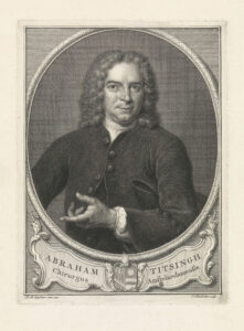
His colleague Giorgio Baglivi (1668-1707) elaborated upon this idea and supposed that the heart pushed the blood into the brain causing it to distend. The dura mater would then contract and push the nervous fluid into the nerves. In this way, he suggested two hearts and named the dura mater cor cerebri.3, p.7 In fact he attributed the highest control center to the dura mater, which he considered a vital structure.1, p. 77 These ideas proved very successful and were accepted by many anatomists and physicians, including Giovanni Maria Lancisi (1654-1720), Friedrich Hoffmann (1660-17420, Giovanni Domenico Santorini (1681-1737), and in particular Georg Ernst Stahl (1659-1734).

Jan Daniel Schlichting

Respiration

The English physician Humphrey Ridley (1653-1708), who graduated from Leiden with a dissertation De lue venerea (1679), may have been among the first to prove that destruction of the dura mater did not cease the movements of the brain. He read his paper at the Royal Society in 1703: “Experimentum anatomicum ad veram durae matris motus causam detegendam institutum” [An anatomical experiment to discover the real cause of the movement of the dura mater].4 Even more convincingly it was proved by the Dutch physician and anatomist Jan Daniel Schlichting (1703-1765), who studied medicine in the Dutch cities of Leiden and Groningen before he settled in Amsterdam. In his “De motu cerebri” he reported on his experiments, carried out upon several kinds of animals (dogs, cats, and rabbits), that brain movements remain after removal of the dura mater. “We solve, therefore, the problem that learned men have created for themselves by saying that the dura mater contracts and relaxes or pulsates”.

Schlichting’s 1750 article

He related the movements with breathing: “Every time a man breathes out, the entire brain rises up, that is, swells, and every time he breathes in, it goes down, that is, it subsides”.5 Thus he mentioned the opposite of what Galen had asserted 1,500 years previously. However, Schlichting did not know whether the swelling up during expiration was due to an increase influx of blood, by the increased air pressure, or by both.1, pp. 82-3 His experiments were repeated and the conclusions largely confirmed by the French physician Anne-Charles Lorry (1726-1783), stating that “The ancients followed their presumptions when they thought that the brain swelled up in time with inspiration: M. Schlichting is indeed supported by observation when he claims that it is synchronous with expiration”.1, p.84 However, Lorry believed the movements were only present in extraordinary conditions, like respiratory obstruction, pathological states, and cranial defects. He was one of several physicians, who denied brain movement to exist in normal conditions.

Jan Daniel Schlichting

The Swiss physician and physiologist Albrecht von Haller (1708-1777) was also among the physicians to refute the Pacchioni-Baglivi concept about the function of the dura mater. He confirmed the elevation of the brain during expiration and sinking down during inspiration. He related this to the venous congestion and outflow. However, it remained a matter of debate how that occurred. Some attributed the movements to the action of the heart, others to that of the influence upon the venous return of blood by breathing, and a third group believed that a combination of the two was responsible.

Anne-Charles Lorry

Brain Proper Moves Synchronously With the Heart

Several physicians observed that the brain moves synchronously with the heart. Among them were some well-known persons including the Italian anatomist and surgeon Realdus Columbus (Matteo Colombo c. 1515-1559), the French anatomist Jean Riolan (1580-1657), the Dutch physician IJsbrand van Diemerbroeck (1609-1674), Ridley, the French anatomist Raymond Vieussens (1641-1715), Haller and Marie-François-Xavier Bichat (1771-1802). However, there were various opinions on how the heart could move the brain. Several, including Bichat, believed it being the effect of the arterial pulse. Others were still denying such a synchronous movement. Marie-Jean-Pierre Flourens (1794-1867), for instance, denied a pulsating movement of the brain, only confirming a synchronous movement with respiration.

Franciscus Cornelis Donders

The German anatomist and anthropologist Johann Alexander Ecker (1816-1887), who summarized the history of brain movements, experimented with animals and concluded that there is a double movement in human beings and mammals, notably with the heart and respiration. The pulsating movement, he believed, was due to the arteries at the base of the brain as well as their branches in the brain parenchyma. He attributed the respiratory movement to the influx into the ventricles of cerebrospinal fluid (CSF) during expiration, although the venous filling in the brain may contribute to it.3, p.123

Ernst von Leyden

Spying on Nature Directly

At the time, some physiologists were more critical about the influence of the CSF. Writing on the results of his experiments, the Dutch physiologist and ophthalmologist Franciscus Cornelis Donders (1818-1889) mentioned that “the CSF is the condition, not the cause of brain movement.” Trying to answer the question whether brain movements exist when the skull is completely closed, he denied any movements of the brain if the skull was closed. The source of the question derived from two observations. Ecker believed the movements continued, whereas Johannes Müller declared that this was physically impossible.6 The French anatomist and physiologist François Achille Longet (1811-1871), based on observations published in Bourgougnon’s 1839 dissertation, also denied brain movements with closed skull. “Sans doute il est curieux de rechercher pourquoi le cerveau se meut chez les animaux trépanés; mais il est, suivant nous, bien plus important de savoir si ces mouvements préexistent à la trépanation” [No doubt it is curious to investigate why the brain moves in trepanned animals; but it is, in our opinion, far more important to know whether these movements pre-exist trepanation]. A comparison was made with the movements of the lungs that are quite extensive when the thoracic cage is opened, in comparison to the much less extensive movements in the physiological situation. Longet considered experiments to research brain movements, during which the skull was opened as useless. Therefore, Donders decided to open the skull of a rabbit. “I managed to spy on nature directly. When the skull is closed I have not only observed immediately and life, what concerns the brain movements, but also the increased and decreased supply of blood, with changes in blood pressure.” After trepanning the skull and removing a small part of the dura mater, he inserted a small glass window. “Through the small glass window one could observe the brain perfectly well, but we saw no trace of movement.7 I now amplified the inhalation and exhalation excursions by keeping the mouth and nose closed. Repeatedly again no movements.” At last, the small glass came loose, after which both types of movement, respiratory as well as circulatory, returned. The experiment was witnessed by several of his colleagues. After bonding the glass in the opening again, he did not observe any movements anymore and was able to see it for more than eleven days, sometimes using a loupe and even a microscope. He repeated the experiment several times and had special watch glasses made for the purpose.

Angelo Mosso

From Brain Movements to Intracranial Pressure

Still working in Königsberg (the present Kaliningrad), the German physician Ernst von Leyden (1832-1910) did important research on brain movements and in particular on intracranial pressure, which he published in 1866.6 He confirmed Donders’ observations, but instead of rabbits he used dogs, in which brain movements are clearer. Next to a small glass window, he inserted a lockable pipe, so that he could easily open and close the hole in the skull. He realized that the pressure waves by respiratory and circulatory movements with the tap locked would be borne by the brain. Like Victor von Bruns (1812-1883), Von Leyden tried to represent the changes in a graph, but encountered too many difficulties to show the movements due the low force that cause them. However, he was able to show the respiratory and circulatory quite nicely, by using a barometer tube filled with water. The rest of his long research paper is about measuring intracranial pressure under normal circumstances as well as with pathologically increased pressure.8

Catherina X (from Mosso10)

Registrations in Patients

About 10 years later in Turin, Carlo Giacomini (1840-1898) in cooperation with the well-known physiologist Angelo Mosso (1846-1910), were able to register brain movements by using an instrument developed by the French physiologist Etienne-Jules Marey (1830-1904; “méthode graphique”). They published their paper in the first volume of Archivio per le Scienze Mediche. The recordings were done in the open skull of the 37-year Catherine X, who suffered from syphilis. Despite years of treatment with potassium iodide and mercury ointment, the skull became affected and the researchers were able to observe the pulsations of the dura mater synchronously with the heart beats.9 In Mosso’s 1881 monograph Kreislauf des Blutes im menschlichen Gehirn [Circulation of the blood in the human brain], we find a drawing of Catherine and her skull.10 Giacomini’s and Mosso’s paper was read at the Académie in Paris by no one less than the famous physiologist Claude Bernard (1813-1878),11 who informed the audience that at the time the patient had been completely cured.

Charles-Emile François-Franck (National Library of Medicine; Digital Collections. Public domain)

Not long after the Italian publication, the French physiologist Charles-Emile François-Franck (1849-1921), a pupil of Marey, was able to register the brain movements of a similar case. He was aided by young Edouard Brissaud (1852-1909; described geste antagoniste in dystonia), who made the registrations and was allowed, at the end of the article, to present the case history. It was about the 34-year old Victoire All… , who suffered from syphilis, for which she was treated with liquor swietenii, a mercury based mixture that was introduced the previous century by Gerard van Swieten (1700-1772). The dosage of mercury was lower than that applied earlier. However, the skull became affected and parts needed to be removed, providing access to the researchers.

At the end of the 19th century, British physiologist Leonard Hill (1866-1952) summarized the literature on the subject, in particular the decades before his book was published. Referring to “Althan,” probably the Estonian born Georg Althann (1839-1898), he noted that “the errors of Donders … arose from the neglect of the fact that the brain pulsates in a direction where resistance is least. Thus when the cranium is trephined and the dura opened, the pulse can scarcely be seen in the occipito-atlantal membrane, for the trephine hole has now become the seat of least resistance. Similarly, when a trephine hole is closed by a glass window, the occipito-atlantal membrane becomes the seat of least resistance, and a pulsation appears there.”12, p.8-15

Today the pulsating brain still is an area of research in particular with respect to the effects on MRI scans and more recently the concept of the glymphatic system. Arterial pulsations are thought to drive flow through perivascular spaces for clearance of metabolic waste.13


References

1. Neuburger M (1897). Die historische Entwicklung der experimentellen Gehirn- und Rückenmarksphysiologie vor Flourens. Stuttgart, Enke. English edition transl. and edited by Edwin Clarke The historical development of experimental brain and spinal cord physiology before Flourens. Baltimore/London, Johns Hopkins University Press.

2. Pacchioni A (1701). De durae meningitis fabrica et usu disquisition anatomica. Rome, Herculis.

3. Ecker A (1843). Physiologische Untersuchunge über die Bewegungen des Gehirns und Rückenmarks : insbesondere den Einfluss der Cerebrospinalflüssigkeit auf dieselben. Stuttgart, Schweizerbart.

4. Ridley H (1703). Experimentum anatomicum ad veram durae matris motus causam detegendam institutum Phil Trans 23, no. 287: 1480-4.

5. Schlichting JD (1750). De motu cerebri. Mémoires de Mathématique et de Physique (Paris) : 113-35.

6. Leyden E (1866). Beiträge und Untersuchungen zur Physiologie und Pathologie des Gehirns. Virchows Arch 37:519–559.

7. Donders FC (1850). De bewegingen der hersenen en de veranderingen der vaatvulling van de Pia Mater, ook bij gesloten onuitzetbaren schedel regtstreeks onderzocht. Nederlandsch Lancet, March/April, 521-53.

8. Koehler PJ, Wijdicks EF (2015). Fixed and dilated: the history of a classic pupil abnormality. J Neurosurg;122:453-63.

9. Giacomini C, Mosso A (1876). Esperienze sui movimenti del cervello nell’uomo. Archivio per le Scienze Mediche 1:245-78.

10. Mosso A (1881). Kreislauf des Blutes im menschlichen Gehirn, Leipzig, Veit, p. 223

11. Giacomini C, Mosso A (1877). Etude graphique des mouvements du cerveau de l’homme. C R Académie Sciences (Paris) 24 : 41-3.

12. Hill L (1896). The Physiology and Pathology of the Cerebral Circulation: An Experimental Research. London: Churchill.

13. Mestre H, Tithof J, Du T, Song W, Peng W, Sweeney AM, Olveda G, Thomas JH, Nedergaard M, Kelley DH (2018). Flow of cerebrospinal fluid is driven by arterial pulsations and is reduced in hypertension. Nat Commun 9:4878.

Multiple Sclerosis DMTs Added to WHO List

Three disease-modifying therapies (DMTs) for multiple sclerosis are now part of the World Health Organization essential medicines list.

By Bassem Yamout1, Tomas Kalincik2,3, Joanna Laurson-Doube4, Bernhard Hemmer5,6, Shanthi Viswanathan7

On July 26, 2023, the World Health Organization (WHO) added three disease-modifying therapies (DMTs) for multiple sclerosis (MS) to its Essential Medicines List (EML) for the first time1. The list is an important tool for achieving universal health coverage, providing guidance to governments, health facilities, and procurers on priority medicines to tackle important public health conditions. The EML includes medicines on the basis of solid evidence for safety and efficacy. Inclusion of any medication in the WHO EML is expected to be associated with increased availability and reimbursement by governmental agencies. The WFN was one of the 15 organizations that endorsed the application to the WHO, submitted by Multiple Sclerosis International Federation and WHO Collaborating Centre Bologna.

With this decision, the WHO formally acknowledged MS as a global health concern as well as the critical importance of making MS treatments available in all health systems at all times. Although most approved MS therapies have been made available in many countries, people with MS living in certain regions of the world, especially in resource-limited settings, do not have access to much-needed therapies, which are either unavailable or unaffordable. In fact, around 70% of countries across the world report that people with MS face barriers accessing DMTs2. The WHO decision to include three MS therapies with different routes of administration, efficacy, and tolerability in their updated list will undoubtedly help increase access of people with MS to high-quality, cost-effective, and evidence-based treatments.

This decision was the culmination of two years of relentless efforts by the MS International Federation (MSIF) and its partner organizations, including several neurological academies, scientific societies, and the regional committees for Treatment and Research in MS (TRIMS). The MSIF set up two independent, multidisciplinary panels and started a comprehensive and rigorous review process in partnership with the WHO Collaborating Centre Bologna, and supported by the Cochrane MS group and McMaster GRADE Centre, both of which are internationally regarded as experts in the field of evidence-based reviews and decision-making3.

The WHO EML Committee decided to add rituximab, cladribine, and glatiramer acetate to the new EML4. Rituximab has been extensively used for treating MS patients especially in low/lower-middle income countries (LLMIC) due to its cost-effectiveness, but its off-label status has curtailed reimbursement by governmental and private insurance systems5. Its inclusion on the WHO EML should help in this respect. The decision to support off-label use of rituximab is supported by strong evidence of its efficacy and safety for this indication.


Dr. Deanna Saylor

“People with MS in lower-resourced settings, including most low- and middle-income countries, often face numerous challenges in their MS journeys. This starts with accessing the specialists and diagnostic tests, including MRIs, required to obtain their diagnosis, and continues with accessing effective treatments for their MS. As a result, people with MS in low- and middle-income countries often are untreated, undertreated, or have to expend significant personal resources in order to obtain treatment. Based on my own experience taking care of people with MS in Zambia, I am confident that if DMTs are available, treatment of MS is feasible, safe, and likely to be highly effective and result in good outcomes among people with MS in low- and middle-income countries.”

Dr. Deanna Saylor, clinical chair of the MSIF Essential Medicines Panel


Large-scale randomized controlled trials (e.g. DELIVER-MS and TREAT-MS) continue to study early use of high efficacy DMTs versus escalation treatment by treatment6,7. Nevertheless, studies have already suggested that early treatment with higher efficacy DMTs in patients with MS can lead to better outcomes compared to treatment initiation with low efficacy DMTs and escalating therapy only upon disease activity or progression8-12.

Intentionally, the treatments listed on the WHO EML are not categorized as first-, second-, or third-line treatments, but encourage clinicians and people with MS to determine the most appropriate course of treatment for the clinical and personal circumstances. Rituximab, cladribine, and glatiramer acetate represent different tiers of treatment effectiveness, modes of administration, and safety in pregnancy and breastfeeding. In particular, glatiramer acetate is known for its favorable safety profile in pregnancy and breastfeeding when compared with the two more potent listed therapies. Pregnancy is a particularly important clinical scenario due to high prevalence of MS in young women and more challenging access to family planning services in LMICs.

Countries can use the WHO decision to shift their treatment algorithms, allowing patients access to higher efficacy DMTs, e.g. rituximab, early in their disease course. Speaking from personal experience treating refugees in the Middle East, the use of rituximab, a high efficacy, safe, and low-cost DMT, made a considerable difference in the lives of people with MS that fled their country and had no means of affording any MS therapy13.

However, despite countries endorsing the concept of the EML over the years, implementing the list into local practice is variable and fraught with challenges, especially in LMICs. We should use this momentous decision to push forward the implementation of the WHO EML through coordinated efforts among national scientific and patient societies, international scientific, and health care organizations, frontline clinicians, and local policymakers.

Patients with MS cannot wait for decades to have this EML implemented: The time for action is now through the growing international momentum to improve care and access to treatment for multiple sclerosis.


1. Yamout is with the Neurology Institute, Harley Street Medical Centre, Abu Dhabi-UAE. 2. Kalincik is with the Neuroimmunology Centre, Department of Neurology, Royal Melbourne Hospital, Melbourne, Australia. 3. Laurson-Doube is with  CORe, Department of Medicine, Melbourne, Australia.4. Hemmer is with Multiple Sclerosis International Federation, London, United Kingdom. 5. Department of Neurology, Klinikum rechts der Isar, School of Medicine, Technische Universität München, Germany. 6. Munich Cluster for Systems Neurology (SyNergy), Germany. 7. Viswanathan is with Department of Neurology, Kuala Lumpur, Hospital, Malaysia.

References

1. WHO endorses landmark public health decisions on Essential Medicines for Multiple Sclerosis [Internet]. [cited 2023 Aug 1]. Available from: https://www.who.int/news/item/26-07-2023-who-endorses-landmark-public-health-decisions-on-essential-medicines-for-multiple-sclerosis

2. MS International Federation. Atlas of MS 2020 – Clinical management report [Internet]. 2020 [cited 2022 Dec 5]. Available from: https://pubmed.ncbi.nlm.nih.gov/35173808/

3. A.10 Cladribine, glatiramer and rituximab – multiple sclerosis – EML [Internet]. [cited 2023 Aug 2]. Available from: https://www.who.int/groups/expert-committee-on-selection-and-use-of-essential-medicines/24th-eml-expert-committee/a10_ms-dmts

4. World Health Organization. Executive summary of the report of the 24th WHO Expert Committee on Selection and Use of Essential Medicines 24 – 28 April 2023 [Internet]. [cited 2023 Aug 1]. Available from: https://apps.who.int/iris/bitstream/handle/10665/371291/WHO-MHP-HPS-EML-2023.01-eng.pdf

5. Laurson-Doube J, Rijke N, Helme A, Baneke P, Banwell B, Viswanathan S, et al. Ethical use of off-label disease-modifying therapies for multiple sclerosis. Mult Scler Houndmills Basingstoke Engl. 2021 Aug;27(9):1403–10.

6. DELIVER-MS – does early treatment with highly effective DMT improve the prognosis for people with Multiple Sclerosis? [Internet]. [cited 2023 Sep 15]. Available from: https://deliver-ms.com/

7. Ontaneda D, Tallantyre E, Kalincik T, Planchon SM, Evangelou N. Early highly effective versus escalation treatment approaches in relapsing multiple sclerosis. Lancet Neurol. 2019 Oct;18(10):973–80.

8. Ontaneda D, Mowry EM, Newsome SD, Naismith RT, Nicholas J, Fisher E, et al. Benefits of early treatment with natalizumab: a real-world study. Mult Scler Relat Disord [Internet]. 2022 Dec 1 [cited 2023 Sep 15];68. Available from: https://www.msard-journal.com/article/S2211-0348(22)00721-0/fulltext

9. Stankiewicz JM, Weiner HL. An argument for broad use of high efficacy treatments in early multiple sclerosis. Neurol – Neuroimmunol Neuroinflammation [Internet]. 2020 Jan 1 [cited 2023 Sep 15];7(1). Available from: https://nn.neurology.org/content/7/1/e636

10. Simonsen CS, Flemmen HØ, Broch L, Brunborg C, Berg-Hansen P, Moen SM, et al. Early High Efficacy Treatment in Multiple Sclerosis Is the Best Predictor of Future Disease Activity Over 1 and 2 Years in a Norwegian Population-Based Registry. Front Neurol [Internet]. 2021 [cited 2023 Sep 15];12. Available from: https://www.frontiersin.org/articles/10.3389/fneur.2021.693017

11. Filippi M, Amato MP, Centonze D, Gallo P, Gasperini C, Inglese M, et al. Early use of high-efficacy disease‑modifying therapies makes the difference in people with multiple sclerosis: an expert opinion. J Neurol. 2022;269(10):5382–94.

12. Brown JWL, Coles A, Horakova D, Havrdova E, Izquierdo G, Prat A, et al. Association of Initial Disease-Modifying Therapy With Later Conversion to Secondary Progressive Multiple Sclerosis. JAMA. 2019 Jan 15;321(2):175–87.

13. Zeineddine MM, Yamout BI. Treatment of multiple sclerosis in special populations: The case of refugees. Mult Scler J – Exp Transl Clin. 2020 Mar;6(1):2055217319848466.

The 2023 International Symposium on Thrombolysis/Thrombectomy and Acute Stroke Therapy, and the Symposium on Collaterals on the Brain

Amir Molaie

David Liebeskind

By Amir Molaie and David Liebeskind

In the rapidly evolving field of stroke care, events that gather global experts to discuss recent trials and innovations are integral to disseminating knowledge and advancing care. Building on the momentum of the 15th World Stroke Congress, the 2023 International Symposium on Thrombolysis/Thrombectomy and Acute Stroke Therapy (TTST) and the 10th annual Symposium on Collaterals on the Brain (Collaterals), proved no exception. The two conferences, held in conjunction at the University of California, Los Angeles (UCLA) Oct. 13-17, 2023, brought together leading experts from around the world to review and deliberate wide-ranging, timely topics in neurovascular care.

Panel discussion on the potential use of generative AI in future vascular and endovascular care, featuring (left to right): Ashutosh Jadhav, MD, PhD; Andrei Alexandrov, MD; Kunakorn Atchaneeyasakul, MD; Alexandra Czap, MD.

The events were hosted by Prof. David Liebeskind in a hybrid virtual and in-person format, assembling 165 attendees. Participants joined from countries on six continents, ranging in background from clinicians and researchers to biomedical engineers and medical students.

The program began with the TTST 2023 portion, with presenters offering their data and input on endovascular thrombectomy for special populations, including late window cases, distal occlusions, and patients with low NIHSS. This was followed by up-to-date summaries of the recently published and newly presented large-core trials. The first day wrapped up with lectures on innovative technologies, including prehospital triage devices and an interactive dialogue on the rising impact of artificial intelligence on stroke care.

Conference dinner hosted by David Liebeskind, MD, pictured here with James Grotta, MD (left) and Alexandra Czap, MD (right).

The following day featured lively discussions regarding the use of alteplase versus tenecteplase, and the overarching role of intravenous thrombolysis prior to mechanical thrombectomy (MT). Sessions also delved into the merits of neuroprotection and the optimization of MT techniques. The day ended with a unique Jeffersonian-style exchange, a forum which fostered interactive, candid, and impassioned conversations involving participants from all levels of training and backgrounds.

The final day of TTST highlighted issues related to stroke systems of care, topics which seamlessly segued into the Collaterals Symposium. These next three days showcased accomplished presenters from throughout the world commenting on their impressive advocacy and contributions to expanding stroke treatment to regions with previously limited access to thrombolysis and thrombectomy. Nearly every geographic region was represented, with speakers converging from afar, including Aida Kondybayeva, MD, PhD, joining from Kazakhstan, and Ossama Mansour, MSc, MD, PhD, from Egypt.

Radoslav Raychev, MD (left) and Aida Kondybayeva, MD, PhD (right) in conversation.

In the spirit of global collaboration and progress in the neurovascular field, TTST and Collaterals embodied the power of shared knowledge and dedication to shaping the future of stroke care on a worldwide scale. We look forward to the advances and work to come in 2024. •


David Liebeskind is professor of neurology at UCLA, director of the Neurovascular Imaging Research Core, and director of the UCLA Comprehensive Stroke Center.

Amir Molaie is a neurology resident at UCLA.