@WFNeurology

By W. Struhal and Prof. P. Engel
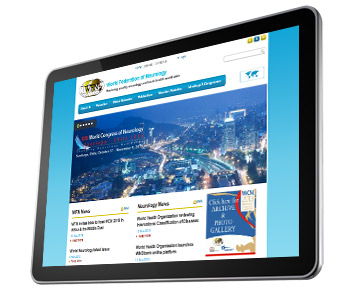
WFNwebsite_tabletThe World Federation of Neurology (WFN) is a huge and complex structure, representing neurologists worldwide. To achieve its aim, many international neurologists collaborate and work on WFN projects, represent the organization as officers or serve as editors or authors for WFN media. These initiatives play an important role in advocating the interests of neurologists on a global scale.

You can follow all of these activities and more at www.wfneurology.org.

Content

A major aim of WFN is supporting educational initiatives and encouraging global networking. The website provides a sound insight into WFN educational activities. These include WFN seminars in clinical neurology, which provide teaching and training materials and patient care guidelines. Exchange among young neurologists is encouraged by WFN through programs such as the Turkish Department Visit, and available grants and awards for young neurologists interested in extending their training internationally. Reports are published on projects such as the Zambia Project, which aims to improve medical care in Zambia. Young neurologists are encouraged to participate in the WFN, and the website lists representatives of young neurologists. A singularly interesting section is neurology for non-neurologists, which provides educational materials for areas where there is a severe shortage of neurologists.
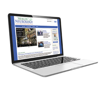
WFNWorldNeuro_ComputerBringing worldwide science  and patient care closer together is a strong objective of the WFN. At  www.wfneurology.org, you will find details on the World Brain Alliance, an umbrella group of international neurological organizations.  WFN applied research groups organize scientific projects and educational activities in neurology subspecialties, and publish their activities on the website on an annual basis.

Some additional important topics presented at www.wfneurology.org:

  • WFN initiatives (e.g. the WFN Africa Initiative)
  • Candidates for 2013 election, including the president of WFN
  • WFN officers, national WFN delegates, WFN regional directors

Do you want to keep up to date?

The WFN website provides insights into our organization, but it offers more than that. Neurology news of major global importance is published in WFN’s publication World Neurology (www.worldneurologyonline.com). Because one aim of the WFN web strategy is to establish direct interaction with its users, social media channels are offered. You may follow WFN updates and actively exchange your thoughts with WFN on Facebook (www.facebook.com/wfneurology), Twitter (www.twitter.com/wfneurology), or the World Federation of Neurology LinkedIn group (linkedin.com). You can use these social networks to interact and get to know other participants of the XXI World Congress of Neurology in Vienna – the first World Congress where social media channels were offered. For Twitter users, please follow our official hashtag (#) and don’t hesitate to use it in your tweets: #WCNeurology.

Aims and vision of the WFN website

The WFN website and WFN digital footprint comprise a platform for neurologists who advocate neurology through WFN initiatives and projects, and inform the public on activities of WFN. Social media platforms offer the prospect of increased online interactivity and the hope that neurologists worldwide will interconnect more. The future vision is that these digital resources will help to build a strong network of neurologists worldwide and strengthen scientific collaboration in neurological research and services.

We warmly invite you to visit www.wfneurology.org.
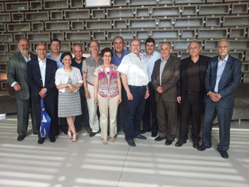
Moroccan Foundation of Neurology

Representatives met in May 2012 to discuss a foundation dedicated to developing neurological

Representatives met in May 2012 to discuss developing neurological sciences in the fight against neurological diseases in Morocco.

The World Congress of Neurology, organized by the Moroccan Society of Neurology in Marrakesh, November 2011, was a great success at both the scientific and organizational level. Moreover, the conference has generated substantial financial benefits for both the Moroccan Society of Neurology and the World Federation of Neurology, which will devote 20 percent of its profits to the development of neurology in Africa.

Moroccan neurologists met in May 2012 and unanimously decided to devote all profits obtained by the Moroccan Society of Neurology to the creation of a foundation dedicated to the development of neurological sciences and the fight against neurological diseases in Morocco.

Indeed, Morocco is experiencing a delay in neurology and care of neurological diseases even compared to countries of the same level of economic development. The number of neurologists remains low: 170 (counting the neurologists in training) for a population of 32 million. Besides, there are few hospital beds dedicated to neurology, and CT scans are only available in large cities.

The country has worrying indicators in public health. Neonatal and perinatal morbidity, which remain high, are causing a large number of children to experience neurodevelopmental disorders responsible for motor and cognitive disabilities that require rehabilitation throughout their lives.

In addition, urbanization, changing lifestyles and increasing life expectancy have caused a rapid epidemiological transition that led to an increase in diseases related to aging. Thus, an epidemiological survey conducted in Rabat and Casablanca in 2009, showed that the prevalence of stroke in people aged 65 years and older was 25 per 1,000. This is almost equal to that found in industrialized countries. As for dementia, the report of the WHO in 2012 estimated the number to be 100,000 cases. Also, Morocco suffers from an unusually high number of road traffic accidents responsible for head injuries and neurological disorders.

Faced with this alarming situation, the foundation is focused on these objectives:

  • To advocate for the increase of the number of neurologists and other caregivers in the neurological field
  • To advocate for a policy of prevention of neurological diseases in both children and adults
  • To promote a better understanding of neurological diseases among the general population
  • To fight against the stigmatization of patients with neurological diseases
  • To inform national policymakers of the urgency to implement a policy of neurological diseases in Morocco
  • To support education and research in neurology
  • To promote the highest standards of ethics and practice in neurological sciences

By its bylaws, the Moroccan Foundation of Neurology is a non-profit organization that can receive donations from persons, associations and governments to accomplish its objectives. The foundation also aims to develop cooperation with other similar institutions in Africa and in the Arab world to improve neurological care in these regions.

The Moroccan Foundation of Neurology, with active support from the World Federation of Neurology, the World Health Organization and other international institutions, will undoubtedly succeed in its mission to improve the lives of patients with neurological diseases.

Nano-Neuromedicine

By Girish Modi and Viness Pillay

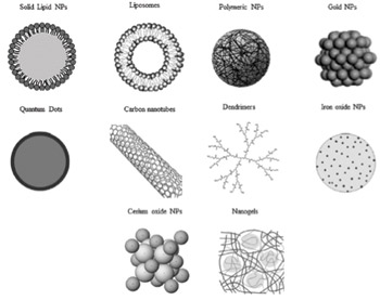
Figure 1. Different type of nanomaterials for biomedical use. Nanomaterials are commonly defined as objects with dimensions of 1-100 nm, which includes nanogels, nanofibers, nanotubes and nanoparticles (NP). In this illustration is represented the morphology of the most commonly used NP for therapy and diagnosis of neurodegenerative diseases (ND). (Ref. 3; reproduced with permission from Elsevier B.V. Ltd. © 2012.)

Figure 1. Different type of nanomaterials for biomedical use. Nanomaterials are commonly defined as objects with dimensions of 1-100 nm, which includes nanogels, nanofibers, nanotubes and nanoparticles (NP). In this illustration is represented the morphology of the most commonly used NP for therapy and diagnosis of neurodegenerative diseases (ND). (Ref. 3; reproduced with permission from Elsevier B.V. Ltd. © 2012.)

Nanotechnology employs engineered materials or devices (nanomaterials) with the smallest functional organization on the nanometer scale (1–100 nm) that are able to interact with biological systems at the molecular level1. They stimulate, respond to and interact with target sites to induce physiological responses while minimizing side effects1. By definition, nanomaterials are natural, incidental or manufactured materials containing particles (nanoparticles), in an unbound state or as an aggregate or as an agglomerate2. There are various methods of preparation of nanoparticles such as emulsion polymerization, interfacial –polymerization, –polycondensation, or –deposition, solvent –evaporation and –displacement, salting-out and emulsion/solvent diffusion, to name a few. There are several different types of nanostructures. These include polymeric nanoparticles, nanocapsules, nanospheres, nanogels, nanosuspensions, nanomicelles, nanoliposomes, carbon nanotubes and nanofibers3  (See Figure 1.)

Nanoparticulate matter and related materials possess an inherent capability to efficiently and effectively cross the blood brain barrier (BBB) leading to an increased concentration of encapsulated bioactives and drugs in the cerebrospinal fluid (CSF). In addition to drug delivery, this BBB crossing ability of the nanoparticles can be employed to develop various real-time diagnostic tools.

Promising features of nanosystems in targeted CNS drug delivery are that

  • their chemical properties can be easily modified to achieve organ-, tissue-, or cell-specific and selective drug delivery
  • the delivery of the drugs can be  controlled
  • they increase the bioavailability and efficacy of incorporated drugs by masking the physicochemical characteristics and thus increase the transfer of drug across the BBB
  • they protect incorporated drugs against enzymatic degradation
  • they have fewer side effects.1
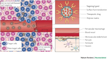
Figure 2. The neurovascular unit (bottom right panel) regulates the dynamic and continuous crosstalk between circulating blood elements and brain cellular components, including perivascular macrophages, astrocytes and neurons. At the interface between blood and brain (the blood-brain barrier), endothelial cells and associated astrocytes are stitched together by structures called tight junctions. The blood-brain barrier hinders the delivery of many potentially important diagnostic and therapeutic drugs to the CNS. The barrier results from the selectivity of the tight junctions between endothelial cells in the blood vessels that restricts the passage of solutes. Astrocyte cell projections called astrocytic feet surround the endothelial cells of the blood-brain barrier, providing biochemical support to those cells. The pericytes shown in this figure are undifferentiated mesenchymal-like cells that also line and support these vessels contributing to the complex layering of cells forming the blood-brain barrier. Nanocarriers (top right panel) are materials that can be configured into several different shapes (tubes, spheres, particles, rods, etc.) and can be loaded with drugs for sustained delivery to specific sites that are targeted by coating the surface of the material with peptides. In this figure, the nanocarrier contains a therapeutic drug, a targeting peptide to increase the penetration of the drug into the brain tissue and a functionalized surface consisting of, for example, an antibody to target a specific antigen or a steric coating made of polyethylene glycol or dextrans. Active targeting (left panel) enhances the biodistribution of drug-loaded nanocarriers in the brain tissue, improving the therapeutic efficacy of drugs. Once particles have extravasated in the target tissue, the presence of ligands on the particle surface facilitates their interaction with receptors that are present on target cells resulting in enhanced accumulation and cellular uptake through receptor-mediated processes (Ref. 4; reproduced with permission from Nature Publishing Group © 2009)

Figure 2. The neurovascular unit (bottom right panel) regulates the dynamic and continuous crosstalk between circulating blood elements and brain cellular components, including perivascular macrophages, astrocytes and neurons. At the interface between blood and brain (the blood-brain barrier), endothelial cells and associated astrocytes are stitched together by structures called tight junctions. The blood-brain barrier hinders the delivery of many potentially important diagnostic and therapeutic drugs to the CNS. The barrier results from the selectivity of the tight junctions between endothelial cells in the blood vessels that restricts the passage of solutes. Astrocyte cell projections called astrocytic feet surround the endothelial cells of the blood-brain barrier, providing biochemical support to those cells. The pericytes shown in this figure are undifferentiated mesenchymal-like cells that also line and support these vessels contributing to the complex layering of cells forming the blood-brain barrier. Nanocarriers (top right panel) are materials that can be configured into several different shapes (tubes, spheres, particles, rods, etc.) and can be loaded with drugs for sustained delivery to specific sites that are targeted by coating the surface of the material with peptides. In this figure, the nanocarrier contains a therapeutic drug, a targeting peptide to increase the penetration of the drug into the brain tissue and a functionalized surface consisting of, for example, an antibody to target a specific antigen or a steric coating made of polyethylene glycol or dextrans. Active targeting (left panel) enhances the biodistribution of drug-loaded nanocarriers in the brain tissue, improving the therapeutic efficacy of drugs. Once particles have extravasated in the target tissue, the presence of ligands on the particle surface facilitates their interaction with receptors that are present on target cells resulting in enhanced accumulation and cellular uptake through receptor-mediated processes (Ref. 4; reproduced with permission from Nature Publishing Group © 2009)

Taking these benefits into consideration, nanotechnology has immense potential such as in the field of neuromolecular diagnostics, discovery of neurodegenerative markers, nano-enabled drug delivery and neuroactive discovery with implications reaching to the prevention, management and treatment of neurological disorders.

In terms of delivery across the BBB, two basic approaches, namely the molecular approach and the polymeric carrier approach can be applied. In the molecular approach, nanonization or ligand attachment of the drugs can be employed to target brain cells, and the drugs can further be enzymatically activated afterward inside the target cell4. However, the availability of such modifiable drugs is low, and only certain drugs with specific functionality can be targeted molecularly. Additionally, a metabolic pathway is always required to activate these drugs inside CNS — further narrowing the options. The second approach of employing polymeric carriers such as drug-loaded nanoparticles can be termed as a universal approach with flexibility of choosing the matrix or polymer system and additionally can be administered by the route of choice: intravenously, intrathecally or as an implantable cerebral device.  (See Figure 2.)

In this way, the BBB can be circumvented via systemic administration or CNS implantation with an ability to control as well as target the release of various bioactive agents used in the treatment of neurodegenerative disorders (NDs). The majority of nanotechnological drug delivery systems for the treatment of NDs are in the form of polymeric nanoparticles. Polymeric nanoparticles are promising for the treatment of NDs as they can pass through tight cell junctions, cross the BBB, achieve a high drug-loading capacity, and be targeted toward the mutagenic proteins.

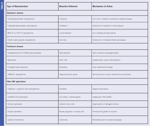
Specific nanosystems explored for advanced experimental treatment of Alzheimer’s disease (AD), Parkinson’s disease and other CNS disorders are listed in Table 1.

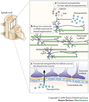
Nano-enabled systems in the form of biodegradable and non-biodegradable templates for regeneration of damaged neurons and peptide-based self-assembling molecules have been employed as scaffolds for tissue engineering, neuroregeneration, neuroprotection and photolithography etching5. Nanoparticulate strategies in the form of bioactive nanoparticles are being employed to provide resealing, repair, regeneration, restoration and reorganization of neural tissue after traumatic spinal cord injury. Nanoparticles are capable of achieving this via the control of secondary injury cascade, reassembly of the tethered membranous structures, creating a neuropermissive microenvironment, rebooting the neuropathophysiologica; connections, and recovery of the sensorimotor responses6,7  (See Figure 3.) Silva and co-workers, 2004, reported the potential of self-assembling peptide nanofibers (SAPN) in neural tissue engineering wherein the nanofibers provided the “neurite-promoting laminin epitope  IKVAV,” thereby inducing a rapid differentiation of cells into neurons via the amplification of bioactive epitope presentation and further restricting the development of astrocytes8.

The current therapeutic paradigms (using conventional drug delivery systems) employed to provide functional recovery in NDs lack adequate cyto-architecture restoration and connection patterns required to overcome the restrictive blood-brain barrier. In addition to the restrictive blood brain barrier; the neuroactive drug therapies present various other challenges such as:

  • higher doses are required to provide significant therapeutic benefit
  • low bioavailability further aggravating the first condition
  • poor absorption even after systemic delivery
  • the unwanted severe side-effects due to preferential uptake of drugs by peripheral cells.
Applications of nanotechnology in clinical neuroscience. Nanotechnology can be used to limit and/or reverse neuropathological disease processes at a molecular level or facilitate and support other approaches with this goal. (a) Nanoparticles that promote neuroprotection by limiting the effects of free radicals produced following trauma (for example, those produced by CNS secondary injury mechanisms). (b) The development and use of nanoengineered scaffold materials that mimic the extracellular matrix and provide a physical and/or bioactive environment for neural regeneration. (c) Nanoparticles designed to allow the transport of drugs and small molecules across the blood-brain barrier (Ref. 7; reproduced with permission from Nature Publishing Group © 2006.)

Applications of nanotechnology in clinical neuroscience. Nanotechnology can be used to limit and/or reverse neuropathological disease processes at a molecular level or facilitate and support other approaches with this goal. (a) Nanoparticles that promote neuroprotection by limiting the effects of free radicals produced following trauma (for example, those produced by CNS secondary injury mechanisms). (b) The development and use of nanoengineered scaffold materials that mimic the extracellular matrix and provide a physical and/or bioactive environment for neural regeneration. (c) Nanoparticles designed to allow the transport of drugs and small molecules across the blood-brain barrier (Ref. 7; reproduced with permission from Nature Publishing Group © 2006.)

The introduction and application of nanotechnological strategies may help in overcoming and even completely removing these neurotherapeutic challenges5.

Although various studies have claimed and demonstrated the potential of nanoparticles for CNS targeting, drug release and even gene delivery, rapidly biodegradable poly(butylcyanoacrylate) (PBCA) nanoparticulate system is the only successful nano-based drug delivery system that is being employed for the  in vivo administration of drugs targeted to the brain. However, the mechanism of performance of this successful system has not been confirmed yet with three different reports stating entirely different mechanisms:

  1. polysorbate 80 coated PBCA nanoparticles reportedly cross the BBB via a carrier based approach by plasma adsorption of apolipoproteins resulting in receptor-mediated endocytosis by brain capillary endothelial cells9
  2. Kreuter and co-workers, 2002, suggested phagocytosis or endocytosis by endothelial cells as the possible transport mechanism through the BBB10
  3. Vauthier and co-workers, 2003, suggested that nanoparticle adhere to the cell membrane with subsequent escape by the P-glycoprotein efflux system to reach the CNS11.

These contrasting reports may lead to delay in regulatory approval of these promising nanosystems, and hence, further research into the exact elucidation of the mechanism of nanoparticle transport across the BBB is required.

Nanoscale classes of neuroactives will widen the scope of therapeutic action beyond merely modifying transmitter function to include stem cell and gene therapies that could offer a more selective mode of targeting. Nanoresearch focused on the regeneration and neuroprotection of the CNS will significantly benefit from the parallel advances in neurophysiology and neuropathology research. Therefore, for nanotechnology applications in neurology and neurosurgery to come to fruition, the following need to be considered:

  • advancements in pharmaceutical chemistry and materials science that produce sophisticated synthetic and characterized approaches
  • advancements in molecular biology, neurophysiology and neuropathology of the nervous system
  • the design and integration of specific nano-enabled applications to the CNS which take advantage of the first two points.
  • If these areas are developed in an integrated and parallel manner, nanotechnology based applications for NDs may begin to reach the clinic. As with all therapeutic approaches, for the treatment of CNS disorders, the challenge of targeting the material, device or drug to the site where it is needed always remains. Therefore, in order for nanotechnology applications directed toward NDs to be fully exploited, it would be
    Table 1. Overview of various nano-enabled neurotherapies and interventions for CNS disorders (Ref. 5; reproduced with permission from Elsevier B.V. Ltd. © 2009.)

    Table 1. Overview of various nano-enabled neurotherapies and interventions for CNS disorders (Ref. 5; reproduced with permission from Elsevier B.V. Ltd. © 2009.)

    important for neurosurgeons, neurologists and neuroscientists to contribute to the scientific process along with pharmaceutical scientists and engineers. True to the highly interdisciplinary nature of this area of research, it is important that technological advancements occur in conjunction with basic and clinical neuroscience advancements5.

Modi is professor and head of Neurology, and the academic head of clinical neurosciences, Faculty of Health Sciences, University of the Witwatersrand. Pillay is professor of Pharmaceutics, Faculty of Health Sciences, University of the Witwatersrand.


References

  1. Modi G, Pillay V, Choonara YE. Advances in the treatment of neurodegenerative disorders employing nanotechnology. Ann NY Acad Sci. 2010;1184:154–172.
  2. Nanomaterials. http://ec.europa.eu/environment/chemicals/nanotech/faq/definition_en.htm (accessed on 20 November, 2013)
  3. Re F, Gregori M, Masserini M. Nanotechnology for neurodegenerative disorders. Nanomedicine: NBMedicine 2012;8:S51–S58.
  4. Orive G, Anitua E, Pedraz JL, Emerich DF. Biomaterials for promoting brain protection, repair and regeneration. Nat Rev Neurosci. 2009;10:682-692.
  5. Modi G, Pillay V, Choonara YE, Ndesendo VMK, du Toit LC, Naidoo D. Nanotechnological applications for the treatment of neurodegenerative disorders. Prog Neurobiol. 2009;88:272–285.
  6. Kumar P, Choonara YE, Modi G, Naidoo D, Pillay V. Nanoparticulate strategies for the 5 R’s of traumatic spinal cord injury intervention: restriction, repair, regeneration, restoration and reorganization. Nanomedicine, Manuscript accepted, In press, To be published in Feb 2014.
  7. Silva GA. Neuroscience nanotechnology: progress, opportunities and challenges. Nat Rev Neurosci.  2006;7:65-74.
  8. Silva GA, Czeisler C, Niece KL, Beniash E, Harrington DA, Kessler JA, Stupp SI. Selective differentiation of neural progenitor cells by high–epitope density nanofibers. Science, 2004;303:1352-1355
  9. Kreuter J, Ramge P, Petrov V, Hamm S, Gelperina SE, Engelhardt B, Alyautdin R, von Briesen H, Begley DJ. Direct evidence that polysorbate-80-coated poly(butylcyanoacrylate) nanoparticles deliver drugs to the CNS via specific mechanisms requiring prior binding of drug to the nanoparticles. Pharm Res. 2003;20: 409–416.
  10. Kreuter J, Shamenkov D, Petrov V, Ramge P, Koch-Brandt C. Apolipoprotein-mediated transport of nanoparticle-bound drugs across the blood–brain barrier. J Drug Target. 2002;10:317–325.
  11. Vauthier C, Dubernet C, Chauvierre C, Brigger I, Couvreur P. Drug delivery to resistant tumors: the potential of poly(alkyl cyanoacrylate) nanoparticles. J Control Release 2003;93:151–160.

Our Brains, Our Future

By Mohammad Wasay MD, FRCP, FAAN

There are many days related to neurological diseases being celebrated by professional organizations in collaboration with the World Health Organization, national organizations and local health ministries, including World Stroke Day, Epilepsy Day and Rabies Day. These days have proved to be extremely helpful in promoting public awareness and generating advocacy throughout the globe including non-developed Asian and African countries.

For example, the World Stroke Organization announced a global competition for public awareness and advocacy campaign focusing on World Stroke Day. In 2012, Brazil, and in 2013, Sri Lanka won the competition creating a huge impact at the national level as well as the regional level. All of the days related to neurology are linked to neurological diseases.

A few years ago, Vladimir Hachinsky, then-WFN president, asked, “Why don’t we celebrate a day for the healthy brain?” The human brain is so fascinating and is so closely linked to the health of the whole human being that we should promote healthy brains. The future of this universe is linked to our brains so we should start a global campaign with the slogan: “Our brains, our future.” This was suggested by the Public Awareness and Advocacy Committee.

Because the World Federation of Neurology was established on July 21, the Public Awareness and Advocacy Committee suggested that World Brain Day should be celebrated on the same date. This proposal was announced at the WCN Council of Delegates meeting in September, and the proposal was received with a warm welcome by delegates; Hachinsky; Raad Shakir, WFN president-elect; Werner Hacke, WFN vice president; and other officials. Our target audience is young brains throughout the world, and we want to promote healthy brain and brain health. Young students and minds are highly interested in knowing how the brain works and how can we make it work better.

We should target to approach one billion people around globe to educate about the brain in 2014. Most activities will focus near World Brain Day but it is a year-long campaign.  National societies should plan activities aimed at young school and college students, and with the help of social and electronic media, the information could go to millions of people. All societies should share their plans and activities, and organizations with the highest impact public awareness activities should be awarded.

We should especially focus efforts on Facebook and Twitter to connect with millions of people. Our Young Neurologists Network on Facebook could be a great resource for this campaign. We should use multiple languages, especially Chinese, Spanish, French, Arabic, Hindi and German. We also could develop a 5-minute promotional video with a brief introduction of WFN in multiple languages. This video could be shared by millions through YouTube and Facebook. We have more than 140 member societies. If we are able to organize a few hundred programs on July 21 in all of those countries, it is bound to create an impact.

Complexity of brain and neurological diseases often becomes a barrier for public awareness. “You should speak plain when you speak brain,” said Keith Newton of WFN. Our message should be simple and easily understandable for lay people. We could design a logo for this purpose which may be a simple global message. WFN and local organizations could start a poster or cartoon design competition to explain brain function and improve public awareness. Best posters, designs or cartoons could be awarded. We expect thousands of entries for this competition and some of these entries could become logos for our future campaigns.

There are many organizations working in this area, including the International Brain Council, the International Brain Research Organization, the AmericanAcademy of Neurology, the International League Against Epilepsy and the World Stroke Organization. We should work with them for this common agenda. Strong liaison and lobbying with WHO is important. If WHO adopts this day in the future, this could be a great success for WFN.

Wasay is the chair of the Public Awareness and Advocacy Committee for the World Federation of Neurology.

Featured Articles from JNS

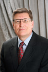
John D. England

John D. England

By John D. England, MD

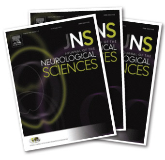
Beginning with this issue of World Neurology, we will feature an “Editor’s Selection” of articles from the Journal of the Neurological Sciences (JNS).  Elsevier, the publisher of JNS, has agreed to provide free access to these articles in PDF format to the members of the World Federation of Neurology.  Simply click on the PDF of the article to access it.

For this issue, we will feature two recent articles:

  • Andreas Steck and members of the Education Committee of the World Federation of Neurology (WFN) provide the results of a survey, which examined the current global state of training in neurology.  These results were compared with a previous survey done in 2006.  The paper outlines the areas where advances have occurred and also indicates areas where improvements are needed.  The findings provide important data which JNS_Magshould be helpful in guiding the future training of neurologists around the world.  The major disappointing aspect of the survey was that only 39 out of the 113 WFN member organizations provided answers to the survey.  Most respondents were from Europe and Asia.  Notable non-responders were Canada, France, India, Italy, Japan, United Kingdom and The United States.

    Manuscript # JNS-D-13-00728
  • Maria Nagel and others from the University of Colorado, U.S. present a remarkable case of a patient suspected of having giant cell arteritis (GCA) whose temporal artery biopsy was initially negative for GCA.  Further analysis demonstrated varicella zoster virus (VZV) antigen and VZV DNA in the temporal artery and adjacent skeletal muscle.  More extensive pathological analysis of temporal artery sections adjacent to those containing VZV antigen showed classic features of GCA.  These findings build upon the previous research from this group, which detected VZV in the temporal arteries of 5/24 patients with clinically suspected GCA.  Taken together, these findings raise important questions about the role of VZV in both suspected and proven GCA.  Manuscript # JNS-D-13-01090R1

We hope that this new feature of highlighted articles from JNS will be a useful addition for readers of World Neurology.

England is Editor-in-Chief, Journal of the Neurological Sciences.

Book Review: Breaking Down Neurophobia

By Sarah Matteson Kranick, MD

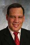
Sarah Matteson Kranick

Sarah Matteson Kranick

A recent study asked medical students and internal medicine residents to rate eight medical subspecialties with regards to the students’ feelings of competency and perceived difficulty.  The 150+ respondents identified neurology as the specialty in which they had least knowledge  (p<0.001) and was most difficult  (p<0.001).  [Winchuk AV BMC Medical Education 2010]

Similar studies in Europe and elsewhere have led to much consternation in the medical education literature over an emerging epidemic of “neurophobia.”  The demand for neurologists is predicted by the Workforce Task Force of the AAN to overtake supply by 2020, making “neurophobia” not just a problem for academic neurologists, but for all of primary medical education.  Primary care doctors will be increasingly called upon to triage, diagnose and treat neurological disorders at a time when subspecialization is increasingly common among neurologists.  How do we prevent “neurophobia” and increase the number of neurophiles among all of our medical students and medicine resident rotators?

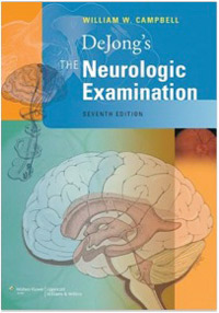
BookReview_CvrThe new edition of “DeJong’s The Neurological Examination,” by William W. Campbell, will appeal to neurophiles.  It has been modernized in many ways — the four-color edition is much easier on the eye, for one thing.  There are more images accompanying the text, with clearer photographs and MRIs to supplement the clinical vignettes.
The text has been reorganized somewhat, but still follows the general neurological encounter as most of us practice it.  This book is longer than the prior edition by almost 200 pages, but the expanded material is primarily clinical in nature, and the neuroscience underlying these observations remain mostly the same.  Most of us in practice will appreciate the balance of anatomy and pathophysiology here, as we are typically consulting such texts when we have just seen a patient with bilateral cortical ptosis, for example, and we are trying to remember whether supranuclear cortical control of the levator muscles has a left or right predominance.  This is the sort of question Campbell answers for us time and time again, with a concise description of the anatomy involved.  The “voice” of this textbook will remind you of a favorite professor from residency, and makes me envious of Campbell’s students at USUHS.

Throughout this edition, links have been inserted to various videos of the neurological exam and clinical examples of abnormal neurological signs.  Having the Kindle edition would certainly make these (hyper)links easier to use, although some reviewers on Amazon have noted that the Kindle edition makes viewing tables less ideal.  Unfortunately, after typing in four examples of links from the print edition, I found non-working videos each time.  Future editions would benefit from a webpage devoted to accompanying videos, or a DVD included in the print version.

Frequently DeJong’s is described as a book for residents, fellows or practicing neurologists.  I agree that the level of detail is likely too much for medical students, and that basic neuroscience must be mastered before attempting to understand clinical neurology.  This textbook, however, can play an important role in medical education at all levels.

For many of us neurophiles, it was the detection of some abnormal neurological sign in a patient we saw as medical students that started our lifelong interest in neurology.  In the short span of a four-week clinical rotation, we cannot expect every student to have enough patient encounters to prevent neurophobia.  Showing them how we practice the neurological exam, and what resources we use to put abnormal findings into context, like this text, may break down some of the intimidation that surrounds our specialty.

Kranick is chief of Neurology Consult Service,  National Institute of Neurological Disorders and Stroke, National Institutes of Health, Bethesda, Md.

DeJong’s The Neurological Examination
By William C. Campbell
830 pages

Parkinson’s Disease: Web-Based Education Program for Health Providers in Cameroon

Camarron_photo

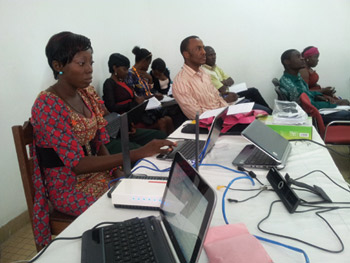
Participants in the web-based program in Cameroon.

Due to rapid demographic changes, the prevalence of Parkinson´s disease (PD) is increasing in sub-Saharan (SS) countries.1  In contrast to developed countries, evidence suggests that most patients with PD are underdiagnosed and untreated, with markedly increased mortality and shortage of qualified personnel.2  In the last years, there has been renewed interest from the World Health Organization, telecomunications companies and medical associations about the use of telemedicine in Africa.3

To help increase access to care and to train providers around the world using technology, the International Parkinson´s disease and Movement Disorder Society (MDS) has sponsored pilot projects in care and education that can lay the foundation for reaching the majority of people with PD. In this regard, a telemedicine program for health providers has been developed at Hospital Laquintinie in Douala, a 3 million inhabitants city of Cameroon.

The course is a web-based program that will provide participants with access to specialized education in the field of movement disorders, which is currently unavailable in their region. Local neurologists and professors at the University of Douala, including Jacques Doumbe, MD, chairman of the Department of Neurology (Hospital Laquintinie), and Erero Njiengwe, PhD (University of Douala) have significantly contributed to the implementation of this course at Hospital Laquintinie in Doula, Cameroon.

The course consists of 12 lectures over the course of a year, which will connect participants with experts in the field of movement disorders using live video, slides, chat and audioconferencing.  The participants will have the opportunity to receive MDS membership and its benefits, including special education to certify them to use the MDS rating scales.  Two courses have been launched — one designed for doctors (neurologists, neurology residents, primary care and internists) and another one for other health professionals (nurses, physiotherapists and psychologists).

The course will be taught by professors from Hospital Universitario de Burgos, Hospital 12 de Octubre, Hospital Clinico San Carlos, Hospital Sant Pau and Hospital Central de Asturias all from Spain; Rush University Medical Center, Columbia University, Rochester University, all in the United States, Parkinson Victoria Association, Health Sciences University all in Australia,Hospital Universitario Asturias, Spain,  North Tyneside General Hospital in Great Britain and Hospital Galway and Parkinson Galway Association in Ireland. The main objectives of this pilot telemedicine education program will be to analyze the feasibility and adherence from participants, as well as satisfaction of users.

However, telemedicine for PD strategy development in Africa is challenging.  It is still expensive, and most of the SS countries have inadequate and communication technologies infrastructure, which creates difficult implementation and little access to the population. Telemedicine also needs to demonstrate success and sustainability, and this type of initiative has to survive beyond the end of the initial funding period.  Therefore, networking provides opportunities to spread the cost of infrastructure of telemedicine development between the local governments, business, foreign education providers and health sectors; therefore the cost burden should not be borne by just one sector.  Telemedicine for PD should be recognized to remove or at least mitigate the barriers that society and physical geography imposes, especially rural areas in Africa, and to be culturally appropriate if they are to be adopted and sustained.


References

  1. Dotchin CL, Msuya O, Walker RW. The challenge of Parkinson’s disease management in Africa. Age Ageing 2007;36:122-7.
  2. Dotchin C, Walker R. The management of Parkinson’s disease in sub-Saharan Africa. Expert Rev Neurother;12:661-6.
  3. Scott RE, Mars M. Principles and framework for eHealth strategy development. J Med Internet Res. 2013 Jul 30;15(7):e155. doi: 10.2196/jmir.2250.


Coordinator

Esther Cubo MD, PhD


Academic Collaborators

Jacques Doumbe, MD; Erero Njiengwe, PhD; Sixto Cubo, PhD


Computer Engineers Team 

Centro Servicios Avanzados (CSA), Burgos (Spain), Paul Onana, Computer Engineer, Douala (Cameroon)


Faculty Members 

Ray Dorsey, MD; Christopher G. Goetz, MD; Katie Kompoliti, MD; Leo Verhagen, MD, PhD; Ellan Louis, MD; Richard Walker, MD; German Moris; Maria José Catalan, MD, PhD; Emily Wang, PhD; Meg Morris, PhD; Victor McConvey, RN; Antonio Campolungo, RN; Marie Cahill; Patrick Browne, RN; Jose Maria Trejo Gabriel y Galán MD, PhD


Sponsor 

International Parkinson´s disease and Movement Disorder Society, through the Telemedicine Task Force


Neurology Training for Non-Neurologists in West Africa Sept. 15-18, 2013

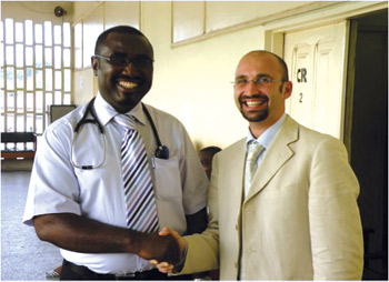
Organizers of the neurology training: Albert Akpalu (left), Korle Bu Teaching Hospital in Accra, Ghana, and Roberto Cilia, Parkinson Institute, Milan, Italy.

Organizers of the neurology training: Albert Akpalu (left), Korle Bu Teaching Hospital in Accra, Ghana, and Roberto Cilia, Parkinson Institute, Milan, Italy.

This course was jointly funded by the Movement Disorder Society (MDS) and the World Federation of Neurology. Additional co-sponsors were the Fondazione Grigioni per il Morbo di Parkinson (Italy) and the Society of Worldwide Medical Exchange (U.S.). This was the first neurology course designed for medical doctors who are not specialized in neurology. It was also open to neurologists interested in training in the field of movement disorders and dementia.

Participants were invited from throughout West Africa. Thirty-six participants attended the course, from Ghana (24), Nigeria (6), Sierra Leone (3), Gambia (2), and Cameroon (1).

The international faculty consisted of 16 international and local members, including neurologists, geriatricians, nurse specialists, a neuropsychologist, a physiotherapist, a nutrition specialist and a general practitioner from rural Ghana. The opening ceremony included guests such as the representative of the Minister of Health and of the nursing system in Ghana, the Italian Ambassador in Ghana, the chairman of Internal Medicine Department of the Teaching Hospital in Accra.

The course was run in English and included slide presentations and practical sessions with assessment of patients. Slide sessions focused on Parkinson’s disease (PD), dementia and other neurodegenerative disorders, covering epidemiology, diagnosis, pharmacological and non-pharmacological therapy. For the practical sessions with patients, participants were divided into small groups of five to six each, under the guidance of a faculty member, aiming to teach how to perform a neurological examination, neuropsychological scales screening for global and frontal-lobe cognitive functions and examples of physical therapy focused on gait and balance. Twelve patients attended these sessions, including PD, Lewy body dementia, Alzheimer’s disease and spino-cerebellar ataxia. In a session titled Bring Your Own Patient,” participants presented a patient from their own clinic and discussed with faculties the most relevant diagnostic and management challenges.

There were joint sessions with the PD Nurse Specialist Course, organized by Richard Walker (chair of the MDS Task Force on Africa).

Doctors and nurses had the opportunity to interact and share experiences about caring for patients. In particular, we included a Beyond the Neurologist session, dealing with the importance of a multidisciplinary approach to patients, especially in settings where medications are limited.

Educational material has been prepared and released for this course, including basic information about motor and non-motor features of PD and/or dementia. These booklets were drafted by different health care professionals (nurses, nutrition specialist, physiotherapists, neuropsychologists) and were full of pictures and photographs to be suitable for patients and caregivers with low education.

The course was a great success and participants’ feedback was positive in large part due to the practical sessions with patients and the interactive sessions. Each participant was given a DVD including all the slide presentations of the course, the UPDRS and the cognitive screening scales, the two educational booklets and photographs of the course. An email list was created including all the participants as well as the Italian and Ghanaian faculty, to share information and help in challenges of everyday clinical practice.

One of our aims was to make this course an opportunity to boost the education in the field of neurodegenerative disorders in developing countries. We promoted two initiatives:

  • The entire course was professionally videotaped to make it available online.
  • We provided two travel grants for the MDS events in 2014, the International Congress in Stockholm and the MDS summer school.

Course Organizers


Roberto Cilia (Italy)
roberto.cilia@gmail.com; roberto.cilia@icp.mi.it

Albert Akpalu (Ghana) 
a_akpalu@yahoo.com

Teaching Center Update

By Wolfgang Grisold and Raad Shakir

Wolfgang Grisold

Wolfgang Grisold

The WFN mission is to foster quality neurology and brain health worldwide. One of the cornerstones of this mission is to sponsor and support worldwide concepts of education. This is the function of the WFN Education Committee. In addition to other valuable tools as congresses, symposia, departmental visits and traveling fellowships.

The WFN has developed the concept of worldwide approved teaching centers. These centers should fulfill standards defined by the WFN in order to fulfill the role for training neurologists and perform this in a standardized internationally recognized manner.  Moreover, these centers will be able to train neurologists from low income countries to further their education and improve their clinical and research abilities.

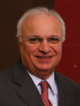
Raad Shakir

Raad Shakir

Once these centers are approved, they can offer training  fellowships, in a neurological subspecialty as well as  a full  training program to produce neurologists who will go back and serve in their own countries. The WFN approval will therefore carry the stamp of international recognition on the status and performance of a training program.

The department visit process has been adapted from the UEMS (UEMS.net)  department visitation process. This process assesses training centers, their hospital resources and equipment, and the training facilities, and has a system of assessing the opinions of all persons involved in the training process using standardized questionnaires. In addition to structured personal interviews, a randomly selected person from all groups is involved in the process. The analysis of the questionnaires and of the structured interviews are all part of the report, which also includes the activities of the hospital, access to other related fields (e.g., neurosurgery) and the completeness of the training program. The report concludes on the status of the present situation and also gives recommendations for further improvement and development.

The WFN Education Committee (Chair Steven Sergay and Co-Chair Wolfgang Grisold) has developed a structured and a well-organized questionnaire, which covers all aspects of a neurological training center.  Prior to any visit, the size, structure as well as the personnel are to be explored. In addition, residents, trainers and hospital staff will be asked to answer standardized questionnaires, aimed at various educational aspects.

The NeurologyCenter in Rabat was the first neurological center worldwide to apply for this status.

The WFN committee consisted of Wolfgang Grisold (trustee, co-chair of the Education Committee), Raad Shakir (then WFN secretary-treasurer general) and Riadh Gouider (president of the Pan African Association of Neurosciences Societies). The visitors were welcomed by the faculty of the hospital and academic members of RabatUniversity.  All information and facts about the hospital and the Moroccan neurology training system were presented.

The Rabat neurological department consists of several sections: an outpatient’s service which is closely associated with neurosurgery, neuroradiology, neuropathology and with the national center of rehabilitation and neurosciences.

The practical visit was guided through the neurological departments, as well as the laboratories, including histopathology, chemistry and genetics. During the visit, patients and trainees, as well as post-graduate and undergraduate students, were met and some questions and discussion were carried out.

The visit also included the electrophysiology laboratories, where practical teaching was observed. The associated departments of neurosurgery and neuroradiology were well equipped, modern CT, MRI, angiography suites and gamma knife were all observed.  There is also a newly adapted rehabilitation unit. The outpatient and emergency as well as the consultation system of the hospital were looked at in detail. The academic research unit of the university provides laboratories including an animal house with space for scientific cooperation, which are open to projects from the neurological department.

The structure of the visit was supplemented by interviews with residents, teaching professors and hospital staff. These interviews were performed in a standardized written format, which was distributed prior to the visit to faculty and current residents. In addition, three teachers and four residents were randomly selected and attended personal interviews. They were asked standardized questions. Also the director of the hospital was personally interviewed. The visiting WFN committee carried out the confidential interviews. A résumé was contained in the report.

Based on the visit, including discussions, there were presentations of the faculty, and detailed analysis of the various aspects of the department.  The visiting committee was fully satisfied that the Hospital Center Ibnou Sina, Rabat, is fully compliant with all aspects of a WFN training center. It will be able and willing to train not only its own national neurologists, but neurologists from Africa and elsewhere and will be providing one year fellowships, as well as a full four year training programs.

The concept of WFN teaching centers will be further developed, and the WFN website will provide a detailed definition of teaching centers, as well as the requirements, questionnaires and application details.  Being a WFN teaching center is a sign of excellence and clearly conveys international recognition of the teaching center in that it fulfills all of the criteria in its structure, equipment and the teaching facilities needed to participate in the important role of neurology education in the future.

Theodore L. Munsat (1930-2013)
An Outstanding Legacy with the WFN

By Marco T. Medina, Dean of the National Autonomous University of Honduras

Marco T. Medina

Marco T. Medina

The World Federation of Neurology (WFN) lost an outstanding leader on Nov. 22, 2013, with the death of Professor Theodore Leon Munsat (“Ted”), in Waltham, Mass., U.S., at age 83.

Munsat was Emeritus professor of Neurology at Tufts University School of Medicine and served the WFN in several capacities as trustee, chairman of the WFN Education and Research Committees, chairman of the WFN ALS Research Group and founding director of the WFN Seminars in Clinical Neurology. He was president of the American Academy of Neurology (AAN) 1989-1991, chairman of the Continuing Educational Committee of the AAN and founding director of AAN’s premier continuing medical education journal Continuum: Lifelong Learning in Neurology1, 2.

Munsat was born in Portland, Maine, in 1930, to Leo and Ethel Munsat. When he was a child, the Munsat family moved to Rutland, Vt. He graduated from Rutland High School in 1948. He received his BA degree in chemistry at the University of Michigan, and in 1957, his MD degree from the University of Vermont, and then completed an internship at Mt. Sinai Hospital in New York, followed by a neurology residency with Houston Merritt at the New York Neurological Institute, Columbia Presbyterian Medical Center. He completed his training with Augustus S. Rose at the University of California in Los Angeles (UCLA). After serving in the Navy for two years, he returned to UCLA in 1963 as assistant professor of Neurology and director of the Muscular Dystrophy Clinic where he worked with Carl Pearson. In 1970, he moved to the University of Southern California, first as associate professor, and then, in 1973, as professor of Neurology. In 1975, he took a 12-month sabbatical in Newcastle upon Tyne, U.K, with Lord Walton. In 1976, he became  chairman of Neurology at Tufts University and the New England Medical Center1.

He was a worldwide leader in ALS research and as chairman of the WFN ALS Research Group that published important international diagnostic guidelines. He authored more than 200 scientific articles and books, including classic texts as Amyotrophic Lateral Sclerosis: A Guide for Patients and Families, Post-Polio Syndrome and Quantification of Neurological Deficits. He received many honors during his long career, including the A.B. Baker Award for Education of the AAM, the Sheila Essey Award for ALS Research, the Lifetime Achievement Award of the WFN Research Group on Neuromuscular Diseases and the degree of Doctor of Medicine, Honoris Causa, by the University of Marseilles1.

Munsat as the WFN chairman of the Education Committee developed a number of successful educational programs around the world:

Ted Munsat

Ted Munsat

  1. A continuing medical education (CME) program  using the journal Continuum, generously donated by the American Academy of Neurology, and  the WFN Seminars in Clinical Neurology.  More than 42 developing countries  participated  in this program, including Argentina, Bangladesh, Brazil, Bulgaria, Cameroon, Chile, Columbia, Congo, Croatia, Cuba, Cypress, Czech Republic, Egypt, Ethiopia, Guatemala, Honduras, Hungary, India, Iraq, Jordan, Kenya, Lebanon, Lithuania, Macedonia, Mexico, Mongolia, Nigeria, Panama, Peru, Romania, Russia, Serbia, Slovenia, Sri Lanka, Syria, Tunisia, Uganda, Uruguay, Vietnam, Venezuela, Yemen, and Zambia  with an extraordinary  local impact3-7.
  2. Support for Neurologic Training program and WFN Certification:  Munsat founded  programs for the WFN to provide assistance in establishing neurology training programs where there were none, helping further develop existing programs and providing a certification process for programs that wished to have an external review. He worked with programs in Ethiopia, Honduras, Guatemala,  Mexico, Peru, etc.3,5,8.

The first pilot program was the Honduras Neurology Training (HNT) Program. Munsat and Professor Alberto Portera Sanchez from the WFN Education Committee visited Honduras in July 1998 for the first time, and after an evaluation, they concluded: “It is our recommendation that a training program in neurology should be established at the Hospital Escuela. We believe that there is a pressing need for more neurologists to address the unmet health needs of the people of Honduras. The relevant members of the medical school faculty have expressed   their strong support of such a program. More than adequate human and structural facilities are currently available. There is no reason why this program could not be a program of unqualified excellence and effectiveness.”

After the initial visit, the National Autonomous University of Honduras established the country’s first Neurology Training Program in 1998. This program was established using a problem- and epidemiological-oriented methodology with oversight by an external WFN review board. By 2013, the program resulted in a 50 percent increase in the national neurologist ratio per inhabitant, significantly improving the quality of patient care and promoted research in the neurosciences. During 10 years, Munsat and Sanchez have visited and evaluated annually the HNT program.  The legacy of Munsat in our program and country is invaluable5,8.

  1. Neurologic care where there is no neurologist. With Gretchen Birbeck, he supported the development of training materials for nonphysician health care providers and established a program for non-physician neurologist clinical officers in Zambia and  Malawi3,9.
  2. The WFN Africa Initiative. Munsat,Johan Aarli, Gretchen Birbeck, Gallo Diopp  and others started this initiative  Six sub-Saharan countries started their participation in the WFN Continuing Education program: Cameroon, Uganda, Zambia, Nigeria, Ethiopia, and Kenya. He helped on the development of the Ethiopia Neurology training program3.
  3. London WFN Education Committee meetings: Since 1993 Munsat organized successful planning meetings in London with members representing WFN regions around the world.
  4. WFN Seminars in Clinical Neurology.  Munsat founded the Seminars in Clinical Neurology because of the lack of educational material specifically designed and directed to neurology health care givers practicing in low resource environments3.

It was a great privilege and honor for me to have met and worked with Munsat. He was gentle, gracious mentor with a warm and sincere personality, who loved helping others every chance he had. He inspired me as well several generations of residents and neurologists all around the world, and he left an outstanding legacy with the WFN.

Munsat  is survived by his  wife, Carla Munsat; his daughter, Amy Munsat,  his son, Peter Munsat  and  six grandchildren. •

References

  1. Rowland TL. Theodore L. Munsat MD: President American Academy of Neurology. Neurology 1987;37:548-9
  2. Munsat TL, Mancall EL, DesLauriers MP. The AAN launches a new education program: CONTINUUM lifelong learning in neurology. Neurology 1994;44:771-2.
  3. Munsat T, Aarli J, Medina M, Birbeck G, Weiss A. International Issues:educational programs of the World Federation of Neurology. Neurology. 2009;10;72(10):e46-9.
  4. Gusev EI, Konovalov AN, Volodin NN, Munsat T, Fedin AI, Khaass A, Gekht AB, Diankina MS, Martynov MIu, Kamchatnov PR, Beliaeva IA, Lebedeva AV, Kovaleva IIu.[Continuing medical education in Russian neurology and neurosurgery]. Zh Nevrol Psikhiatr Im S S Korsakova. 2009;109(5):5-9.
  5. Medina MT, Munsat T. Neurology education in Latin America and the World Federation of Neurology. J Neurol Sci. 2010; 15;298:17-20.
  6. Medina MT, Munsat T. Continuing medical education in developing countries. J Neurol Sci. 2003; 15;190:1-2.
  7. Correale J, Allegri RF, Pelli-Noble RF. Background of the Sociedad Neurologica Argentina: current state and concerns about neurologic education. Neurology. 2013;80:1978-80
  8. Medina MT, Munsat T, Portera-Sánchez A, Durón RM, Becker CA, Holden KR; WFNEducation Committee. Developing a neurology training program in Honduras: a joint project of neurologists in Honduras and the World Federation of Neurology. J Neurol Sci. 2007;253:7-17.
  9. Birbeck GL, Munsat T. Neurologic services in sub-Saharan Africa: a case study  among Zambian primary healthcare workers. J Neurol Sci. 2002;200:75-8.