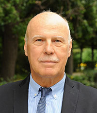
Multidisciplinary Strategies to Prevent and Combat Brain Diseases-Moscow

By Wolfgang Grisold

An international conference, focusing on “multidisciplinary strategies to prevent and combat brain diseases,” took place in Moscow on November 27-28, 2019, and followed the concept of integration and multidisciplinarity. The organization was smoothly run by Prof. Alla Guekht and her team and linked neurology to neurosurgery, internal medicine, and psychiatry. The meeting was held in the Buyanov’s city hospital, and needless to say was equipped with all facilities, including press briefings before the meeting.

Wolfgang Grisold

The opening ceremony gave greetings from the Russian health care minister, Veronica Skvortsova, the Moscow head of the health care department, Alexey Khripun, the president of the All-Russian Society of Neurologists, Y.I. Gusev, and from the leading international neurological organizations — WFN, EAN, and ELAE. A short presentation about the WFN activities in regard to education, global networks, and cooperation with the WHO was made.

Dr. J. Breda, as the chair of the European regional NCD bureau of WHO, gave an initial presentation on the work of the WHO, primary care, and also aspects of environment and nutrition, which are eminently important for many non-communicable diseases (NCDs). This talk and statements are important for the mutual understanding of the specialization of neurology and the activities of the WFN. This was followed by a talk focusing on the historic role of Russian neurology. The impressive figure of the beginning of the 20th century, Bekhterev, was discussed, including his longing for innovation and his successful achievements in neurology, psychiatry, and health care organization and education.
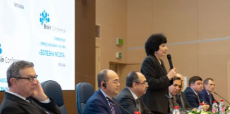
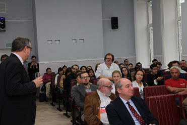
Opening ceremony
From left to right: Prof. M .Hilz (Germany), Prof. D. Muresanu (Romania), V.N. Buzin (Ministry of Health of the Russian Federation), Prof. A.B.Guekht (Russia), Dr. J.Breda (Portugal), Prof. W. Grisold (Austria), Prof. N.A. Shamalov (Russia), Dr. A.V.Salikov (Russia).

During the first day of the conference, several other topics, such as stroke, rehabilitation, the autonomic nervous system, and neurooncology, were presented. Although these topics have defined contents being discussed at many occasions, the keynote speakers were able to pick out exciting developments and add valuable information.

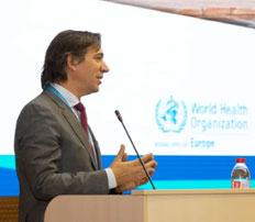
J. Breda, the head of the European Office for the Prevention and Control of Noncommunicable Diseases (NCDs) in Moscow.

The second day entered the often-neglected chapter of neuropsychiatry, which reemerges in international fields and undergoes a metamorphosis from conversion into functional disorders. This lecture by Prof. W. Curt LaFrance Jr. (U.S.) made two important points: functional disorders exist and may be more frequent than we admit, and several concepts of explanation and treatment were available. Another message was that a society for functional disorders has been recently established.

A complex of lectures discussed epilepsy and seizures. Epidemiology and the complex meaning of incidence and prevalence were beautifully and carefully explained by Prof. W. Allen Hauser (U.S.), who shared his insights into this important basis of his work and considerations. Several other lectures addressed epilepsy and seizures, and were supported by excellent videos of patients, EEG recordings, and imaging.

Prof. Hilz and Prof. Mkrtchan discussing the study on autonomic dysfunction in epilepsy.

The end of the second day was devoted to carefully prepared case presentations, which focused on several topics, such as complicated cerebrovascular disease, genetic diseases, complex seizures, neuroinfections, and rare neuropathies. In several cases, speakers from other disciplines were invited to make valuable comments and give diagnostic clues. The spirit of these case presentations was enthusiastic and also made it clear that neurology is deeply rooted in multidisciplinary and multiprofessional work. From the engagement of speakers and the audience, these short case presentations seemed to become a well-accepted format.

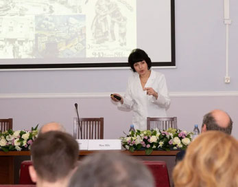
Advisory board meeting: Prof. A. Guekht presenting the achievements of the Moscow Research and Clinical Center for Neuropsychiatry.

On the third day, there was a meeting in the Moscow Research and Clinical Center for Neuropsychiatry within the international advisory board of the center. The members of international and Russian faculty discussed ongoing collaborative projects on comorbidity of epilepsy and mental disorders, self-harm behavior, the role of neuroinflammation in stress, and some others. Junior researchers of the center presented their papers and interesting cases.

Research labs and clinical facilities of the center were presented. The members of the board appreciated the high level of the technologies in the labs and the clinical wards of the center.

The size and the spirit of the meeting also allowed many personal and professional interactions, and faculty members interacted with local neurologists, allowing the platform for advice and cooperation to continue.

Having attended these meetings for brain diseases for several years, the interaction of attendees with the faculty is increasing, the magnitude of interaction and discussion during the meeting is developing, and the impression is that young neurologists and psychiatrists are increasingly fluent in English, which helps increase cooperation and communication. These types of integrative meetings, with a strong educational accent, are valuable not only from the point of content, but also from communication.

Wolfgang Grisold is Secretary General of the WFN.


WFN/FINE Neuroinfection Series

BY CHANDRASHEKHAR MESHRAM

Neuroinfections form a large group of disorders commonly seen by neurologists as well as other physicians. Old, new, and emerging infections, their patterns of presentations, and imaging features as well as therapies pose challenges in practice. There are not many meetings that address these issues. Therefore, the Tropical and Geographical Neurology Specialty Group of World Federation of Neurology (WFN) in collaboration with the Forum for Indian Neurology Education (FINE) and under the aegis of the World Federation of Neurology has organized a neuroinfection webinar series. This program is dedicated to Prof. Jagjit Singh Chopra.

The webinar-based meeting began on July 18, 2020, and will run every Saturday 7-9 p.m. Indian Standard Time (IST)/1:30-3:30 p.m. Coordinated Universal Time (UTC). There will be a weekly lecture by a renowned neuroinfection expert, covering many aspects followed by presentation and discussion of three cases.

Registration is free, and the link for registration can be found here: http://neurologycourses.com/user/registration.

The program can be accessed at https://us02web.zoom.us/j/801815712 using the password “ilovefine” with no punctuation.

The event was formally inaugurated on July 18 by Prof. Raad Shakir, former president of the WFN and chair of the specialty groups. Prof. Hector Garcia, from Peru, delivered the first talk in the series on neurocysticercosis. Participants had the opportunity to interact with the speaker through a question and answer session after the talk. This was followed by presentation and discussion on three interesting and challenging cases.

Subsequent talks will be on Saturday, July 25, by Prof. Erich Schmutzhard (Austria) on cerebral malaria, on Aug. 1 by Prof. Marco Tulio Medina (Honduras) on Zika and other arbovirus infections, on Aug. 8 by Prof. Riadh Gouider (Tunisia) on neurobrucelloisis, on Aug. 15 by Prof. Serefnur Ozturk (Turkey) on encephalitis, on Aug. 22 by Prof. Jeremy Day (Vietnam) on cryptococcosis, on Aug. 29 by Prof. Avindra Nath (USA) on the approach to neuroinfections, and on Sept. 4  by Prof. Joseph Berger (USA) on fungal infections of the CNS.

Interesting and challenging cases of dengue, chikungunya, rabies, ebola virus, nipah encephalitis, neuroschistosomiasis, neurognathostomiasis, trypanosomiasis, onchocerciasis, neuroangiostrongylosis, amoebic encephalitis, sarcocystosis, CNS TB, HIV, and other diseases will be presented and discussed during the series.

We welcome delegates from different countries to participate and present the cases. Those interested in presenting cases should write to the moderators of the program. Chandrashekhar Meshram, president of the Tropical and Geographical Neurology Specialty Group, is the program director. Gagandeep Singh and Rahul Kulkarni are moderators, while Sudhir Kothari and Roop Gursahani are advisors.

Long-Haul COVID

By Avindra Nath and B. Jeanne Billioux

By Avindra Nath and B. Jeanne Billioux

It is becoming increasingly apparent that many patients who recovered from the acute phase of the SARS-CoV-2 infection have persistent symptoms. This includes clouding of mentation, sleep disturbances, exercise intolerance, and autonomic symptoms. (See Tables 1 and 2 below) Some also complain of temperature dysregulation and lymphadenopathy. Although there are no peer-reviewed papers at the moment on these patients, many news articles have been written about this phenomenon1-4 and apparently there are Facebook pages where there are several thousand patients with these symptoms. They describe themselves as the “Long-Haul-COVID” or “Long-Tail COVID.” Many of these patients are health care workers who had massive exposure to the virus early in the pandemic and describe having symptoms for 100+ days. Since many of the symptoms are neurological in nature, these patients are seeking us out for help. It is important that we characterize these patients and try to document the objective findings and then determine how best to study their pathophysiology to develop proper guidelines for treatment.

Most of these patients were in excellent health prior to getting infected with SARS-CoV-2. They all had myriad symptoms during the acute phase; however, as the fever and respiratory symptoms improved, they are left with persistent systemic symptoms, some of which are gradually improving, but not all are following that course. While some were admitted to the hospital due to pulmonary symptoms, the majority were isolated at home. Many of these symptoms overlap with those of patients with myalgic encephalomyelitis/chronic fatigue syndrome (ME/CFS). The cause of ME/CFS remains unknown despite decades of research of the syndrome. Many of these patients similarly report a viral infection as a trigger, but since they come to our attention months and years after symptom onset, it is impossible to know what may have triggered the symptoms.

Symptoms of Long-Haul COVID

  • Insomnia or frequent awakenings
  • Inability to concentrate
  • and think clearly
  • Easy fatiguability despite normal lung function
  • Anorexia or increased appetite
  • Temperature dysregulation
  • Lymphadenopathy
  • Dysautonomia

Table 1. Symptoms.

Long-Haul COVID thus represents an excellent opportunity to study the pathophysiology of ME/CFS that may have broader implications. It is unclear at present if these patients may have endocrine abnormalities, but certainly with SARS-CoV-1, hypothalamic/pituitary dysfunction and adrenal insufficiency has been reported5. It would, of course, be equally important to determine if there might be any premorbid conditions or medications that might contribute to these symptoms.

Autonomic symptoms in Long-Haul COVID

  • Tachycardia upon mild exercise or standing
  • Night sweats
  • Gastroparesis
  • Constipation
  • Peripheral vasoconstriction

Table 2. Autonomic symptoms.

Even at this early stage, it might be important to determine what the potential pathophysiological mechanisms might be. So far there is no convincing evidence for widespread infection of the brain with the virus. The virus has been detected in CSF and the brain in very rare cases; however, there is evidence for widespread glial cell activation that may be related to metabolic dysfunction or to the massive immune activation in the periphery. Other possibilities are specific immune responses targeted against specific regions of the brain and autonomic nervous system. Depending on the predominant underlying pathophysiological mechanism at play, targeted treatment might be possible.

Unfortunately, it looks like the pandemic has gotten out of control. It is spreading rapidly across the globe, and even if we have an effective vaccine, we might never have enough dosages to vaccinate the eight billion inhabitants on this planet. And we also have a subset of the population that is opposed to any kind of vaccination. So COVID-19 is here for the long haul, and neurologists are going to play a critical role in the management of these “Long-Haulers.” Several efforts are underway to prospectively follow these patients with persistent symptoms, including studies at the intramural program at the United States National Institutes of Health in Bethesda, Maryland. Hopefully we can get to the bottom of these manifestations soon and find solace for these patients in a timely manner. •

Avindra Nath, MD, is chief of the Section of Infections of the Nervous System and Clinical Director, National Institute of Neurological Disorders and Stroke (NINDS), National Institutes of Health in Bethesda, Maryland.

Jeanne Billioux, MD, is staff clinician and head of the program in International Neuroinfectious Diseases within NINDS. Her research focus is on emerging infectious diseases and conducting research on the neurological consequences of infections in an International setting.

References:

  1. https://www.wsj.com/articles/three-months-in-these-patients-are-still-ravaged-by-covids-fallout-11593612004
  2. https://www.washingtonpost.com/health/could-covid-19-cause-long-term-chronic-fatigue-and-illness-in-some-patients/2020/05/29/bcd5edb2-a02c-11ea-b5c9-570a91917d8d_story.html
  3. https://www.bloomberg.com/news/videos/2020-06-10/the-lingering-symptoms-of-covid-19-video
  4. https://www.washingtonpost.com/health/2020/06/11/coronavirus-chronic/?arc404=true
  5. Leow MK-S, Kwek DS-K, Ng AW-K, Ong K-C, Kaw GJ-L, Lee LS-U. Hypocortisolism in survivors of severe acute respiratory syndrome (SARS). Clin Endocrinol. 2005;63(2):197-202.

New JNS Metrics Released

By John D. England, MD

John D. England, MD

John D. England, MD

I am pleased to announce that new metrics have been released for the Journal of the Neurological Sciences, the official journal of the World Federation of Neurology (WFN). The widely cited Impact Factor (IF) has risen to 3.115, which is a +17.5% increase compared to 2018. This is an all-time high for the journal’s Impact Factor. The Cite Score, which is the average citations per published peer-reviewed document, has risen to 4.9 compared to 4.4 in 2018. Moreover, the worldwide penetration, usage, and number of downloads from the journal have increased steadily. The Journal of the Neurological Sciences is now truly an international and well-respected journal.

Many individuals are responsible for the success of our journal. I especially wish to recognize our Managing Editor, Dr. Nicole Villemarette-Pittman, and our Associate Editors, Drs. Carmel Armon, Andreas Charidimou, Hamilton Farris, Daniel Truong, and Donald Silberberg. The continued support of Elsevier and the World Federation of Neurology (WFN) has allowed our journal to prosper and improve. I am grateful to the leadership of the WFN for its continued encouragement and support. Drs. William Carroll, Raad Shakir, Wolfgang Grisold, Marco Medina, Walter Struhal, and Steven Lewis have all provided notably important contributions to the Journal of the Neurological Sciences.

In addition, I wish to thank our Editorial Board Members, authors, reviewers and readers for their support and encouragement. With the help of so many, I am confident that our journal will continue to serve the global community of neurology and neuroscience. •

John D. England, MD, is editor-in-chief of the Journal of the Neurological Sciences, the official journal of the WFN.


Diseases of Civilization and Nervous Disorder in the Age of Enlightenment

by Peter J. Koehler

Figure 1. George Cheyne

The relation between civilization and disease has been known for centuries. Researchers found evidence that in the pre-history period, small hunter-gatherer populations tended to be less troubled by disease. With growing population density following the settlement of farmers around 12,000 years ago (in the Neolithic period), and later the start of commercial traveling and increasing mobility, pathogens could spread easier. Yersinia pestis (bubonic plague in the Late Middle Ages), Treponema pallidum (syphilis, starting around 1500), and Variola (smallpox, in particular during the 17th and 18th century) are well-known examples. Sometimes whole nations were associated with a disease, the best example being the “Spanish disease” and “morbus Gallicus,” referring to syphilis. In the Renaissance period, there was already some knowledge about the spread, and quarantine protocols were promoted in some places upon the arrival of ships (40 days, hence the word quarantine, from the French “quarant”). In periods of plague, markets and city gates were closed.

Not Only Contagious Diseases

Another type of disease of civilization was recognized in the 18th century and associated with the “secularized revamping of the Christian legend of the Fall, wherein Original Sin and the expulsion from Paradise had inaugurated the regime of hard labor, disease, suffering, and death in the temporal world.”1 Several physicians in this period, including the Scottish physician George Cheyne (1672-1743; Figure 1; not to be confused with John Cheyne [1777-1837]), who practiced in London and Bath, and the Swiss physician Samuel August Tissot (1728-1797; Figure 2), and also philosopher Jean-Jacques Rousseau (1712-1778) pointed to the relatively healthy hunter-gatherer and peasant people of the early days, who were engaged in physical labor, exercise in the open air, and taking healthy foods. Town life in the 18th century with its commerce, wealth, indoor work, and bad habits, including taking too much food and alcohol and too little exercise, could easily lead to health problems, which often resulted in the use of opiates, tobacco, more alcohol, and remedies.1 According to Tissot, “gens des lettres” were particularly prone, and he even dedicated a book to the subject in 1768, De la santé des gens des lettres [On the health of men of learning], which was published in several editions at least up to 1826 and considered an important publication in the history of occupational disease. In his preface, the author indeed referred to the pioneer Bernardino Ramazzini (1633-1714, author of De Morbis Artificum Diatriba / Diseases of Workers).

Tissot’s Santé des gens des lettres

Figure 2. Samuel-Auguste Tissot (Collection de l’université de Lausanne

In his De la santé des gens des lettres (Figure 3), Tissot referred to numerous cases from medical literature described by well-known physicians. The association was already known ages ago, as for instance Galen of Pergamon (129-c210) described the history of the peripatetic philosopher Premigenes, who lived a life of reading and writing, thereby not perspiring well, for which he had to take a bath every day, to avoid accumulation of the sharps humours.2 More recent physicians included Jean Fernel (1497-1558), Johann Jakob Wepfer (1620-1695), and Giovanni B. Morgagni (1682-1771), who associated mental efforts and exhaustion, which led to severe or even mortal disease. Wepfer, for instance, mentioned the case of a young man (22 years old), who studied day and night, became delirious, hurt several people, and killed his guard.2 Tissot also referred to Gerard van Swieten (1700-1772) and his Commentaria in Hermanni Boerhaave aphorismos de cognoscendis et curandis morbis (5 vols. 1742-72), in which he wrote about “Les Gens de Lettres qui mènent une vie sédentaire, & qui pâlissent sur leurs livres, sont souvent exposés à une apoplexie.” [“Men of learning, who live a sedentary life, and turn pale above their books, are often exposed to apoplexia.”)2 Hypochondria was also a well-known ailment, Tissot believed. “Parmi les maux que la vie sédentaire de Hommes des Lettres produit presqu’inévitablement en dérangeant la circulation dans les viscères du bas-ventre & y produisant un principe d’obstructions, on doit comptant l’hypocondrie.” (“Among the ailments that are almost inevitably caused by the sedentary life of men of learning by deranging the circulation of the bowels of the abdomen and there producing a source of obstructions, one has to reckon hypochondria.”)2 Tissot not only wrote on men, but also mentioned women, for instance a 50-year-old woman, who had read books at night in her youth and then started suffering from insomnia and fluxions. With respect to the causes, he mentioned the air around these persons that was rarely freshened, in contrast to the pure and fresh air from the fields, continuation of the work during a part of the night, and lack of exercise leading to “cardialgia,” which had already been noted by Aretaeus of Cappadocia (first century CE) and Aetius of Amida (mid-fifth and mid-sixth century).2

Rousseau

Figure 3. Title page of Tissot’s book [On the health of men of learning

At the time, a well-known critic of civilization was Rousseau. An interesting section of Tissot’s book deals with the preface of Rousseau’s play Narcisse ou l’amant de lui-même (“Narcissus or the lover of himself;” 1753). Reading the original text, Rousseau indeed wrote about his concerns with respect to “Le goût des lettres, de la philosophie & des beaus arts amollit les corps & les ames.” (“The fondness of letters, philosophy, and fine arts, weaken the body and soul.) He continued, noting that “Le travail du cabinet rend les hommes délicats, affoiblit leur tempéramment, & l’ame garde difficilement sa vigueur quand le corps a perdu la sienne. L’étude use la machine, épuise les esprits, détruit la force, énerve le courage, et cela seul montre assez qu’elle n’est pas faite pour nous : c’est ainsi qu’on devient lâche et pusillanime, incapable de résister également à la peine & aux passions.” (“Office work makes people delicate, weakens their temperament, and the soul can keep its strength more difficult, when the body has lost it. Study wears out the machine, exhausts the spirits, destroys the power, weakens courage, and just that sufficiently shows that it has not been made for us: This is how one becomes unresisting and faint-hearted, incapable to resist distress as well as passions.”)

Seated in the Nervous System

Several physicians, including the mentioned Cheyne and Tissot, believed that these modern diseases from abundance and sedentary life had their seat in the nervous system, writing about a nervous tone. These physicians did not always apply contemporary humoral pathophysiological theories that still reigned, at the time (although Cheyne was still writing in terms of “interruption or interception of the vibrations of the nerves by the viscidity of juices”). The excessive food and alcohol, in combination with a lack of exercise, would result in obstruction of nervous fibers that led to all kinds of complaints that were not observed in people who led a healthier life in the countryside. The term “nervousness” became fashionable for physicians as well as patients. Physicians came to believe that many of the current diseases had a nervous origin. Remarkably, nervous disorders had a positive connotation as it was believed to be observed in particular in intelligent and rich people, the beau monde. Interestingly, gout, at the time, was another disease that had the same connotation and was associated with wealth, food, and alcohol.

In this way a psychosomatic paradigm seemed to play an important role in the age of Enlightenment. Not only organic causes, but also ideas, imagination, and emotions were taken into consideration. Much more could be said about the further development of this concept during the 19th century — degeneration ideas, social Darwinism, nervous exhaustion, and neurasthenia with the work of George Beard (1839-1883) and Silas Weir Mitchell (1829-1914) — and 20th century — eugenics starting with Francis Galton (1822-1911) and ending in the Nazi cruelties — but let us keep to the 18th century here.

Cheyne’s English Malady

Figure 4. Title page of Cheyne’s book.

Not only the mentioned part of society was susceptible for these diseases, but even whole nations were associated with it. Cheyne’s book was named The English Malady (Cheyne, 1733; Figure 4). In the preface he noted that “all our Neighbors on the Continent, by whom nervous Distempers, Spleen, Vapors, and Lowness of Spirits, are in Derision called [it] the English Malady. And I wish there were not so good Grounds for this Reflection.” He continued to mention the reasons for this, including “the Moisture of our Air, the Variableness of our Weather … the Rankness and Fertility of our Soil, the Richness and Heaviness of our Food, the Wealth and Abundance of the Inhabitants (from their universal Trade), the Inactivity and sedentary Occupations … and the Humor of living in great, populous and consequently unhealthy Towns.”4 Furthermore, it is of interest to write out the whole title of the book, as it occurs on the title page, The English Malady: or, a Treatise of Nervous Diseases of all Kinds, as Spleen, Vapours, Lowness of Spirits, Hypochondriacal, and Hysterical Distempers, as well as the titles of the three divisions of the book, notably Part I. Of the Nature and Cause of Nervous Distempers; Part II. Of the Cure of Nervous Distempers; Part III. Variety of Cases that illustrate and confirm the Method of Cure; to which Cheyne added “With the Author’s own case at large.”

In chapter six, “of the Frequency of Nervous Disorders, in later Years, beyond what they have been observed in former Times,” we find information on the three main causes, including 1) luxury; 2) an inactive, sedentary, or studious life; and 3) living in great and populous cities. Most, if not all, of the cases described came from the higher classes, indeed finishing with an extensive description of his own condition, with intermittent fever and “vertiginous Paroxysm, so extremely frightful and terrible, as to approach near to a Fit of an Apoplexy … I found after this, some small Returns of my Vertigo (in Bed especially) on lying on a particular Side…” (This episode seems to have been benign paroxysmal positional vertigo.) Later he suffered from headache, depression, and anxiety, as he “went about like a Malefactor condemned, or one who expected every Moment to be crushed by a ponderous Instrument of Death”. 4

Figure 5a. From Classification of diseases from William Cullen’s Synopsis Nosologiae Methodicae (American edit. of 1783).

In general, the nervous distempers were usually treated by medicaments, diet, and exercise. The author himself resorted to omitting suppers, eating less animal food at dinners, “and drinking very little fermented Liquor,” which resulted in losing several of his friends. He retired to the countryside “into the fine Air,” took medicaments, had setons made in the neck, and took gentle purges. He suffered from melancholy, lost weight, and called it a “Nervous and Scorbutical Disorder.”4 In his Essay on Regimen (1740), which is often quoted by vegetarians, as he advised vegetables rather than animal food, he even provided a number of aphorisms “to instruct and ease the Memory of the Valetudinarian.” Among the regimens, he emphasized horseback riding in aphorism no. 25: “Rideing is the best of all Exercises to get Health, and to promote the Digestions, especially in nervous Distempers”.5 He believed “Hypochondres are thereby most shaken and exercised.”4 In a letter to writer and printer Samuel Richardson (1689-1761), Cheyne advised “riding on the new Chamber Horse so well known in London. It is certainly an excellent Contrivance for the Sedentary, Studious, and Thinking Part of Mankind except the fresh Air.” He was using the device himself an hour every day and “do more when the Weather will not permit me to walk in the Garden or ride my Coach.”

Nosological Systems and Neurosis

Figure 5b. From Classification of diseases from William Cullen’s Synopsis Nosologiae Methodicae (American edit. of 1783).

The importance of the nervous system as the origin of diseases was also recognized by Robert Whytt (1714-1766), professor of the theory of medicine at the University of Edinburgh, although he had a different approach. He noticed that nervous diseases may mimic somatic conditions and can be strongly influenced by emotions, sometimes being triggered by intense imagination. His successor Wiliam Cullen (1710-1790), who drew up an important nosological system, believed that the category of nervous diseases — he coined the term neurosis — was the most important among four categories (although historian of science Dyde pointed to the differences of view with this respect between Whytt and Cullen). In this age of nosology, in which diseases were classified in a similar way as Carl Linnaeus (1707-1778) did with the plant world (he also made a nosological system of diseases), Cullen classified diseases into 1) pyrexiae (fevers); 2) neuroses ; 3) cachexiae; and 4) locales (Figure 5). His classification influenced the French physician Philip Pinel (1745 – 1826), who recognized neuroses of the senses, cerebral function, locomotion and voice, nutrition, and sexual function. It may be regarded a mixture of diagnoses from modern neurology and psychiatry, although other diseases were still classified within this category. The class of neurosis mainly included diseases that Pinel considered “functional,” and this contained more diseases than we would call psychiatric nowadays (and the term cannot be equated with its later use by Freud). This functional orientation of neurosis remained during almost the whole 19th century, though the category of neuroses became smaller in the course of the century as more diseases were found to have a neuropathological substrate. In the 1880s, for example, William Gowers (1845-1915) mentioned the following functional disorders: chorea, paralysis agitans, tremor, tetanus, tetany, occupation neurosis, etc.

Conclusion

In conclusion, the 18th century disease of civilization, associated with abundance and sedentary life, and its treatment looks familiar to us today. The association these physicians made with the nervous system is of interest against the background of the psychosomatic perspective. The concept of nervous disorder or neurosis at the time, however, was much broader than today, a kind of repository, and cannot be compared with its use in the early 20th century (psychoneurosis).


C. Miller Fisher: Stroke in the 20th Century

by Louis R. Caplan, Oxford University Press 2020

by Vladimir Hachinski

Who was the greatest contributor to the field of stroke in the 20th century?” If you answer the question, you risk displeasing somebody somewhere with a different opinion. On being asked that question after a lecture in Moscow, I answered: “The greatest contributor to the understanding of the common causes of stroke is C. Miller Fisher.” Few would disagree. He established the relationship of carotid disease to ischemic stroke, characterized the clinical and pathological feature of lacunar syndromes, and proved that atrial fibrillation can cause stroke in the absence of cardiac valvular disease.

Lou Caplan is one of Miller Fisher’s most distinguished pupils and colleagues. He describes these and others of Miller Fisher’s contributions in historical context.

Carotid Disease

Hans Chiari in 1905 described the relationship of carotid stenosis to stroke. If the implications were ever appreciated, they were soon forgotten. Transient ischemic attacks (TIAs) were recognized, but most physicians accepted the explanation offered by the great Canadian/American/British physician William Osler, who attributed them to “vasospasm.” The explanation of vasospasm as a cause of TIAs proved plausible, convenient, and wrong. Miller Fisher observed the transient platelets and other materials through the retinal arteries during a TIA and concluded that embolism from the extracranial arteries was the common cause of TIAs. It was MiIler Fisher who established that carotid disease could lead to stroke and laid the scientific bases for carotid endarterectomy.

Lacunar Syndromes

Miller Fisher described five classical lacunar syndromes based on extensive clinical observations and meticulous brain dissections. As atherosclerotic large vessel disease decreases with better treatment, small vessel disease gains greater prominence.

Atrial Fibrillation

When Miller Fisher began his career, atrial fibrillation as part of rheumatic heart disease was recognized as being associated with stroke, but not as a cause by itself. In 1977 he published a study that left no doubt that atrial fibrillation by itself could cause stroke though cardiac embolism. Even before his definitive study, he had been advocating prophylactic anticoagulation in atrial fibrillation, still the mainstay of prevention.

Caplan’s own clear writing is enhanced by quotes from Miller Fisher’s detailed memoirs spanning a long life. The main headings make for easy reading and scanning.

Caplan’s direct knowledge of the field, of the main players and his own contributions allow him to state: “This book is also in many ways a biography of a disease (stroke) as it evolved during the 20th century.” This is not to deny the contribution of others, as Caplan has documented himself in his co-edited book on stroke syndromes, nor the fact that advances in the field increasingly are made by contributions from all around the world.

Caplan manages to convey a sense of the times and how a field dominated by therapeutic nihilism became one of a growing array of treatments and interventions. Caplan’s scene-setting illustrated by Miller Fisher’s own words also convey a sense of the man: Miller Fisher as a doctor, naval officer, survivor of a torpedoed ship, prisoner, prison physician, neurologist, neuropathologist, teacher, mentor, and thought leader. He, along with his many pupils and the pupils of pupils, has played a major role in transforming the field. These include Robert Ackerman, Lou Caplan, Steven Cramer, Stephen Davis, Geoffrey Donnan, Phillip Kissler, Walter Koroshetz, JP Mohr, Alan Ropper, Martin Samuels, and Philip Wolf, among many others.

Seldom has a medical story been told so well, so credibly, and so accurately by someone who has been a partner and participant in the advances that he describes. It is a delight to read by anyone with a health sciences background, and a must for those in the field of stroke. George Santayana wrote, “Those who cannot remember the past are condemned to repeat it.” We do want all those involved in stroke to remember the past, so that we can have many repeats of Miller Fisher’s inspiring example in the multiplying subfields of stroke.

Vladimir Hachinski, MD DSc is Distinguished University Professor, University of Western Ontario London, Canada, and a past president of the WFN.


WFN/AFAN e-Learning Day

The World Federation of Neurology and the African Academy of Neurology are delighted to announce to you their first virtual one-day teaching course, the WFN/AFAN e-Learning Day, which will be held on Saturday, October 10, 2020.

This educational event will include speakers from the European Academy of Neurology (EAN), the American Academy of Neurology (AAN), and the World Stroke Organization (WSO). It will bring together neurologists and residents in neurology in Africa and around the globe.

The main topic will be “Stroke: A Treatable and Preventable Disease.” It will be treated in four scientific sessions.

Through this initiative we will discuss stroke from bench to bedside with a collection of educational lectures that will be presented by a group of experts capable of providing deep insight into the latest developments in their field.

More details regarding the first WFN/AFAN e-Learning Day and registration will be announced soon on the WFN website.


COVID-19: A Neurologist’s Perspective

By Avindra Nath and B. Jeanne Billioux

The crisis we are currently facing is unprecedented in every way. Just a few months ago, we were talking about developing targeted gene therapies for a spectrum of diseases, including ultrarare diseases. Only a few weeks later, the health care system finds itself overburdened and undersupplied to the point where we are talking about rationing health care1. Maintenance care has been pushed to telemedicine clinics and elective procedures have ground to a halt. Many patients sick with respiratory symptoms are being sent home to isolate themselves, and some are dying at home. There is an acute shortage of ventilators to the point that in some hospitals one ventilator is being shared by multiple patients2.

Figure 1. Distribution of comorbidities in patients requiring inpatient care due to COVID-19.

Several basic medicines are in limited supply. Although many hospitals and institutions recognized the need to stockpile personal protective equipment, several hospitals have run out of masks and gowns due to a limited supply chain. This crisis has tested community-based ingenuity, and in some hospitals, personal protective equipment is being fashioned by staff and community volunteers out of plastic visors and trash bags. Many doctors on the front line have succumbed to the infection, and many others are quarantined, a sobering reminder of these dire circumstances. The words here just a few months prior would read as a work of fiction, but this is the unfortunate reality of the crisis we face – COVID-19. Nearly every country and every major city in the world has been affected by the infection. On April 12 alone, there were over 10,000 new infections and nearly 1,000 deaths in a single day in New York. What started in Wuhan in November 2019 has become a global pandemic necessitating drastic changes in our way of life.

About COVID-19

COVID-19 is caused by the virus SARS-CoV2, a single-stranded RNA virus. Merely 60 nm in size, the virus that can only be visualized by an electron microscope has caused massive devastation. Although many pandemics have occurred in the past several decades, SARS-CoV2 has an array of features that have made it incredibly effective in spreading through the population. Perhaps the most important among these features is that asymptomatic and pre-symptomatic hosts can spread it. These asymptomatic carriers can infect large populations without knowing that they are infected with the virus. In fact, every time we speak, we release droplets into the air that can carry the virus a few feet.3 This property is the reason that distancing of at least six feet from one another and use of masks even made of cloth by the general public can be effective at lowering the degree of spread. Evidence suggests the virus can be easily inactivated by a wide variety of cleansing and disinfecting agents, including proper use of soap and water4.

Since the virus is spread through the respiratory passages, it manifests predominantly with respiratory symptoms. Most patients develop fever, dry cough, and fatigue/malaise, with many also reporting headache, myalgias, rhinorrhea, and anosmia with ageusia. Gastrointestinal symptoms occur in some patients. The symptoms may last for one to two weeks with nearly full recovery. Some symptoms such as fatigue may take longer to recover. However, nearly 20% to 30% patients may develop much more severe pulmonary symptoms. Toward the end of the first or second week, when other symptoms are improving, these patients develop dyspnea due to massive inflammation in the lungs caused by a viral pneumonia resulting in an acute respiratory distress syndrome. These patients require ventilatory support, and mortality rates are high. However, those patients who manage to survive the ordeal can recover with few residual symptoms, although the long-term consequences of the pulmonary damage are currently not known5. Patients who require hospitalization shed virus for an average of 20 days (range 8 to 37 days) from the time of symptom onset. The possibility that the virus may get reactivated has been raised. The Korean CDC is following 51 such patients who were thought to be cured but became positive again after leaving quarantine. If the virus is capable of reactivation, and whether reactivated virus is capable of infection, remains an open question. However, the findings in the Korean patients are likely related to false negative PCR testing.

Complications and Risk Factors

Multiple systemic complications may occur in patients who have severe respiratory symptoms. This may include myocarditis, which can be fatal in nearly 50% of those who develop it. A coagulopathy may occur in others resulting in both venous and/or arterial occlusions. Renal failure is a late complication of the disease.

Several risk factors have been identified for the severe manifestations of the illness. (See Figure 1). Interestingly, children only develop a mild illness and generally recover fully. Older adults have the highest risk. The complications seem to be more common in males. Hypertension and diabetes are also major risk factors which account for nearly 50% of the comorbidities in hospitalized patients6. The reasons for this are not entirely clear. One hypothesis suggests that since angiotensin converting enzyme 2 (ACE2) is the receptor for SARS-CoV-2, the use of ACE inhibitors to treat hypertension or diabetes can induce the expression of the receptor making the cells more vulnerable to infection with the virus. Clinical studies are underway to test this hypothesis. Current recommendations are to keep patients who are already on ACE inhibitors and ACE receptor blockers on their medications, as the risk of adverse events of discontinuing these medications may outweigh the minimization of risk for COVID-19.

As neurologists, we worry about our patients who have a chronic neurological illness. Can the illness itself or the medications that they are on put our patients at greater risk of severe illness? These questions are particularly important in the context of nursing homes, where neurologic comorbidities are common, and the virus has displayed rapid spread. Most certainly, patients with diseases such as Parkinson’s disease, stroke, myasthenia gravis, or other diseases that can impair mobility may also impair lung function. Patients with immune-mediated disorders such as multiple sclerosis, neuromyelitis optica, and myasthenia gravis who are on immunosuppressant drugs may be at risk for more severe complications of the illness. Various organizations such the National Multiple Sclerosis Society are collecting data on patients who develop COVID-19. These data repositories are going to be helpful in determining what medications pose greater risk of complications from the infection. In the meantime, recommendations and guidelines are emerging from various societies based on our current knowledge for the management of patients with stroke7, multiple sclerosis (nationalmssociety.org), epilepsy (ilae.org) and myasthenia gravis8.

Neurological complications are rare but are being increasingly recognized9. These complications can involve the entire neuro-axis. They may occur during active viral infection and as a post-viral syndrome. (See Table 1). Some patients may present with altered mental status in the absence of respiratory or other typical COVID-19 symptoms as their sole initial presenting feature of SARS-CoV2 infection10. Anosmia is a common symptom of any upper respiratory tract infection. But anosmia with COVID-19 has received special attention. It seems to be one of the most common symptoms and often occurs in the absence of rhinorrhea. This suggests involvement of the olfactory nerve or pathway by the virus. As the majority of patients with anosmia recover their sense of smell and taste after the acute phase of the illness, the nerve endings or the cells surrounding the nerves may be affected, allowing for regeneration to occur. In a case report of a patient with sudden anosmia due to COVID-19, it was found that the olfactory clefts were inflamed, with relative sparing of the olfactory bulb11. In a mouse model of coronavirus infection, the virus can be transmitted via olfactory pathways trans-synaptically to the brain and to the brainstem12. This has raised concern about the potential long term consequences of anosmia in COVID-19. However, the mouse coronavirus uses a different receptor and hence may not replicate the human disease. Nevertheless, it is important to prospectively monitor the patients to make sure they do not develop any long-term sequelae since we know that anosmia is a recognized early symptom of neurodegenerative diseases such as Parkinson’s disease and Alzheimer’s disease.

Strokes with COVID-19

Presentation

Venous sinus thrombosis
Ischemic strokes in multiple arterial distributions
Small blood vessel occlusions
Watershed Infarcts

Pathophysiology

Coagulopathy: elevated D-dimer, PT, aPTT
Antiphospholipid antibodies
Cardioembolic
Hypoperfusion
Risk factors
Myocarditis
Known vascular risk factors
ARDS and multiorgan impairment

Table 2.

Myalgia frequently accompanies the illness. Most viral illnesses can cause body aches and pains. However, in some patients with COVID-19, the muscle aches can be quite severe. Muscle tenderness may last for several days after all other symptoms have resolved. They can involve the back muscles. A case of rhabdomyolysis13 was reported similar to what was also seen with the SARS14, although this patient was also on lopinavir/ritonavir which may have contributed to the myolysis. Since the onset of these symptoms is early in the course of the illness, it is possible that the virus invades the muscle to cause myositis, however, pathological findings have not yet been described. Importantly, these patients need proper hydration to prevent kidney damage. Also, it should be noted that potential medications used in the treatment of COVID-19 (including some protease inhibitors) may cause patients to be predisposed to muscle damage.

Meningitis and encephalitis are rare. Dull headaches are common and typically occur at the onset of the illness and resolve within a few days. They are not accompanied by any signs of meningeal irritation. However, a classical presentation of a viral meningitis has been described with COVID-19 and virus can be detected in the CSF. Encephalitis is harder to diagnose. Most patients who become comatose do so after development of ARDS and multi-organ failure, hence the CNS symptoms are attributed to hypoxia and metabolic abnormalities. Fever itself can cause delirium. However, a few cases of encephalitis where patients developed generalized seizure and coma are now being described. In one such patient from Japan, the patient had mild pleocytosis and detectable virus in the CSF. An MRI showed lesions in the temporal lobe and adjacent ventriculitis15. Few neuropathological findings have been published, but one study found low levels of SARS-CoV-2 RNA in the brain by PCR of 4 different COVID-19 patients at autopsy15a. Another case study found evidence of betacoronaviral infection of the brain with postmortem electron microscopic evaluation15b. From the earlier SARS epidemic in 2003, autopsy findings showed that the virus could be detected in the brain by multiple techniques in all patients evaluated (n=8)16. Spread of SARS-CoV-2 into the brain could involve an array of mechanisms. The virus can spread via the vasculature and enter the brain carried by infected leukocytes. Transneuronal spread has been hypothesized to also occur from the lung via the vagal nerve or from the nasal passages via the olfactory nerve.

Strokes are being increasingly recognized in this population and occur as the presenting symptom of the infection or any time during the illness. (See Table 2.) In a study from China, 5% (n=11) of 211 patients admitted with COVID-19 had acute ischemic strokes, 0.5% (n=1) had cerebral venous thrombosis, and 0.5% (n=1) had cerebral hemorrhage17. While most often these patients have underlying vascular risk factors, there are several patients where nothing other than the SARS-CoV-2 infection can be identified as a cause of the stroke. The virus is known to invade endothelial cells and can also cause a coagulopathy. Elevated D-dimer levels and increased PT and activated PTT have been described. Antiphospholipid antibodies have also been detected18. Some may develop disseminated intravascular coagulation. The virus can also cause a cardiac myositis19 which could also cause a stroke by hypoperfusion or embolism. Some patients may simultaneously develop deep vein thrombosis or vascular occlusions in other organs.

Atypical Acute Respiratory Distress Syndrome is the major cause of death in patients with COVID-19. What is atypical is that these patients have severe hypoxemia even when the lung capacity and mechanics are well preserved20. Even when the pCO2 is rising the patients are not hyperventilating and lose their respiratory drive. They develop what seems like an Ondine’s Curse. However, these patients do not have any other brainstem signs so the pathophysiology of this condition remains unclear at the present time. However, it is critical that these patients be treated with oxygen, and prone positioning also seems to help. Early ventilatory support should also be considered.

Post-viral syndromes occur when the patient is seemingly improving from the viral syndrome at about a week to three weeks after the onset of the viral prodrome. An isolated case of acute necrotizing hemorrhagic encephalopathy has been described21. This patient had bilateral thalamic lesions and other lesions in the temporal lobes which are typical of the syndrome. It is thought to be mediated by cytokine storm. A patient with transverse myelitis with quadriparesis, a sensory level and bowel and bladder involvement has been described22. However, MRI or CSF evaluation was not reported. A single case of Guillain Barre Syndrome (GBS) has been published in a patient from China23. A case series from Italy of five COVID-19 patients who developed GBS described three patients with an axonal form of GBS and two with a demyelinating process24. We recently communicated with a patient who had a sensory variant of GBS. The illness was self-limiting and did not require intervention. Acute disseminated encephalomyelitis has been recently described in an adult patient with SARS-CoV-224a; similarly, several cases have been described with the human coronavirus-OC4325 and with MERS26.

However, multiple challenges in the evaluation of patients with neurological complications exist. It is difficult to get neuroimaging when patients are acutely infected for fear of contamination of the scanners. Performing surgery or autopsies are also challenging due to the production of aerosols and lack of proper safety measures.

Therapeutic Debate

Anti-virals: Even though currently there is no proven antiviral therapy for the human coronaviruses, several drugs are being considered for clinical trials and empirical treatment of patients. (See Table 3.) There are 287 studies on coronavirus registered on www.clinicaltrials.gov. In vitro studies have shown some efficacy with chloroquine and hydroxychloroquine. These drugs cause acidification of the endosome-lysosomes and prevent viral replication. They have an anti-inflammatory effect. However, it requires pretreatment of cells prior to infection and has only a minimal effect post infection. While clinical trial results remain unpublished, these drugs have been utilized in clinic off-label in COVID-19 patients at several institutions. Well-controlled studies are necessary to know whether these drugs are efficacious against the virus. There is now a scarcity of the drug, and some countries have banned its export. Several HIV protease inhibitors have been shown to bind to the SARS-CoV-2 protease but clinical experience in small numbers of humans infected with the virus have failed to show clinical efficacy with lopinavir/ritonavir combination. Many clinical trials are currently underway that include nucleoside analogs such as remdesivir, and convalescent serum or intravenous immunoglobulin. Although the ability of most of these agents to enter the CNS is unknown, animal studies of remdesivir (GS-5734) have shown evidence of CNS penetrance, albeit at lower levels than other tissues27. Interestingly, a few drugs used to treat patients with multiple sclerosis such as teriflunomide and beta-interferons are considered to have anti-viral effects. But their effect on SARS-CoV-2 is still unknown.

Anti-inflammatory drugs: The most common cause of death is the massive immune response in the lungs leading to consolidation of the lungs with inflammatory infiltrates. Several immune suppressive medications are being used empirically. These include corticosteroids and IL-6 blockers. A case for the use of methotrexate has been made due to its broad anti-inflammatory properties and good CNS penetration.

What the Future Holds

As we continue to face the ongoing crisis, early results show reasons for optimism. In several states in the U.S., exponential growth trends have tapered. Distancing and preventive measures seem to be effective in flattening of the curve and helping institutions lower concomitant caseloads. The number of new infections and deaths are not rising as rapidly. Optimistically, we will soon face a new challenge of when and how to reopen our clinics and operating rooms. But what will this clinical environment look like? Social distancing is likely to play a role, and providers may see patients and enter any public spaces with masks on and maintain a distance of six feet from each other. Telemedicine will likely continue to play a much larger role in routine health care. A safe and effective vaccine could solve many of these issues, though development and testing of such a vaccine prior to administration to the general populace will take significant time. Another possibility is host adaptation. Most viruses are cyclical in nature. Mutations may occur that make the virus less virulent. Early signals suggest this might be the case with SARS-CoV-2. A 382 nucleotide deletion in open reading frame 8 has been identified in some circulating strains. A similar deletion also emerged in the SARS virus in 2003 that was associated with poor replication fitness28. However, until then, we will continue to see patients with COVID-19, and as neurologists we need to be vigilant for potential complications that require our attention and intervention. It is our duty to protect and advocate for the most vulnerable. •

Avindra Nath, MD, is chief of the Section of Infections of the Nervous System and Clinical Director, National Institute of Neurological Disorders and Stroke (NINDS), National Institutes of Health in Bethesda, Maryland.

B. Jeanne Billioux, MD, is staff clinician and head of the program in International Neuroinfectious Diseases within NINDS. Her research focus is on emerging infectious diseases and conducting research on the neurological consequences of infections in an International setting.

Correspondence
Avindra Nath, MD; Room 7C-103; Bldg. 10;
10 Center Drive, Bethesda, MD, 20982; U.S.
301-496-1561; e-mail: natha@ninds.nih.gov

References

1. Emanuel EJ, Persad G, Upshur R, et al. Fair Allocation of Scarce Medical Resources in the Time of Covid-19. N Engl J Med 2020.
2. Truog RD, Mitchell C, Daley GQ. The Toughest Triage – Allocating Ventilators in a Pandemic. N Engl J Med 2020.
3. Lewis D. Is the coronavirus airborne? Experts can’t agree. Nature 2020;580:175.
4. Kampf G, Todt D, Pfaender S, Steinmann E. Persistence of coronaviruses on inanimate surfaces and their inactivation with biocidal agents. J Hosp Infect 2020;104:246-251.
5. Guan WJ, Ni ZY, Hu Y, et al. Clinical Characteristics of Coronavirus Disease 2019 in China. N Engl J Med 2020.
6. Zhou F, Yu T, Du R, et al. Clinical course and risk factors for mortality of adult inpatients with COVID-19 in Wuhan, China: a retrospective cohort study. Lancet 2020;395:1054-1062.
7. Leadership AASC. Temporary Emergency Guidance to US Stroke Centers During the COVID-19 Pandemic. Stroke 2020.
8. International MGC-WG, Jacob S, Muppidi S, et al. Guidance for the management of myasthenia gravis (MG) and Lambert-Eaton myasthenic syndrome (LEMS) during the COVID-19 pandemic. J Neurol Sci 2020;412:116803.
9. Mao L, Jin H, Wang M, et al. Neurologic Manifestations of Hospitalized Patients With Coronavirus Disease 2019 in Wuhan, China. JAMA Neurol 2020.
10. Filatov A SP, Hindi F, et al. . Neurological Complications of Coronavirus Disease (COVID-19): Encephalopathy. Cureus 2020;12:e7352. doi:7310.7759/cureus.7352.
11. Eliezer M, Hautefort C, Hamel AL, et al. Sudden and Complete Olfactory Loss Function as a Possible Symptom of COVID-19. JAMA Otolaryngol Head Neck Surg 2020.
12. Dube M, Le Coupanec A, Wong AHM, Rini JM, Desforges M, Talbot PJ. Axonal Transport Enables Neuron-to-Neuron Propagation of Human Coronavirus OC43. J Virol 2018;92.
13. Jin M, Tong Q. Rhabdomyolysis as Potential Late Complication Associated with COVID-19. Emerg Infect Dis 2020; 26.
14. Tsai LK, Hsieh ST, Chang YC. Neurological manifestations in severe acute respiratory syndrome. Acta Neurol Taiwan 2005;14:113-119.
15. Moriguchi T, Harii N, Goto J, et al. A first Case of Meningitis/Encephalitis associated with SARS-Coronavirus-2. Int J Infect Dis 2020.
15a. Menter, JD; Haslbauer, R; Nienhold, S, et. al. Post-morten examination of COVID-19 patients reveals diffuse alveolar damage with severe capillary congestion and variegated findings of lungs and other organs suggesting vascular dysfunction. Histopathology. 2020 May 4.
15b. Paniz-Mondolfi A, Bryce C, Grimes Z, et al. Central Nervous System Involvement by Severe Acute Respiratory Syndrome Coronavirus -2 (SARS-CoV-2). J Med Virol. 2020 Apr 21.
16. Gu J, Gong E, Zhang B, et al. Multiple organ infection and the pathogenesis of SARS. J Exp Med 2005;202:415-424.
17. Li YW, W.; Zhou, Y.; et al., . Acute Cerebrovascular Disease Following COVID-19: A Single Center, Retrospective, Observational Study. Lancet 2020:https://ssrn.com/abstract=3550025.
18. Zhang Y, Xiao M, Zhang S, et al. Coagulopathy and Antiphospholipid Antibodies in Patients with Covid-19. N Engl J Med 2020.
19. Kim IC, Kim JY, Kim HA, Han S. COVID-19-related myocarditis in a 21-year-old female patient. Eur Heart J 2020.
20. Gattinoni L, Coppola S, Cressoni M, Busana M, Rossi S, Chiumello D. Covid-19 Does Not Lead to a “Typical” Acute Respiratory Distress Syndrome. Am J Respir Crit Care Med 2020.
21. Poyiadji N, Shahin G, Noujaim D, Stone M, Patel S, Griffith B. COVID-19-associated Acute Hemorrhagic Necrotizing Encephalopathy: CT and MRI Features. Radiology 2020:201187.
22. Zhou KH, J.; Dai, D.; Feng, Y.; Liu, L.; Nie, S. Acute myelitis after SARS-CoV-2 infection: a case report. https://wwwmedrxivorg/content/101101/2020031620035105v1fullpdf 2020.
23. Zhao H, Shen D, Zhou H, Liu J, Chen S. Guillain-Barre syndrome associated with SARS-CoV-2 infection: causality or coincidence? Lancet Neurol 2020.
24. Toscano G, Palmerini F, Ravaglia S, et al. Guillain-Barre Syndrome Associated with SARS-CoV-2. N Engl J Med 2020.
24a. Zhang T, Rodricks MB, Hirsh E. COVID-19-Associated Acute Disseminated Encephalomyelitis: A Case Report. medRxiv 2020.04.16.20068148; doi: https://doi.org/10.1101/2020.04.16.20068148
25. Yeh EA, Collins A, Cohen ME, Duffner PK, Faden H. Detection of coronavirus in the central nervous system of a child with acute disseminated encephalomyelitis. Pediatrics 2004;113:e73-76.
26. Arabi YM, Harthi A, Hussein J, et al. Severe neurologic syndrome associated with Middle East respiratory syndrome corona virus (MERS-CoV). Infection 2015;43:495-501.
27. Warren TK, Jordan R, Lo MK, et al. Therapeutic efficacy of the small molecule GS-5734 against Ebola virus in rhesus monkeys. Nature 2016;531:381-385.
28. Su YCF, Anderson, D. E., Barnaby, Y. E, et al.,. Discovery of a 382-nt deletion during the early evolution of SARS-CoV-2. Biorxiv 2020;
https://doi.org/10.1101/2020.03.11.987222.

It’s All About COVID-19

William Carroll, MD

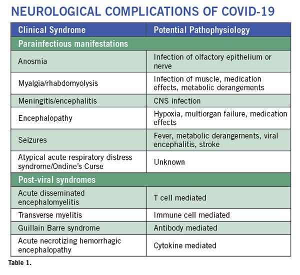
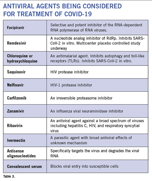
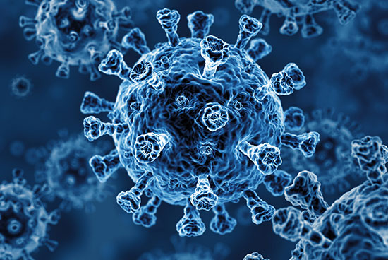
This is the most important event in the last century. Arguably, aside from the tragic loss of life, the widespread and virtually simultaneous shutdown of global and regional economies, and the restriction of individual movements and travel with the enforcement of social distancing has no precedent. Wars, nuclear bombs, AIDS, global financial crises, and the threat of global warming have not done so much, so rapidly, to so many. Countries, governments, peoples, and individuals are all learning, some the hard way, that without methods to prevent the mutation of new zoonotic viruses and vaccines to combat them, public health measures are all we have to reduce the toll of human lives and the economic consequences which, in turn, also impact human life and livelihoods. The constraints effected by the implementation of public health restrictions and the unparalleled rise in the use and need for health, hospital, and ICU resources affects all aspects of health care globally and at all levels of socioeconomic standing.

In doing so, it increases the risk, both directly and indirectly, to those vulnerable and those not so vulnerable with varying levels of disability at all ages and to those who are in need of care, particularly neurological care. The dislocation of livelihoods and risk to health is greatest for the vulnerable. This also includes those who are in foreign countries for a variety of economic reasons, but usually to support themselves or their families in their home country as well as those seeking refuge from conflict and strife. I believe the WFN has a role to play at this time. As bad as the rapidity of infection and rise in the death toll have been to date, even worse is to contemplate the ravaging effects upon countries without the resources of Europe, North America, and the advanced nations of Asia. Africa, India, parts of central and east Asia, South America, and a host of smaller less-resourced countries around the world are at risk over the coming year or more.

The WFN Has an Important Role in This Pandemic.

First, we need to support the WHO. Despite being criticized for being slow to call COVID-19 a pandemic and for bias in its assessment of the situation in the Wuhan province of China early on, it is the only global public health organization. In its daily updates during the executive board meeting of the WHO in Geneva in February, which I attended, it was active, alert to the need for resources in Africa and in Iran, and successful in its procurement and deployment. It cannot mandate actions nor enter countries without permission. The WHO continues to operate amid a growing distrust of globalized intervention and previous criticism. Nevertheless, the advice from Geneva and the regional WHO bodies has been consistent and remains appropriate. Countries must institute and relax measures as they see fit for their particular circumstances.

Second, it is important to support national and regional endeavors through each of our national health services to both meet the impact of COVID-19 on health and life, control the rate of spread of the virus, and ensure patients with neurological disorders are not overlooked as well as endeavors to mitigate the economic burden of the pandemic. This is no easy task. Although principles to guide countries and populations are emerging, they will undoubtedly require local and national interpretation to fit the circumstances. It is clear that different countries will have different tolls at different times and some, if not all, will experience similar differences with successive waves of the infection.

Third, the WFN and its members have a dual role neurologically over and above assisting health services to meet the pandemic. First, as I have already mentioned, the importance of maintaining the high standard of neurological care for those with neurological illnesses and the mitigation wherever possible of the effects of the pandemic on them, whether it be on support services, infusion centers, clinics, or diagnostic services. It is also important to be alert to the possibility of neurological injury caused by COVID-19 and to papers and publications claiming the same prematurely and creating unnecessary alarm. In this time of fear and heightened sensitivity, it behoves us all to analyze carefully reports purporting to identify neurological injury as part of the pandemic. While the SARS-CoV-2 virus has not been proven to be neurotropic, there have been several letters and reports, some in reputable journals, of small series of SARS-CoV-2 patients with severe infections having neurological signs and deficits. To my knowledge, many of these reports to date have not involved trained neurologists nor have they adequately assessed the effects of comorbidities or been followed up. Furthermore, there have been none claiming neurological disease from those with milder infections. At this time, there have been at least two reports totaling a handful of cases with Guillain Barre Syndrome in association with COVID-19 infection (including one who tested negative for COVID-19 after the onset of neuropathy) and two of acute necrotizing encephalopathy, neither of which could be described as a significant risk for COVID-19 patients. It is up to us to see clearly in this “fog of war.”

The WFN has adopted the policy on its website to post material only from peer-reviewed journals and if it has not been peer-reviewed optimally to warn readers to assess the material even more carefully. The WFN is alert to the need for all neurologists to be vigilant and to look for evidence of neurological injury during this pandemic. All of us are aware of the Von Economo-Cruchet disease of encephalitis lethargica described in 1917 and merging then among the 1918-1919 “Spanish flu” pandemic. The WFN has supported its Environmental Neurology Specialty Group chaired by Prof. Gustavo Roman to establish a registry of WFN member societies who are collecting country-wide or center-based data and is aware of moves to establish a “COVID-19 Neuro Research Coalition.” On a more individual note, the WFN now has a bulletin board on its COVID-19 page for member societies to post brief reports (c-19@wfneurology.org) on the situation in general or on specific aspects in their country or region to apprise the federation and the membership. It goes without saying that such sharing has a number of advantages, not the least in maintaining the feeling of community while we are all separated.

Effects of COVID-19 on the WFN

Briefly, I should address the WFN timetable in this new world. First, you will have seen the cancellation of many important meetings already and the possibly optimistic postponement of others. The Annual General meeting of the WFN Council of Delegates remains scheduled for 9 a.m. to Noon, Sept. 9,

during the ECTRIMS/ACTRIMS meeting in Washington. But it may well be changed to a virtual meeting. The WFN was already moving to enhance its electronic activities for a number of reasons before the pandemic so it will have two new video productions available shortly on its website. The first is about the WFN now and the second about the 2021 World Congress of Neurology in Rome. The World Brain Day program on Parkinson’s disease and shared with the International Parkinson Disease and Movement Disorders Society is proceeding well, and a new initiative on brain health will run in parallel. I will have more to say on both in my next column.

Finally, on behalf of the Trustees and staff of the WFN, I wish you and your members and their families all safe passage through this crisis. •

William Carroll
WFN President

From the Editors

By Steven L. Lewis, MD, Editor, and Walter Struhal, MD, Co-Editor

We would like to welcome all neurologists from around the globe to this issue of World Neurology, and at the start would like to again wish you, your families, and your patients all the best of health and safety at this time.

Steven L. Lewis, MD, Walter Struhal, MD

Much of this issue is devoted to an update on COVID-19. We are honored that the cover story on this issue is written by Drs. Avindra Nath and B. Jeanne Billioux from the Section of Infections of the Nervous System at the United States National Institute of Neurological Disorders and Stroke (NINDS). They provide an up-to-the moment summary of the evolving knowledge about the interface of COVID-19 on neurology and our patients. In the President’s column, Dr. William Carroll reminds us of the importance of the pandemic on our patients and the role the WFN plays in, among other important aspects, advocating for maintaining the highest standard of care for neurological patients worldwide. Dr. Carroll also updates us on the enhancements to the WFN website as a clearinghouse for accurate and up-to-date information about neurological involvement in COVID-19 as well as its effects on neurological societies and neurological patients and services worldwide.

Dr. John England, editor-in-chief of the Journal of Neurological Sciences (JNS), provides his Editor’s Update on the journal, informing us of the efforts to invite and expeditiously publish the accepted papers in JNS relating to the neurological aspects of COVID-19. This issue also features an obituary, reprinted from JNS, of Prof. Alberto Portera-Sánchez, a pioneer of Spanish neurology and former vice president of the WFN, along with additional heartfelt words from Vladamir Hachinski, former president of the WFN.

Drs. Tissa Wijeratne, Claudia Trenkwalder, president of the International Parkinson and Movement Disorders Society (IPMDS), Wolfgang Grisold, and Dr. Carroll, announce and update us on the efforts and ongoing plans for this year’s World Brain Day. This year’s World Brain Day focuses on ending Parkinson’s disease as a collaborative effort between the WFN and the IPMDS. Drs. Dafin Muresanu, Selevan Ovidiu, Cristian Andriescu, and Stefan Strilciuc, describe the history of neurological meetings in Transylvania, particularly the annual international events organized by the Society for the Study of Neuroprotection and Neuroplasticity (SSNN).

With this issue, we are also pleased to introduce a new column, written by WFN Secretary-General Wolfgang Grisold, who will update us with each issue about the many Committees of the WFN and their critical roles, beginning with the Standards and Evaluation Committee.

This issue also features a number of reports from the recipients of Junior Traveling Fellowships (JTFs) to attend the World Congress of Neurology (WCN) in October 2019 in Dubai, United Arab Emirates. These heartfelt reports are reminders of the wonderful “in person” congress that so recently occurred (and yet now seems so distant for many reasons). Equally importantly, these reports serve as a reminder of the upcoming WCN 2021 we are so actively planning and looking forward to attending in Rome, Italy in October 2021.

Finally, as Dr. Carroll reminds us in his column, the Annual General Meeting (AGM) of the WFN Council of Delegates (COD) remains scheduled for Sept. 9, 2020 during the ECTRIMS/ACTRIMS meeting in Washington. We look forward to seeing all of the delegates at this meeting, whether it is virtual or in person.

We hope you enjoy this issue of World Neurology, and look forward to receiving your contributions, especially updating all of our colleagues on how the current pandemic has (or has not) affected neurologists, our societies, and our patients around the globe. •