WFN Neurosonology Specialty Group: Teaching Workshops

Workshops discussed clinical use of transcranial Doppler ultrasonography for patients with different diseases, including its application for wartime traumatic brain injury.

By Kateryna Potapova, Larisa Sokolova, Dmitro Andreichenko, Svetlana Dudukina, and Alex Razumovsky

The Neurosonology Specialty Group (NSG) of the WFN is dedicated to the promotion of science and research as well of education and training in the field of ultrasonic techniques and its clinical utilization. Therefore, international cooperation and the dissemination of scientific information within the field of neurosonology is part of the NSG activities.

Since the start of the Russia-Ukraine conflict, it has become imperative and clear that Ukraine needs highly skilled and advanced medical training given the magnitude of civilian deaths and injuries.

Monitoring of the brain after acute injury is central to the practice of neurocritical care for patients with a wide range of disorders, including subarachnoid hemorrhage, traumatic brain injury (TBI), ischemic and hemorrhagic strokes, and infectious disease of the central nervous system as well as encephalopathy of various etiologies. The WFN NSG endorsed a transcranial Doppler (TCD) workshop program where there was a description of fundamentals of TCD and advantages relevant to the clinical utilization of TCD in the neurocritical care environment with emphasis on diagnosis and monitoring for cerebral vasospasm and intracranial hypertension in patients after wartime TBI.

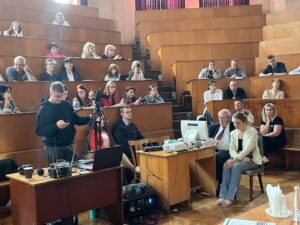
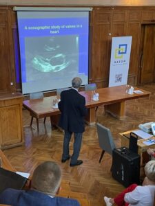
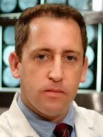
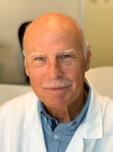
In mid-September 2023, NGO Razom Co-Pilot Project assisted in organizing medical training in Kyiv at the O.O. Bogomolets National Medical University. Specialists from the U.S. conducted a three-day workshop there on TCD diagnostics for doctors from all over Ukraine. In Kyiv, there were 72 attendees from six regions of Ukraine and Kyiv. Training was led by Dr. Alex Razumovsky (Specialty Care Consultant, President of TCD Global, Inc. and NSG WFN Advisory Board member) and Dr. Kenneth Green (Retired U.S. Navy commander, managing director of CounteRisk Technologies, Inc. and co-chair for Military Medicine and Veterans Affairs Subcommittee at the Society for Brain Mapping and Therapeutics).

The high professionalism of the instructors provoked great interest among attendees; many practical questions were asked, to which the lecturers gave complete and comprehensive answers. Everyone was given the opportunity to conduct TCD studies independently, under the guidance of faculty. Most of the participants successfully answered test questions after the workshop and received continuing medical education certificates. In addition, as part of these efforts, a modern TCD instrument was donated by DWL company (Compumedics Germany GmbH/DWL USA, Inc) to the University’s Neurology Department in order to conduct detailed examination of cerebral vessels, which can detect strokes due to the thrombosis, stenosis, vasospasm and a host of other causes.

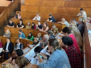
Further learning on TCD clinical utilization took place at I. I. Mechnikov Dnipropetrovsk Region Clinical Hospital in Dnipro where 15 neurologists, anesthesiologists, and ultrasound specialists also practiced their new diagnostic skills and already started performing TCD examinations for their patients. Today, Mechnikov Hospital in Dnipro is receiving a large number of military personnel with war-time TBI. Conducted workshop and TCD clinical utilization provided opportunity to doctors of the Mechnikov Hospital to improve diagnosis, prevent complications, and plan treatment management for patients in neurointensive care, endovascular center, and the neurosurgery department.

All participants expressed great gratitude to the faculty and organizers of the workshop during such a difficult period and expressed hope for further cooperation. •

Epilepsy Care in the Democratic Republic of Congo

by Dr. Prince Kazadi,  Dr. Moise Lubendo, Dr. Daniel Tshiyuk, and Dr. Nazaire Kimponto

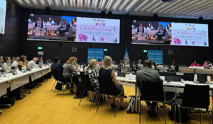
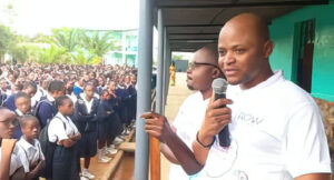
The ASLEK Epilepsy Association in the Democratic Republic of Congo (DRC) is the unique national non-profit organization in the DRC, working in the field of research, staff training, community education, and patient care in neurology in general and epileptology
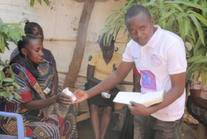
  specifically since 2008.

ASLEK take cares of 2,500 patients suffering with epilepsy in the DRC, of whom 1,002 patients have at least one psychiatric entity (anxiety, depression, etc.)

The DRC currently has about 105 million inhabitants. With a minimum prevalence of epilepsy of 1%, or 1,050,000 patients, suffering with epilepsy whose total number of neurologists is 10.

Our areas of action:

  • Epilepsy education
  • Epilepsy management
  • Epilepsy research
  • Medical training
  • Social support

Epilepsy Education

Aslek Epilepsy in DRC organizes awareness campaigns in the community, schools, churches, and public squares to combat the stigma and social discrimination suffered by people suffering from epilepsy in the DRC.

Management of Epilepsy

ASLEK had the privilege of being among the associations supported in Africa by ROW Foundation; we received after six months, a supply of drugs, allowing us to cover the care of 74 patients eligible for levetiracetam and 26 patients eligible for lamotrigine and other patients receive medicines from members’ contributions.

We work with TeleEEG UK, a non-profit organization helping resource-limited countries without many neurologists like the DRC, with EEG interpretations and we also work in collaboration with the Federal University of Rio de Janeiro in Brazil, which supports us with interpretations of EEGs  from rural areas of the DRC.

We are working on the implementation of IGAP in the DRC.

Research on Epilepsy in the DRC

ASLEK works in epilepsy research in the DRC in collaboration with University College London (UCL) for studying the impact of inbreeding on the risk of genetic disorders in three local communities in the DRC.

This perspective is the most unique and has shown a desire to study genetic susceptibility that can potentially explain the high incidence and prevalence of epilepsy in communities with high consanguineous marriage rates. These factors have not yet been explored in resource-limited country contexts due to the very high cost of genetic sequencing, as well as the absence of a laboratory, and much more emphasis has been placed on parasitic diseases associated with seizures.

ASLEK is also involved in research on the study of early detection of cognitive degeneration in collaboration with Neurosteer.

Medical Training

The Democratic Republic of Congo currently has only 10 neurologists for 105 million inhabitants and all of them are in the country’s large cities, and there are no neurologists in rural areas.

This lack of neurologists in the Democratic Republic of Congo remains a major challenge to overcome because it is the basis of erroneous diagnoses and inadequate care.

ASLEK Epilepsy in DRC is the only non-governmental organization training doctors and nurses in rural areas of the DRC on epilepsy, with a view to improving the management of epilepsy through the use of a smartphone which generates a summary to share with neurologists around the world for remote therapeutic guidance (telemedicine) with some challenges related to the internet connection for sharing data from rural areas of the DRC.

There is great interest in training primary doctors in neurology in the DRC through the granting of comprehensive training scholarships.

Social Plans

Epilepsy is considered in several regions of the DRC as a contagious disease. This makes this pathology more stigmatized in the DRC, thus making the lives of people suffering with it very difficult and even unlivable, leading some patients to commit suicide and adopt a life of isolation as a means of defense.

ASLEK Epilepsy in DRC thus fights against the stigmatization of epilepsy in schools, while using students as a bridge to convey the message to parents, churches and public awareness campaigns.

We train and inform the Congolese community about epilepsy by promoting sketches and videos on local and national television channels, referring to the slogan of our association which says epilepsy is not contagious.

In addition to awareness campaigns against stigma and social discrimination, ASLEK, within the limits of its means, also provides material support in kind and in cash to children suffering from epilepsy, most of whom come from very poor families and live with less than one American dollar per day.

Challenges

Challenges we face include the following:

  • Medical staff
  • The remarkable deficit of personnel specialized in neurology.
  • Lack of infrastructure, including lack of clinics specialized in epilepsy ; added to this, is a problem with medical equipment, a notable deficiency in EEG, CT, and MRI for the causal diagnosis of certain forms of epilepsy, and the absence of biochemical laboratories drug deficiencies and the high costs of drugs available on the DRC market such as sodium valproate, levetiracetam, topiramate, etc.
  • Many patients do not accept the diagnosis for fear of stigmatization, social discrimination and rejection, barrier to marriage among young women, lack of cooperatives for food self-sufficiency, problem of social reintegration, and the abandonment medication due to lack of food.

Conclusion

Epilepsy is a treatable neurological condition; however, in resource-limited countries this pathology still remains a heavy burden.

This being said, in this fight, we at ASLEK Epilepsy are seeking partnerships with other international organizations to meet these significant challenges. •


Dr. Prince Kazadi is the director of ASLEK Epilepsy in DRC.

The Study of Brain Movements


Given the relative simplicity of the conditions, which did not require a complicated experimental set-up, the obstacle lay only in the unfortunate predominance of rigid doctrines, which distorted the bare facts and arbitrarily remodeled them in favor of vague speculations. Only in this way could it come about that for a time the brain could be denied any movement and that the dura mater was elevated to the most important of all parts of the body, the meninges to the seat of all ability to move and to erapture.

Max Neuburger, 1897


Italian anatomist Antonio Pacchioni

By Peter J. Koehler, MD, PhD, FAAN

This citation is from a chapter on “Versuche an der harten Hirnhaut und über Hirnbewegung” [Experiments on the Dura Mater and on Brain Movement] in the book Die historische Entwicklung der experimentellen Gehirn- und Rückenmarksphysiologie vor Flourens [The historical development of experimental brain and spinal cord physiology before Flourens] by Austrian physician and medical historian Max Neuburger (1868-1955).1 Naturalists have thought about and done experiments with respect to brain movements at least since Galen of Pergamon (129-216 CE). They wondered about the relation between the function of the brain and its movements. These movements could be observed in cranial wounds, as was described in the Edwin Smith surgical papyrus almost three millennia BCE, but also in children while the fontanel was still open, which was probably already noted by Pliny (23-79 CE). Most neurologists will have observed the movements indirectly, when doing a lumbar puncture and measuring the pressure in a plastic tube. In this essay, I will provide some historical information about this subject.

Pacchioni’s publication (1701)

Galen

Galen opined that the whole brain moved and had a systole as well as diastole. The movement was believed to be related to respiration. Air was thought to enter the ventricular system by the nose and cribriform plate of the ethmoid bone during inhalation, thereby expanding the brain. During exhalation it would leave the brain again. This had a dual function, including the driving of animal spirits from the ventricles into the nerves and the perception of smells. Interestingly, several physicians related the swelling and collapse of the bulging brain in cranial trauma to the phases of the moon, an idea that seems to have lasted for a long period.1, p.71 As is true for so many of Galen’s ideas, the dual function continued to be relied upon by physicians for many centuries, including Bauhinus (Gaspard Bauhin 1560-1624) and Carolus Fracassatus (Carlo Fracassati 1630-1672).

Giorgio Baglivi

Muscular Dura Mater or Brain Proper?

Two lines of ideas developed with respect to the origin of brain movements. One group of naturalists believed it was caused by the movements of the dura mater, the brain itself moving only secondarily. Others believed it was due to movement of the brain proper. Indeed a contractile power was assigned to the dura mater by Italian anatomist Antonio Pacchioni (1665-1726), who was interested in particular in the dura mater (De durae meningis fabrica et usu disquisitio anatomica [An anatomical discussion of the structure and use of the dura mater]).2 He became well-known by the eponym Pacchioni’s or arachnoid granulations.

Report of Ridley’s paper at the Royal Society (1703)

When studying the dura mater, after boiling it, he was able to demonstrate a radiating arrangement of its fibers in the form of pyramids. He thought this structure consisted of three muscles and four tendons.3, p. 77 He compared the arrangement of the fibers with the chambers of the heart and recognized a double movement. At one moment, the cerebrum was compressed and the cerebellum became free. At the following moment, the situation would be the reverse. In this way, these muscles were responsible for transport of blood to the brain and the circulation of the nervous juice, like the heart had its function for the circulation of blood.

His colleague Giorgio Baglivi (1668-1707) elaborated upon this idea and supposed that the heart pushed the blood into the brain causing it to distend. The dura mater would then contract and push the nervous fluid into the nerves. In this way, he suggested two hearts and named the dura mater cor cerebri.3, p.7 In fact he attributed the highest control center to the dura mater, which he considered a vital structure.1, p. 77 These ideas proved very successful and were accepted by many anatomists and physicians, including Giovanni Maria Lancisi (1654-1720), Friedrich Hoffmann (1660-17420, Giovanni Domenico Santorini (1681-1737), and in particular Georg Ernst Stahl (1659-1734).

Jan Daniel Schlichting

Respiration

The English physician Humphrey Ridley (1653-1708), who graduated from Leiden with a dissertation De lue venerea (1679), may have been among the first to prove that destruction of the dura mater did not cease the movements of the brain. He read his paper at the Royal Society in 1703: “Experimentum anatomicum ad veram durae matris motus causam detegendam institutum” [An anatomical experiment to discover the real cause of the movement of the dura mater].4 Even more convincingly it was proved by the Dutch physician and anatomist Jan Daniel Schlichting (1703-1765), who studied medicine in the Dutch cities of Leiden and Groningen before he settled in Amsterdam. In his “De motu cerebri” he reported on his experiments, carried out upon several kinds of animals (dogs, cats, and rabbits), that brain movements remain after removal of the dura mater. “We solve, therefore, the problem that learned men have created for themselves by saying that the dura mater contracts and relaxes or pulsates”.

Schlichting’s 1750 article

He related the movements with breathing: “Every time a man breathes out, the entire brain rises up, that is, swells, and every time he breathes in, it goes down, that is, it subsides”.5 Thus he mentioned the opposite of what Galen had asserted 1,500 years previously. However, Schlichting did not know whether the swelling up during expiration was due to an increase influx of blood, by the increased air pressure, or by both.1, pp. 82-3 His experiments were repeated and the conclusions largely confirmed by the French physician Anne-Charles Lorry (1726-1783), stating that “The ancients followed their presumptions when they thought that the brain swelled up in time with inspiration: M. Schlichting is indeed supported by observation when he claims that it is synchronous with expiration”.1, p.84 However, Lorry believed the movements were only present in extraordinary conditions, like respiratory obstruction, pathological states, and cranial defects. He was one of several physicians, who denied brain movement to exist in normal conditions.

Jan Daniel Schlichting

The Swiss physician and physiologist Albrecht von Haller (1708-1777) was also among the physicians to refute the Pacchioni-Baglivi concept about the function of the dura mater. He confirmed the elevation of the brain during expiration and sinking down during inspiration. He related this to the venous congestion and outflow. However, it remained a matter of debate how that occurred. Some attributed the movements to the action of the heart, others to that of the influence upon the venous return of blood by breathing, and a third group believed that a combination of the two was responsible.

Anne-Charles Lorry

Brain Proper Moves Synchronously With the Heart

Several physicians observed that the brain moves synchronously with the heart. Among them were some well-known persons including the Italian anatomist and surgeon Realdus Columbus (Matteo Colombo c. 1515-1559), the French anatomist Jean Riolan (1580-1657), the Dutch physician IJsbrand van Diemerbroeck (1609-1674), Ridley, the French anatomist Raymond Vieussens (1641-1715), Haller and Marie-François-Xavier Bichat (1771-1802). However, there were various opinions on how the heart could move the brain. Several, including Bichat, believed it being the effect of the arterial pulse. Others were still denying such a synchronous movement. Marie-Jean-Pierre Flourens (1794-1867), for instance, denied a pulsating movement of the brain, only confirming a synchronous movement with respiration.

Franciscus Cornelis Donders

The German anatomist and anthropologist Johann Alexander Ecker (1816-1887), who summarized the history of brain movements, experimented with animals and concluded that there is a double movement in human beings and mammals, notably with the heart and respiration. The pulsating movement, he believed, was due to the arteries at the base of the brain as well as their branches in the brain parenchyma. He attributed the respiratory movement to the influx into the ventricles of cerebrospinal fluid (CSF) during expiration, although the venous filling in the brain may contribute to it.3, p.123

Ernst von Leyden

Spying on Nature Directly

At the time, some physiologists were more critical about the influence of the CSF. Writing on the results of his experiments, the Dutch physiologist and ophthalmologist Franciscus Cornelis Donders (1818-1889) mentioned that “the CSF is the condition, not the cause of brain movement.” Trying to answer the question whether brain movements exist when the skull is completely closed, he denied any movements of the brain if the skull was closed. The source of the question derived from two observations. Ecker believed the movements continued, whereas Johannes Müller declared that this was physically impossible.6 The French anatomist and physiologist François Achille Longet (1811-1871), based on observations published in Bourgougnon’s 1839 dissertation, also denied brain movements with closed skull. “Sans doute il est curieux de rechercher pourquoi le cerveau se meut chez les animaux trépanés; mais il est, suivant nous, bien plus important de savoir si ces mouvements préexistent à la trépanation” [No doubt it is curious to investigate why the brain moves in trepanned animals; but it is, in our opinion, far more important to know whether these movements pre-exist trepanation]. A comparison was made with the movements of the lungs that are quite extensive when the thoracic cage is opened, in comparison to the much less extensive movements in the physiological situation. Longet considered experiments to research brain movements, during which the skull was opened as useless. Therefore, Donders decided to open the skull of a rabbit. “I managed to spy on nature directly. When the skull is closed I have not only observed immediately and life, what concerns the brain movements, but also the increased and decreased supply of blood, with changes in blood pressure.” After trepanning the skull and removing a small part of the dura mater, he inserted a small glass window. “Through the small glass window one could observe the brain perfectly well, but we saw no trace of movement.7 I now amplified the inhalation and exhalation excursions by keeping the mouth and nose closed. Repeatedly again no movements.” At last, the small glass came loose, after which both types of movement, respiratory as well as circulatory, returned. The experiment was witnessed by several of his colleagues. After bonding the glass in the opening again, he did not observe any movements anymore and was able to see it for more than eleven days, sometimes using a loupe and even a microscope. He repeated the experiment several times and had special watch glasses made for the purpose.

Angelo Mosso

From Brain Movements to Intracranial Pressure

Still working in Königsberg (the present Kaliningrad), the German physician Ernst von Leyden (1832-1910) did important research on brain movements and in particular on intracranial pressure, which he published in 1866.6 He confirmed Donders’ observations, but instead of rabbits he used dogs, in which brain movements are clearer. Next to a small glass window, he inserted a lockable pipe, so that he could easily open and close the hole in the skull. He realized that the pressure waves by respiratory and circulatory movements with the tap locked would be borne by the brain. Like Victor von Bruns (1812-1883), Von Leyden tried to represent the changes in a graph, but encountered too many difficulties to show the movements due the low force that cause them. However, he was able to show the respiratory and circulatory quite nicely, by using a barometer tube filled with water. The rest of his long research paper is about measuring intracranial pressure under normal circumstances as well as with pathologically increased pressure.8

Catherina X (from Mosso10)

Registrations in Patients

About 10 years later in Turin, Carlo Giacomini (1840-1898) in cooperation with the well-known physiologist Angelo Mosso (1846-1910), were able to register brain movements by using an instrument developed by the French physiologist Etienne-Jules Marey (1830-1904; “méthode graphique”). They published their paper in the first volume of Archivio per le Scienze Mediche. The recordings were done in the open skull of the 37-year Catherine X, who suffered from syphilis. Despite years of treatment with potassium iodide and mercury ointment, the skull became affected and the researchers were able to observe the pulsations of the dura mater synchronously with the heart beats.9 In Mosso’s 1881 monograph Kreislauf des Blutes im menschlichen Gehirn [Circulation of the blood in the human brain], we find a drawing of Catherine and her skull.10 Giacomini’s and Mosso’s paper was read at the Académie in Paris by no one less than the famous physiologist Claude Bernard (1813-1878),11 who informed the audience that at the time the patient had been completely cured.

Charles-Emile François-Franck (National Library of Medicine; Digital Collections. Public domain)

Not long after the Italian publication, the French physiologist Charles-Emile François-Franck (1849-1921), a pupil of Marey, was able to register the brain movements of a similar case. He was aided by young Edouard Brissaud (1852-1909; described geste antagoniste in dystonia), who made the registrations and was allowed, at the end of the article, to present the case history. It was about the 34-year old Victoire All… , who suffered from syphilis, for which she was treated with liquor swietenii, a mercury based mixture that was introduced the previous century by Gerard van Swieten (1700-1772). The dosage of mercury was lower than that applied earlier. However, the skull became affected and parts needed to be removed, providing access to the researchers.

At the end of the 19th century, British physiologist Leonard Hill (1866-1952) summarized the literature on the subject, in particular the decades before his book was published. Referring to “Althan,” probably the Estonian born Georg Althann (1839-1898), he noted that “the errors of Donders … arose from the neglect of the fact that the brain pulsates in a direction where resistance is least. Thus when the cranium is trephined and the dura opened, the pulse can scarcely be seen in the occipito-atlantal membrane, for the trephine hole has now become the seat of least resistance. Similarly, when a trephine hole is closed by a glass window, the occipito-atlantal membrane becomes the seat of least resistance, and a pulsation appears there.”12, p.8-15

Today the pulsating brain still is an area of research in particular with respect to the effects on MRI scans and more recently the concept of the glymphatic system. Arterial pulsations are thought to drive flow through perivascular spaces for clearance of metabolic waste.13


References

1. Neuburger M (1897). Die historische Entwicklung der experimentellen Gehirn- und Rückenmarksphysiologie vor Flourens. Stuttgart, Enke. English edition transl. and edited by Edwin Clarke The historical development of experimental brain and spinal cord physiology before Flourens. Baltimore/London, Johns Hopkins University Press.

2. Pacchioni A (1701). De durae meningitis fabrica et usu disquisition anatomica. Rome, Herculis.

3. Ecker A (1843). Physiologische Untersuchunge über die Bewegungen des Gehirns und Rückenmarks : insbesondere den Einfluss der Cerebrospinalflüssigkeit auf dieselben. Stuttgart, Schweizerbart.

4. Ridley H (1703). Experimentum anatomicum ad veram durae matris motus causam detegendam institutum Phil Trans 23, no. 287: 1480-4.

5. Schlichting JD (1750). De motu cerebri. Mémoires de Mathématique et de Physique (Paris) : 113-35.

6. Leyden E (1866). Beiträge und Untersuchungen zur Physiologie und Pathologie des Gehirns. Virchows Arch 37:519–559.

7. Donders FC (1850). De bewegingen der hersenen en de veranderingen der vaatvulling van de Pia Mater, ook bij gesloten onuitzetbaren schedel regtstreeks onderzocht. Nederlandsch Lancet, March/April, 521-53.

8. Koehler PJ, Wijdicks EF (2015). Fixed and dilated: the history of a classic pupil abnormality. J Neurosurg;122:453-63.

9. Giacomini C, Mosso A (1876). Esperienze sui movimenti del cervello nell’uomo. Archivio per le Scienze Mediche 1:245-78.

10. Mosso A (1881). Kreislauf des Blutes im menschlichen Gehirn, Leipzig, Veit, p. 223

11. Giacomini C, Mosso A (1877). Etude graphique des mouvements du cerveau de l’homme. C R Académie Sciences (Paris) 24 : 41-3.

12. Hill L (1896). The Physiology and Pathology of the Cerebral Circulation: An Experimental Research. London: Churchill.

13. Mestre H, Tithof J, Du T, Song W, Peng W, Sweeney AM, Olveda G, Thomas JH, Nedergaard M, Kelley DH (2018). Flow of cerebrospinal fluid is driven by arterial pulsations and is reduced in hypertension. Nat Commun 9:4878.

Multiple Sclerosis DMTs Added to WHO List

Three disease-modifying therapies (DMTs) for multiple sclerosis are now part of the World Health Organization essential medicines list.

By Bassem Yamout1, Tomas Kalincik2,3, Joanna Laurson-Doube4, Bernhard Hemmer5,6, Shanthi Viswanathan7

On July 26, 2023, the World Health Organization (WHO) added three disease-modifying therapies (DMTs) for multiple sclerosis (MS) to its Essential Medicines List (EML) for the first time1. The list is an important tool for achieving universal health coverage, providing guidance to governments, health facilities, and procurers on priority medicines to tackle important public health conditions. The EML includes medicines on the basis of solid evidence for safety and efficacy. Inclusion of any medication in the WHO EML is expected to be associated with increased availability and reimbursement by governmental agencies. The WFN was one of the 15 organizations that endorsed the application to the WHO, submitted by Multiple Sclerosis International Federation and WHO Collaborating Centre Bologna.

With this decision, the WHO formally acknowledged MS as a global health concern as well as the critical importance of making MS treatments available in all health systems at all times. Although most approved MS therapies have been made available in many countries, people with MS living in certain regions of the world, especially in resource-limited settings, do not have access to much-needed therapies, which are either unavailable or unaffordable. In fact, around 70% of countries across the world report that people with MS face barriers accessing DMTs2. The WHO decision to include three MS therapies with different routes of administration, efficacy, and tolerability in their updated list will undoubtedly help increase access of people with MS to high-quality, cost-effective, and evidence-based treatments.

This decision was the culmination of two years of relentless efforts by the MS International Federation (MSIF) and its partner organizations, including several neurological academies, scientific societies, and the regional committees for Treatment and Research in MS (TRIMS). The MSIF set up two independent, multidisciplinary panels and started a comprehensive and rigorous review process in partnership with the WHO Collaborating Centre Bologna, and supported by the Cochrane MS group and McMaster GRADE Centre, both of which are internationally regarded as experts in the field of evidence-based reviews and decision-making3.

The WHO EML Committee decided to add rituximab, cladribine, and glatiramer acetate to the new EML4. Rituximab has been extensively used for treating MS patients especially in low/lower-middle income countries (LLMIC) due to its cost-effectiveness, but its off-label status has curtailed reimbursement by governmental and private insurance systems5. Its inclusion on the WHO EML should help in this respect. The decision to support off-label use of rituximab is supported by strong evidence of its efficacy and safety for this indication.


Dr. Deanna Saylor

“People with MS in lower-resourced settings, including most low- and middle-income countries, often face numerous challenges in their MS journeys. This starts with accessing the specialists and diagnostic tests, including MRIs, required to obtain their diagnosis, and continues with accessing effective treatments for their MS. As a result, people with MS in low- and middle-income countries often are untreated, undertreated, or have to expend significant personal resources in order to obtain treatment. Based on my own experience taking care of people with MS in Zambia, I am confident that if DMTs are available, treatment of MS is feasible, safe, and likely to be highly effective and result in good outcomes among people with MS in low- and middle-income countries.”

Dr. Deanna Saylor, clinical chair of the MSIF Essential Medicines Panel


Large-scale randomized controlled trials (e.g. DELIVER-MS and TREAT-MS) continue to study early use of high efficacy DMTs versus escalation treatment by treatment6,7. Nevertheless, studies have already suggested that early treatment with higher efficacy DMTs in patients with MS can lead to better outcomes compared to treatment initiation with low efficacy DMTs and escalating therapy only upon disease activity or progression8-12.

Intentionally, the treatments listed on the WHO EML are not categorized as first-, second-, or third-line treatments, but encourage clinicians and people with MS to determine the most appropriate course of treatment for the clinical and personal circumstances. Rituximab, cladribine, and glatiramer acetate represent different tiers of treatment effectiveness, modes of administration, and safety in pregnancy and breastfeeding. In particular, glatiramer acetate is known for its favorable safety profile in pregnancy and breastfeeding when compared with the two more potent listed therapies. Pregnancy is a particularly important clinical scenario due to high prevalence of MS in young women and more challenging access to family planning services in LMICs.

Countries can use the WHO decision to shift their treatment algorithms, allowing patients access to higher efficacy DMTs, e.g. rituximab, early in their disease course. Speaking from personal experience treating refugees in the Middle East, the use of rituximab, a high efficacy, safe, and low-cost DMT, made a considerable difference in the lives of people with MS that fled their country and had no means of affording any MS therapy13.

However, despite countries endorsing the concept of the EML over the years, implementing the list into local practice is variable and fraught with challenges, especially in LMICs. We should use this momentous decision to push forward the implementation of the WHO EML through coordinated efforts among national scientific and patient societies, international scientific, and health care organizations, frontline clinicians, and local policymakers.

Patients with MS cannot wait for decades to have this EML implemented: The time for action is now through the growing international momentum to improve care and access to treatment for multiple sclerosis.


1. Yamout is with the Neurology Institute, Harley Street Medical Centre, Abu Dhabi-UAE. 2. Kalincik is with the Neuroimmunology Centre, Department of Neurology, Royal Melbourne Hospital, Melbourne, Australia. 3. Laurson-Doube is with  CORe, Department of Medicine, Melbourne, Australia.4. Hemmer is with Multiple Sclerosis International Federation, London, United Kingdom. 5. Department of Neurology, Klinikum rechts der Isar, School of Medicine, Technische Universität München, Germany. 6. Munich Cluster for Systems Neurology (SyNergy), Germany. 7. Viswanathan is with Department of Neurology, Kuala Lumpur, Hospital, Malaysia.

References

1. WHO endorses landmark public health decisions on Essential Medicines for Multiple Sclerosis [Internet]. [cited 2023 Aug 1]. Available from: https://www.who.int/news/item/26-07-2023-who-endorses-landmark-public-health-decisions-on-essential-medicines-for-multiple-sclerosis

2. MS International Federation. Atlas of MS 2020 – Clinical management report [Internet]. 2020 [cited 2022 Dec 5]. Available from: https://pubmed.ncbi.nlm.nih.gov/35173808/

3. A.10 Cladribine, glatiramer and rituximab – multiple sclerosis – EML [Internet]. [cited 2023 Aug 2]. Available from: https://www.who.int/groups/expert-committee-on-selection-and-use-of-essential-medicines/24th-eml-expert-committee/a10_ms-dmts

4. World Health Organization. Executive summary of the report of the 24th WHO Expert Committee on Selection and Use of Essential Medicines 24 – 28 April 2023 [Internet]. [cited 2023 Aug 1]. Available from: https://apps.who.int/iris/bitstream/handle/10665/371291/WHO-MHP-HPS-EML-2023.01-eng.pdf

5. Laurson-Doube J, Rijke N, Helme A, Baneke P, Banwell B, Viswanathan S, et al. Ethical use of off-label disease-modifying therapies for multiple sclerosis. Mult Scler Houndmills Basingstoke Engl. 2021 Aug;27(9):1403–10.

6. DELIVER-MS – does early treatment with highly effective DMT improve the prognosis for people with Multiple Sclerosis? [Internet]. [cited 2023 Sep 15]. Available from: https://deliver-ms.com/

7. Ontaneda D, Tallantyre E, Kalincik T, Planchon SM, Evangelou N. Early highly effective versus escalation treatment approaches in relapsing multiple sclerosis. Lancet Neurol. 2019 Oct;18(10):973–80.

8. Ontaneda D, Mowry EM, Newsome SD, Naismith RT, Nicholas J, Fisher E, et al. Benefits of early treatment with natalizumab: a real-world study. Mult Scler Relat Disord [Internet]. 2022 Dec 1 [cited 2023 Sep 15];68. Available from: https://www.msard-journal.com/article/S2211-0348(22)00721-0/fulltext

9. Stankiewicz JM, Weiner HL. An argument for broad use of high efficacy treatments in early multiple sclerosis. Neurol – Neuroimmunol Neuroinflammation [Internet]. 2020 Jan 1 [cited 2023 Sep 15];7(1). Available from: https://nn.neurology.org/content/7/1/e636

10. Simonsen CS, Flemmen HØ, Broch L, Brunborg C, Berg-Hansen P, Moen SM, et al. Early High Efficacy Treatment in Multiple Sclerosis Is the Best Predictor of Future Disease Activity Over 1 and 2 Years in a Norwegian Population-Based Registry. Front Neurol [Internet]. 2021 [cited 2023 Sep 15];12. Available from: https://www.frontiersin.org/articles/10.3389/fneur.2021.693017

11. Filippi M, Amato MP, Centonze D, Gallo P, Gasperini C, Inglese M, et al. Early use of high-efficacy disease‑modifying therapies makes the difference in people with multiple sclerosis: an expert opinion. J Neurol. 2022;269(10):5382–94.

12. Brown JWL, Coles A, Horakova D, Havrdova E, Izquierdo G, Prat A, et al. Association of Initial Disease-Modifying Therapy With Later Conversion to Secondary Progressive Multiple Sclerosis. JAMA. 2019 Jan 15;321(2):175–87.

13. Zeineddine MM, Yamout BI. Treatment of multiple sclerosis in special populations: The case of refugees. Mult Scler J – Exp Transl Clin. 2020 Mar;6(1):2055217319848466.

The 2023 International Symposium on Thrombolysis/Thrombectomy and Acute Stroke Therapy, and the Symposium on Collaterals on the Brain

Amir Molaie

David Liebeskind

By Amir Molaie and David Liebeskind

In the rapidly evolving field of stroke care, events that gather global experts to discuss recent trials and innovations are integral to disseminating knowledge and advancing care. Building on the momentum of the 15th World Stroke Congress, the 2023 International Symposium on Thrombolysis/Thrombectomy and Acute Stroke Therapy (TTST) and the 10th annual Symposium on Collaterals on the Brain (Collaterals), proved no exception. The two conferences, held in conjunction at the University of California, Los Angeles (UCLA) Oct. 13-17, 2023, brought together leading experts from around the world to review and deliberate wide-ranging, timely topics in neurovascular care.

Panel discussion on the potential use of generative AI in future vascular and endovascular care, featuring (left to right): Ashutosh Jadhav, MD, PhD; Andrei Alexandrov, MD; Kunakorn Atchaneeyasakul, MD; Alexandra Czap, MD.

The events were hosted by Prof. David Liebeskind in a hybrid virtual and in-person format, assembling 165 attendees. Participants joined from countries on six continents, ranging in background from clinicians and researchers to biomedical engineers and medical students.

The program began with the TTST 2023 portion, with presenters offering their data and input on endovascular thrombectomy for special populations, including late window cases, distal occlusions, and patients with low NIHSS. This was followed by up-to-date summaries of the recently published and newly presented large-core trials. The first day wrapped up with lectures on innovative technologies, including prehospital triage devices and an interactive dialogue on the rising impact of artificial intelligence on stroke care.

Conference dinner hosted by David Liebeskind, MD, pictured here with James Grotta, MD (left) and Alexandra Czap, MD (right).

The following day featured lively discussions regarding the use of alteplase versus tenecteplase, and the overarching role of intravenous thrombolysis prior to mechanical thrombectomy (MT). Sessions also delved into the merits of neuroprotection and the optimization of MT techniques. The day ended with a unique Jeffersonian-style exchange, a forum which fostered interactive, candid, and impassioned conversations involving participants from all levels of training and backgrounds.

The final day of TTST highlighted issues related to stroke systems of care, topics which seamlessly segued into the Collaterals Symposium. These next three days showcased accomplished presenters from throughout the world commenting on their impressive advocacy and contributions to expanding stroke treatment to regions with previously limited access to thrombolysis and thrombectomy. Nearly every geographic region was represented, with speakers converging from afar, including Aida Kondybayeva, MD, PhD, joining from Kazakhstan, and Ossama Mansour, MSc, MD, PhD, from Egypt.

Radoslav Raychev, MD (left) and Aida Kondybayeva, MD, PhD (right) in conversation.

In the spirit of global collaboration and progress in the neurovascular field, TTST and Collaterals embodied the power of shared knowledge and dedication to shaping the future of stroke care on a worldwide scale. We look forward to the advances and work to come in 2024. •


David Liebeskind is professor of neurology at UCLA, director of the Neurovascular Imaging Research Core, and director of the UCLA Comprehensive Stroke Center.

Amir Molaie is a neurology resident at UCLA.

Bridging the Treatment Gap

National drug regulatory authorities should prioritize essential medicines to ensure availability.

By Brhlikova, P., Walker, R., Fothergill–Misbah, N., Pollock, AM.

The Intersectoral Global Action Plan on Epilepsy and other Neurological Disorders (IGAP) 2022-2031 recognizes the gap in availability of essential medicines to treat neurological disorders1 and the need for health system strengthening to ensure access to them. Access to essential medicines is fundamental to the human right to health and is enshrined in the United Nations Sustainable Development Goals.

On July 26, 2023, the World Health Organization (WHO) published the 23rd edition of its Model List of essential medicines. This important concept deserves wider attention from policymakers and physicians worldwide. First launched by WHO in 1977 in response to concerns raised by low- and middle-income countries (LMICs) about the need to prioritize medicines for use in their underresourced health systems in the face of an avalanche of new drugs being brought to market, it is now used to guide the development of national Essential Medicine Lists (EMLs) in 137 countries to promote access to appropriate medicines. It is also used as the basis for government procurement, pricing, and underpins standard treatment guidelines and rational prescribing.

Although essential medicines are intended to address the priority health care needs of populations and should be available at all times, many LMICs struggle to make them available, and affordable, and many millions of people must go without the medicines they need. Some therapeutic classes are affected more than others. The main reasons for this are perceived demand, restricted budgets for government procurement, lack of health professionals, and/or because the medicine is not licensed for use in the country.

Table 1. Number and proportion of all, anti-epileptics and antiparkinsonism essential medicines without a registered product based on National Drug Registers (February 2018) and Essential Medicines List (EML) 2016 for Kenya, 2017 for Tanzania, and 2016 for Uganda.
(Source: Green et al. 2023)

Even when medicines are listed on national EMLs, a fundamental problem is the disconnect between EMLs and drug registers. National drug registers (NDRs), or national registers of authorised medicines, are government lists containing information on medicinal products authorised for use in the relevant country. A medicine cannot be marketed and made generally available in a country unless it is authorised for use in the country by the national medicines regulatory authority. However, the registration process does not prioritize registration of essential medicines for public health need. Rather, the applications for marketing authorizations are made by the manufacturers on the basis of market potential and profit. As essential medicines are usually generic, low-cost medicines, they may not be available because the manufacturers have not applied for a licence.

Curiously, there has been little research to date on how registration in relation to the availability of essential medicines contributes to the treatment gap: most work has focused downstream on availability of medicines in pharmacies, hospitals, and health centers.

In a recent study linking data from drug registers to national EMLs in three countries in East Africa, between over a quarter to a half of essential medicines were not registered: Kenya 28% (175/632), Tanzania 50% (400/797) and Uganda 40% (266/663) 2.

In the anti-epileptics/anticonvulsants class, Kenya lists 18 essential medicines, Tanzania lists 17, and Uganda has 15. However, only up to two-fifths of these were registered at country level (5 in Kenya, 7 in Tanzania, 5 in Uganda). Levodopa/carbidopa remains the gold-standard treatment for Parkinson’s and is on the national EMLs of all three countries: Tanzania’s also includes biperiden as an alternative and Kenya’s includes pramipexole. However, at the time of the study, no EM products were registered in any of the three countries.

In contrast, over-registration of non-essential medicines was found. Of the thousands of registered products on the NDRs, more than half are not essential; in Kenya 71% (4350/6151), Tanzania 64% (2278/3590) and Uganda 58% (2268/3896).  High numbers of registered products for specific medicines suggests high market potential and sales for these medicines. This is of concern for several reasons. First, over-registration of non-essential medicines diverts regulatory resources away from public health need toward registering non-priority and, often clinically sub-optimal medicines. Second, the risk of mis-prescribing and inappropriate use and harms is also greatly increased as these medicines will not have standard treatment guidelines (STGs). Third, most medicines are paid for out-of-pocket in these countries and costs of non-priority and sub-optimal medicines overburdens households.

Table 2. Antiparkinsonism medicines listed as essential Kenya (2016), Tanzania (2017), and Uganda (2016) and their registration status (NDR, 2018).

For instance, diclofenac is a non-steroidal anti-inflammatory drug (NSAID) used as an analgesic. It has been removed from the WHO Model List due to significant cardiovascular risks and because safer alternatives are available. Despite this, diclofenac remains on the national EML of almost 90% of countries globally and forms one-third of the market for NSAID use in low-, middle- and high-income countries. Of the 219 diclofenac products registered for use across Kenya, Tanzania, and Uganda 127 (58%) do not meet strength and dosage form specified in the national EMLs. Pregabalin, an analgesic, is on none of the national EMLs but has 77 registered products across the region.

The essential medicines concept, predominantly used in LMICs, is increasingly being considered by high income countries including Canada. It is crucial to note that the United States, once a consistent opponent of the essential medicines concept, has recently acknowledged its importance3,4.

LMICs could once again lead the way in bridging the treatment gap with essential medicines if they were to prioritize their registration, restrict registration of non-essential medicines and review the registration of the top selling non-essential medicines to ensure rational and appropriate use. •


The authors are from the Population Health Sciences Institute at Newcastle University, United Kingdom.

References

1. World Health Organization. Intersectoral global action plan on epilepsy and other neurological disorders 2022-2031. Geneva: World Health Organization; 2023 (https://www.who.int/publications/i/item/9789240076624, accessed 02/10/2023).

2. Green A, Lyus R, Ocan M, Pollock A, Brhlikova P. Registration of essential medicines in kenya, tanzania and uganda: A retrospective analysis. Journal of the Royal Society of Medicine. 2023:01410768231181263. doi: 10.1177/01410768231181263.

3. Taglione MS, Persaud N. Assessing variation among the national essential medicines lists of 21 high-income countries: A cross-sectional study. BMJ Open. 2021;11:e045262. doi: 10.1136/bmjopen-2020-045262.

4. Brhlikova P, Persaud N, Osorio-de-Castro CGS, Pollock AM. Essential medicines lists are for high income countries too. BMJ. 2023;382:e076783. doi: 10.1136/bmj-2023-076783.

WCN 2023 and WFN Activities

Here’s a preview of the upcoming World Congress of Neurology (WCN) 2023 in Montreal as well as an update on some WFN activities.

By Wolfgang Grisold

Wolfgang Grisold

The preparation for a WCN starts four years before the congress, when a new site is elected by the Council of Delegates (COD). The WFN is aware of how much effort goes into these site applications and bidding procedures and is thankful to all societies who make this effort. Yet, only one site is selected based on the needs of the WFN.

The WCN is also the site for the COD meeting, which is the highest decisive WFN body. The delegates this year will decide on two positions within the trustees: treasurer and one trustee. The present term of the current office holders ends and cannot be prolonged. I want to thank Richard Stark, WFN treasurer, and Morris Freedman, elected trustee, for their efforts and contributions to the WFN.

During the Montreal meeting, the congress site for 2027 will be selected. This congress meeting will be held in the African region.

All voting will be electronic, and information on the dates and voting procedures will be on the website.

As we have experienced in recent years, the face-to-face meetings are important despite technology, and the WCN is a unique platform for taking all parts of neurology on board, as well as providing the platform for international participants and personal exchange, which are important elements in communication. Important ideas and developments are often generated in personal contact “over a cup of coffee.” Using this idea, we have generated a few “coffee meetings” at the WCN. These will be public coffee spaces, where important topics such as the IGAP and WFN education will be presented by experts, and participants can ask questions or make comments. We will also have the three past presidents and myself available for thoughts on the WFN in regard to the past, present, and future during one of these sessions. The coffee meetings will be arranged in the exhibition area and will be open. No registration will be necessary. We hope you will be able to participate in this new initiative.

We are introducing other elements to WCN 2023, including debates and meet the lecturers of plenary sessions. We will continue the AAN/WFN advocacy course as well as feature two sessions created by our group of young neurologists. We are grateful for initiatives of our Canadian hosts, which will include a painting session, Yoga session, and a WCN run.

Image during WHO Rehabilitation Meeting. “Disability is part of human diversity” from the WHO meeting (from Third Global Rehabilitation Meeting.)

The scientific program is of high quality and the result of a long preparation time of the Scientific Committee, which was chaired by Mathew Kiernan and Alex Henri-Bhargava, and the Teaching Course Committee, which was chaired by Riadh Gouider and Morris Freedman. We are honored that the WHO will participate in a plenary lecture and in a joint session, which will give information and transparency on the important cooperation with the WHO.

We are looking forward to the plenary lectures and scientific sessions and to the hopefully lively exchange at the guided poster tours. Needless to say, the educational program and teaching courses will be an excellent opportunity to update and receive important information from an excellent faculty.

The social program will consist of the welcome reception, a president’s reception for delegates and committee members, a ticketed networking event, and a final reception by the host society. On Oct. 17, we will host a reception 4:15 to 5:30 p.m. for young neurologists (Young Neurologist Networking Event) at the congress venue. Montreal is a vibrant city and will provide many additional opportunities for networking.

Because of all of our experiences from COVID-19, we have all become much more comfortable with electronic media, and we will offer the WCN virtually. This has at least two reasons: first, to enable persons from low-income countries to participate (also with electronic posters), and second, to offer the possibility of participation for countries that cannot obtain visas, or where the burden of travel and congress costs are too high. We are aware of visa issues and have added a list to the WCN website where we will try to help.

We have the traditional congress bursaries, which lift a significant part of the costs, and we will give a number of free virtual congress participations to a number of low-income countries, selected areas of crisis, and to students.

One of the core activities of the WFN is to provide education in all parts of the world, and we are glad that our concept of the educational days, one for Africa, one for Asia, and a joint event with IHS /GPAC for Africa are well-attended, and we can keep the principle of permanent education and information at a constant level, despite two years between the World Congresses.

As the permanent stream of information and development in neurology increases, the need for additional worldwide education will grow, and the WFN will have to make efforts to fill this gap.

It is important to continue WFN educational projects, and we want to thank all hosting participants in the WFN Department Visit programs, as well as all WFN Training Centers for their ongoing work. The concept of the Training Centers started in Rabat in 2013, and we thank this location and its chair, Prof. El Alaoui for the initiating spark. Rabat was followed by Cairo, Senegal, Mexico, and Cape Town. We will take this opportunity to thank the chairs of the WFN Training Centers in a special session at the WCN in Montreal. The activities to permanently sustain the Training Centers is almost entirely carried out by the WFN, and is a long-term commitment based on the financial capacity of the WFN.

We realize that all of this is not enough for global education, but we add piece by piece, such as hopefully a Training Center in Asia devoted to education for low-income Asian countries.

We are also glad on the support for the department visit program, and India will join offering department visits for Asian low-income countries. The overarching theme is “to empower regions” and help to create centers of excellence that can train and promote neurology at the WFN level.

We are also convinced that science, knowledge, and skills in neurology are not enough, and we will need to invest in advocacy skills and leadership. The future of neurology will depend on engaged leaders in neurology. In addition to our permanent AAN-WFN advocacy course at the WCN, the WFN is aware of the need of more comprehensive training.

The WFN had a successful World Brain Day (WBD), themed “Brain Health and Disability,” with 470 registrants at the webinar and several reports from successful local WBD days on the website. We think that the topic “Brain Health and Disability” has initiated worldwide interest in the important aspect of disability. (J Neurol Sci 2023 Aug 15;451:120720). As we know from the Global Burden of Disease, neurological diseases are the main cause for disability, and the WFN needs to make efforts to keep the interest growing, as many of our patients are affected and often permanently and chronically. The increased access to rehabilitation and care, and disability as a right and not depending on charity, are important. Further engagement following disability in neurology will be needed.

We also want to make readers aware of the WHO rehabilitation meeting and formation of the WHO rehabilitation alliance and the package of interventions for rehabilitation: Module 3: Neurological Conditions, which can be downloaded.

Please continue to follow our website and social media for ongoing events, and if you or your society want to report on activities or an important event, do not hesitate to submit an article to World Neurology. The editors Profs. Lewis and Struhal will carefully look at it for possible publication.

Looking forward to meeting you in Montreal, either in person or virtually, serving the same theme: “Promote Neurology Worldwide!” •

Join Us at the XXVI World Congress of Neurology

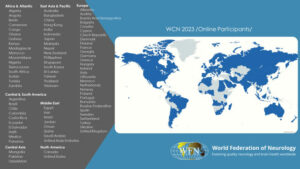
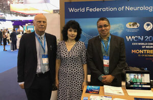
Montreal plays host to the WCN 2023 Oct. 15-19.

  The XXVI World Congress of Neurology (WCN 2023) will allow us to collaborate with thousands of peers from across the world, witness the best scientific and teaching programs and ground-breaking research. Taking place in Montreal, Canada, and accessible online, between Oct. 15-19, 2023, this event will offer many opportunities to broaden your knowledge and exchange ideas.

A Diverse Array of Sessions

WCN 2023 will feature an impressive lineup of sessions that cater to the diverse interests and expertise of neurology professionals. Attendees can look forward to engaging in the following exciting and novel sessions:

  • Presidential Symposia: Delve into cutting-edge topics and insights presented by leading experts in the field.
  • Regional Symposia: Explore regional perspectives and unique challenges in neurology from across the world.
  • Plenary Lectures: Gain valuable insights from distinguished speakers who will share their expertise and research findings.
  • Teaching Courses: Enhance your knowledge and skills through specialized teaching courses.
  • GNA Meeting: Connect with the Global Neurology Alliance and participate in discussions shaping the future of neurology.
  • Free Communications: Present and discuss your own research and discoveries with fellow neurology professionals.
  • Joint Sessions With Other Societies: Collaborate and exchange ideas with professionals from related fields.
  • Coffee Talks Sessions: Engage in candid conversations with current and past WFN leaders as they discuss topics like collaboration between WFN and WHO.
  • Young Neurologist Sessions: A dedicated space for emerging talents in neurology to network and learn from experienced peers.
  • Meet With Plenary Speakers: Interact with keynote speakers and engage in thought-provoking discussions.
  • Patient Day & Patient Forum: Gain insights into patient perspectives and participate in discussions about patient care.
  • AI Session and More: Stay updated on the latest advancements in artificial intelligence applied to neurology.

Tournament of the Minds

The 10th Tournament of the Minds, a highlight of WCN, will once again blend education and entertainment. This unique event will explore various aspects of neurology in an engaging and interactive manner, offering attendees an opportunity to test their knowledge and problem-solving skills.

Meet Us at the WFN Booth

Meet WFN staff and trustees at the WFN booth in the exhibition hall.

As seen in this chart, the WCN is truly a global conference that touches every corner of our world. With participants from more than 80 countries, WCN2023 will bring together thousands of neurologists from around the world, thereby fulfilling one of the WFN’s primary goals: to foster quality neurology worldwide by promoting global neurological education and training.

Five Days of Scientific Excellence

WCN 2023 promises five exhilarating days filled with inspiring scientific experiences. Attendees will have the privilege of interacting with a global community of neurology professionals, fostering meaningful connections, and sharing their expertise.

With more than 2,300 abstracts submitted, the XXVI World Congress of Neurology stands as a testament to the enthusiasm and dedication of neurology professionals worldwide. Whether you are a seasoned expert or an emerging talent, WCN 2023 offers a remarkable platform to collaborate and learn.

Mark your calendars for Oct. 15-19, 2023, and join us in Montreal and online for this exceptional gathering of minds in the world of neurology. •

New Workplaces, New Opportunities to Mitigate Neurological Disability

Workplace disease-specific education and management programs to increase awareness, diagnosis, management, and productivity.

By Dr. Olivia Begasse de Dhaem

Olivia Begasse de Dhaem

Mitigating the impact of the disability related to neurological diseases goes beyond medical management and rehabilitation. Workplace accommodations and workplace disease education and management programs must be included in the continued efforts of the 2023 World Brain Day campaign.

As stated by Prof. David Dodick on World Brain Day 2023, disability is associated with developing chronic conditions; earlier mortality; disparities in terms of access to care, education, job; stigma; and poverty. Supporting people with neurological disorders in the workplace is crucial to mitigate the downstream impact of disability. Neurological disability interferes with occupational potential and earning, which in turn can negatively impact mental health and access to medical care and worsen the level of disease state and its related disability. Being in the workforce contributes to general and mental health.

Let’s take the example of migraine, the second leading cause of disability in the world according to the Global Burden of Diseases. Migraine is the second cause of presenteeism (being present at work but not as productive as usual due to the symptoms of a disease) in the United States, estimated to account for 16% of total workplace presenteeism and to cost $240 billion (USD) annually. Although the migraine-related productivity loss increases with the frequency of migraine attacks, there is significant productivity loss interictally as 41.5% of workers have moderate to severe interictal symptoms.

Thankfully, there are evidence-based ways to help workers with migraine, such as social support (between colleagues and by supervisor), job satisfaction, a sense of autonomy, and workplace accommodations (such as natural lights, noise reduction, scent-free areas, regular breaks, air quality). Small interventions such as raising awareness in the workplace and helping a supervisor understand the disease can have a huge impact. One patient of the author was about to lose her job as a cashier at a supermarket due to the numerous work absences she had because of her chronic migraine. After discussions and a doctor’s note, her supervisor helped her change her job from cashier to bagger, so instead of constantly rotating her neck to look at a bright screen while scanning, she was placing the groceries in bags. She was also allowed to take regular breaks to lay down on a mat in a dark, quieter closet. These accommodations enabled her to continue working full time with rare absences.

Migraine workplace education and management programs offered to all employees and supervisors increase understanding of the disease, decrease stigma, improve diagnosis and treatment, and increase productivity. It is crucial to educate the entire employee population and not target a specific group.

The Fujitsu Headache Project enrolled 73,432 (91%) of its employees in Japan with a migraine prevalence of 17%. This workplace education and management program improved:

• the understanding of headache disorders (73% of participants)

• the attitude toward colleagues with headache disorders (83% of participants without headache) – productivity

It reduced absences and presentee days by 1.2 and 14 days per employee per year, respectively, with a 32-fold positive return-on-investment.

As workplaces are being redesigned after the COVID-19 pandemic, now is the time for neurologists to advocate for equitable work opportunities for people with neurological diseases and to promote workplace education programs like the International Headache Society Global Patient Advocacy Coalition (IHS-GPAC) Migraine Fitness modules. •

For additional information:

• Begasse de Dhaem O. Migraines are a Serious Problem. Employers Can Help. Harvard Business Review. February 2021.

• Begasse de Dhaem O, Sakai F. Migraine in the workplace. eNeurologicalSci. 2022;27:100408. June 2022.

• Sakai F, Igarashi H, Yokoyama M, Begasse de Dhaem O, et al. Diagnosis, knowledge, perception, and productivity impact of headache education and clinical evaluation program in the workplace at an information technology company of more than 70,000 employees. Cephalalgia. 2023;43(4):3331024231165682.

• IHS-GPAC website.

Dr. Olivia Begasse de Dhaem is on the executive committee of the International Headache Society Global Patient Advocacy Coalition (IHS-GPAC) and co-chair of the IHS-GPAC Summit III in Seoul in 2023.

From the Editors

We’d like to welcome all readers to the August 2023 issue of World Neurology.

The issue begins with the President’s Column, where WFN President Dr. Wolfgang Grisold updates us on the upcoming World Congress of Neurology (WCN) Oct. 15-19 in Montreal and the many exciting new initiatives of the WCN, as well as other global activities of the WFN.  Many initiatives and opportunities at the upcoming WCN are also well described in an accompanying article.

Next, Dr. Olivia Begasse de Dhaem describes innovative workplace initiatives intended to mitigate neurologic disability from migraine. In this issue’s History column, Dr. Peter J. Koehler delves into the differences and similarities of the theories of brain and mind as proposed John Hughlings Jackson and Thomas Laylock.

Dr. Dafin Muresanu then reports on two recent successful conferences that occurred in Romania: the EFNR-WFNR Eastern Europe Regional Meeting and the National Neurology Forum.

WFN Past President Dr. Raad Shakir, who is current Chair of the WFN Nominating Committee. reports on the activities and important responsibilities of the WFN Nominations Committee. Dr. Riadh Gouider, Dr. Grisold, Dr. Steven Lewis, and Dr. Imen Kacem describe the recent history of the WFN Educational Days and current planning for exciting upcoming Educational Days. Finally, the statements of all of the current candidates for WFN Treasurer and WFN Elected Trustee are republished/published in this issue, in anticipation of the upcoming voting by the members of the WFN Council of Delegates.

In closing, we look forward to seeing so many of you at the WCN in Montreal (in person or virtually) this October! And don’t forget to sign your department up for the exciting Tournament of Minds! •