by Peter J. Koehler
Some years ago, I presented a lecture on the history of CT and MR at the History of Neurology course of the American Academy of Neurology (2018). Soon after the invention of X-rays by Wilhelm Conrad Röntgen (1845-1923) in 1895, two steps needed to be made to improve brain imaging, notably the introduction of contrast enhancement and the production of 3D images.
The first step was taken by the use of air contrast in the ventricles of the brain, inserted directly (1918) or by lumbar puncture (1919). Arterial contrast followed in the 1927. The technique was called arterial encephalography as it was the brain rather than the arteries, in which physicians were interested at the time.
The second problem was, at least temporarily, solved by the introduction of planigraphy, better known as tomography, which was introduced in the early 1930s1. For many decades, a combination of air contrast X-rays and analogous tomography with small amounts of air was applied (PEG or pneumoencephalography). The procedure was quite uncomfortable for the patients. After the introduction of the air directly in the ventricle or by lumbar punction, the patients were rotated in a somersault chair to bring the air into the place of interest in the head. I remember having seen such a patient returning to the ward suffering from severe headache and vomiting. A short film clip of the procedure can be found here: ((46) The Scanner Story (Part 1 of 2 of documentary covering early CT development) – YouTube and go to the section 3.04-4.00 minutes).
Mathematics: The Inverse Radon Transform
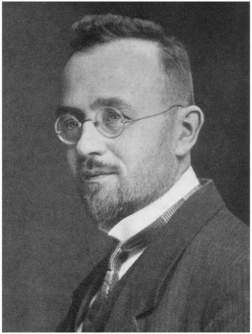
Johann Radon (ca. 1920; Wikimedia Commons, public domain).Johann Radon (ca. 1920; Wikimedia Commons, public domain).
In the late 1950s and early 1960s, the first attempts to look for a more patient-friendly method were conducted. Interestingly, the mathematical method for the reconstruction of an object from multiple projections had already been discovered by the mathematician Johann Radon (1887-1956) in 1917. Born in Tetschen, Bohemia (now Děčín in the Czech Republic), he studied at the University of Vienna. Due to his near-sightedness, he was exempt from the draft. He was active as a professor of mathematics at several universities in central Europe. His 1917 publication did not have immediate practical applications and was not used by subsequent CT pioneers.
Pioneers in Kiev: Rediscovering the Inverse Radon Transform
What is less well-known is that in the late 1950s, Ukraine pioneers were working on a reconstruction project. Doing research at the Igor Sikorsky Kyiv Polytechnic Institute (KPI) in Kiev and named after the Kiev born aircraft designer Igor Sikorsky (1889-1972), two men tried to make the experimental arrangement for reconstruction. Semyon Isaakovich Tetelbaum (1910-1958) studied at the KPI, after which he became electrical design engineer working on projects involving radio and television. In 1940, he became professor at the KPI. Boris Isakovich Korenblum (1923-2011) was born in Odessa, a city in the south of Ukraine, on the Black Sea, and changed careers from the violin to one in mathematics after winning a mathematics competition. He studied mathematics in Kiev. Being of Jewish descent, he escaped from being killed by the Nazis in Babi Jar. Losing his job during Stalin’s anti-Semitic campaign of 1952-1953, he got a position at the Construction Engineering Institute in Kiev, where he stayed until his emigration to Israel and later to the United States.
In 1956, Tetelbaum published “About the Problem of Improvement of Images Obtained With the Help of Optical and Analog Instruments” in Bulletin of the Kiev Polytechnic Institute2. The subsequent year another article by him followed in the same periodical, notably “About a Method of Obtaining Volumetric Images by Means of X-ray Radiation.3” (See Figure 1.) In 1958, Korenblum, Tetelbaum, and Tyutin published an article titled “About a Scheme of Tomography” in the Proceedings of Higher Educational Institutions – Radiophysics4.
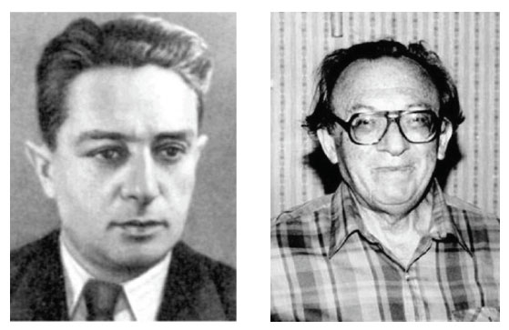
Semyon I. Tetelbaum (left) and Boris I. Korenblum.
The articles remained unnoticed in the Western world until Harrison H. Barrett (Department of Radiology and Optical Sciences Center), William G. Hawkins (Department of Mathematics), and Michael L.G. Joy (Institute of Biomedical Engineering) of the University of Arizona published a “Historical Note on Computed Tomography” in 1983. The authors wrote that Tetelbaum “formulated the tomography problem in terms of line integrals and found the exact solution, which is now referred to as the inverse Radon transform.” Korenblum, Tetelbaum, and Tyutin “gave a detailed account of the theory, including a method of fan-beam correction, equivalent to reordering the data, and a discussion of a practical way of handling the singularity in the integrand of the inverse Radon transform.” Furthermore, in their paper, the Ukraine scientists “presented a block diagram of television-based analog computing system for implementing the reconstruction” and estimated the system should be capable of “reconstructing a 100 x 100 image in five minutes.” (See Figure 2.)
One of the authors of the historical note (Hawkins) programmed the reconstruction algorithm and concluded that it performed satisfactorily, although “because it was not computationally efficient, only a 32 x 32 image was reconstructed.”5 As the papers were published in the Russian language, it was difficult to access the contents. More recently, two of the three articles have been translated by Alex Gustschin, chair of biomedical physics in the Department of Physics and Munich School of Bioengineering, Technical University of Munich.6,7 He also provided comments and biographical data on Tetelbaum and Korenblum, but was unable to find information about Tyutin.
So why didn’t Korenblum and his colleagues build the machine in 1958? In 1983, Barrett and colleagues wrote that they “continue to search unsuccessfully for later references to the reconstructed images.” Gustschin suggested that the unexpected death at age 48 of Tetelbaum in 1958 may have caused the end of the project.
Second Rediscovery of the Radon Transform
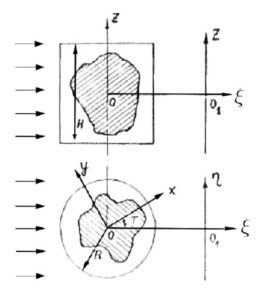
Figure 1. From Tetelbaum’s 1957 publication.3
Unaware of the publications of Radon or the Ukraine scientists, William Henry Oldendorf (1925-1992), a neurologist working at UCLA, built a prototype to transmit a beam of X-rays through the head and reconstruct its image in 1959-1960. Although the method was patented, he was unable to get it funded.
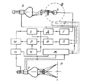
Figure 2. Functional block diagram of the television-based computing device from Korenblum et al. 1958.4
Working in Cape Town, South Africa, approximately in the same period, and later moving to Tufts University, Allan McLeod Cormack (1924-1998) formulated mathematics of reconstruction for simple attenuating objects with certain symmetry and applied a reconstruction technique (1957) with some success. He constructed an experimental scanner (1963) and only in 1970 he became aware of the solution found by Johann Radon in 1917. The story of Godfrey Newbold Hounsfield (1919-2004) is probably well-known. Working at EMI in London since 1949, he led a design team for building the first all-transistor computer (1958) to be constructed in Britain. Then he did early tests with a laboratory CT using a gamma ray source and single detector.
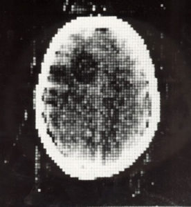
Figure 3. First clinical CT-scan (October 1971).
In October 1971, the first clinical scan was made. High-resolution images of a 41-year-old woman with a frontal tumor were produced in cooperation with neuroradiologist James Ambrose (1923-2006) at Atkinson Morley Hospital. (See Figure 3.) Cormack and Hounsfield received the Nobel Award for Medicine or Physiology in 1979. A 15-minute documentary on Hounsfield and Ambrose can be found at (46) The Scanner Story (Part 1 of 2 of documentary covering early CT development) – YouTube. •
References
-
Lutters B, Koehler PJ. Cerebral pneumography and the 20th century localization of brain tumours. Brain. 2018 Mar 1;141(3):927-933.
-
Tetelbaum SI. About the problem of improvement of images obtained with the help of optical and analog instruments” Bulletin of the Kiev Polytechnic Institute 1956;21:222.
-
Tetelbaum SI. About a method of obtaining volumetric images by means of X-ray radiation. Bulletin of the Kiev Polytechnic Institute 1957:22:154-60.
-
Korenblum BI, Tetelbaum SI, Tyutin AA. About a scheme of tomography Proceedings of Higher Educational Institutions – Radiophysics 1958;1:151-7.
-
Barrett HH, Hawkins WG, Joy ML. Historical note on computed tomography. Radiology. 1983 Apr;147(1):172
-
Gustschin A. Translation of Tetelbaum SI: About a Method of Obtaining Volumetric Images by Means of X-ray Radiation. arXiv:2001.03806.
-
Gustschin A. Translation of Korenblum et al. About a scheme of tomography. arXiv:2004.03750.
-
Webb S. From the Watching of, 1990. Shadows: The Origins of Radiological Tomography Bristol & New York. Hilger
