Three Steps to European Neurology Harmonization: Core Curriculum, Visitation Program, European Board Examination

By Wolfgang Grisold, Walter  Struhal and Svein Ivar Mellgren

European neurology is in the process of increasing harmonization. This is a consequence of the right of European neurologists to practice freely in Europe. This implies practically and scientifically based needs to approach the issue with care and preparation. Presently the UEMS/EBN (European Union of Medical Specialists-Section of Neurology/European Board of Neurology) provides a core curriculum for neurologic training, a definition of the neurology speciality, a training center visitation program and a European Board examination.

The UEMS (www.uems.net) is the union of European medical specialists, and is constituted of representatives from national societies and sections. The European Board of Neurology has representatives from European UEMS members and has biannual meetings. The website of the UEMS/EBN contains information of present activities, including that on examination and visitation of departments. One of the main activities of the UEMS is education. The UEMS/EBN also has provided a core curriculum, a definition of neurology within the UEMS, in addition to the department visitiation program and board examination, and is active in the accreditation of CME activities.

Content of Neurology

The content of neurology training for residents varies in different European countries of Europe.1 This is not only depending on national traditions, but mainly on the way neurology is practiced and how health system structures are used. In addition, local and national health systems have different professional relations with related overlapping medical fields. Pediatric neurology is in most countries attached to pediatrics. The definition of European minimal standards of content and structures of neurology are available in the UEMS/EBN chapter 6.2 The Core Curriculum of Neurology has also been published previously3 and is now (2013) in the process of revision.

Practical skills are trained mostly within the core competencies of neurology such as stroke, extrapyramidal diseases, multiple sclerosis and epilepsy.4 Some disciplines, such as clinical neurophysiology, may not be part of the neurology curriculum in several countries, but are still practiced by neurologists or specially trained neurophysiologists (in some countries with a separate specialty in clinical neurophysiology). Skills and particular competences will become more important as future stroke therapies with interventions also need the participation of neurologists.5 Rotation and exchange of trainees are also encouraged by the UEMS.

European Board Examinations

Many European sections, like urology, anesthesiology, ophthalmology and others have developed a  European board examination, which in some countries in part or as a whole has replaced the national examination. The attendees of these examinations have to prove that their knowledge is at a European level, and can use the examinations a sign of quality and excellence. The UEMS-SN/EBN decided to develop a European Board examination in 2004, and after several years of development the first examination took place in Milan 2009 during the ENS congress.

The choice of the instruments of the examination is difficult and much debated within the medical disciplines. This concerns the source of questions, the format of the examination, the examination format and the evaluation of results. At present, no universally applicable format of examinations is offered by the UEMS/Council for European Specialist Medical Assessments (CESMA),6 and the development of questions often depends on the individual section constructing these examinations according to their needs and requests.

Elements of the European board examination

It was decided that an instrument for reliable and objective testing of skills and practical issues would not be feasible within the neurology examination format, and therefore only candidates were accepted from EU/European Economic Area (EEA) countries, who had already been trained, or were declared qualified for the examination by their national society.  These European candidates are conferred the title “Fellow of the UEMS/EBN.” By 2013 also applicants from non-European countries were admitted to the examination provided they could present proof of similar qualifications.

In more detail, for the European Fellow of Neurology, a three-step approach was designed:

1: The national certification of trainees by their national society is considered as part of the examination. This certificate proves the person to be qualified for the examination. It may be based on a certain number of years of training, a national examination or status as an already certified neurologist. This mode of acceptance would ensure that practical training is confirmed by the national society. This step can practically only be accepted for UEMS states. Also for non-European states, certification of training, or national speciality, or board acceptance need to be provided at application for the UEMS/EBN examination

2: A set of 120 multiple-choice questions (MCQs) with one single best answer has been the main part of the examination. The MCQs were provided by members of the UEMS/EBN, the large European neurological societies (EFNS (European Federation of Neurological Soceties) and ENS (European Neurological Society (ENS), other neurological societies (Movement Disorder Society, European Stroke Society)) and recently also e-brain.

Before the examination all questions were graded independently by an examination board in a specific database. Only questions that obtained an average score over 5 (range 0-10) were accepted. The quality of questions and options of answers were then assessed by members of the Department of Medical Education, Ege University, Turkey, chaired by Prof. Ayhan Caliskan.7 This examination quality assurance concentrated on question stem, clarity, language, ambiguousness, and flaws. The final editing of the accepted questions was performed by the chairmen of the examination committee, who also distributed the number of MCQs to different topics.

3: Until 2013, the examinees also had to sit at an oral examination which consisted of four case vignettes with structured questions/answers. The oral examination was replaced from 2013 (examination in Barcelona) by EMQ (extending matching questions).  The major advantage of EMQs over MCQs is that this format not only evaluates knowledge, but also clinical reasoning. The EMQ case scenarios have been developed and written by experts in various topics on a given problem. 100 scenarios were prepared for Barcelona.

The passing limit of the MCQ questions, oral examination, and of the case presentations has been set to 75 percent in the first series of examinations. From 2012 onward, a passing limit was determined by an evaluation panel the day before the examination, based on the Nedelsky and Angoff  methods. This represents an additional effort to optimize the procedures according to educational standards.

Finally, the trainees need to orally present a case, and they thus earn extra points. This is a strictly oral presentation lasting 5 minutes, which is judged by two jurors according to a scheme.

Practical and Financial Issues

In the creation of the EBN examination, several practical questions had to be solved. The organization, including a database of questions, communication and practical aspects of the examination were handled by a professional company, the Vienna Medical University-affiliated Vienna Medical Academy.8 The examinations have taken place during the European neurological congresses and the societies (EFNS and ENS) have provided the necessary rooms. This has served the purpose that the examination could be combined with a congress visit. The cooperation with the societies has demonstrated their interest and engagement in the UEMS/ENB and the European board examination. In addition to sufficient rooming also secretarial staff from the ViennaMedicalAcademy have been present, and in addition handled the computer analysis of the MCQs and EMQs. As a routine, a full report of the recent examination is presented to the board of UEMS/EBN with regard to participation, questions, feedback of participants, the examination committee, assistance from the EgeUniversity and ViennaMedicalAcademy, and economical aspects.

In recent years, the development of European board examinations as a sign of quality and toward a move to harmonization has been encouraged. The UEMS group called CESMA (Council for European Specialist Medical Assessments)6 has been established, and has regular meetings to work on the format and quality of European board examinations.

Funding

The UEMS/EBN received initially two grants from the ENS and EFNS as a contribution to develop the EBN examination. All other costs have been covered from the resources of the UEMS/EBN, which has its income mostly from member fees (national societies) and by fees paid by the candidates, which do not cover by far the organizational expenses.

The largest academic input and development was done by volunteers of the UEMS/EBN and many European specialist, who wrote questions, helped to prepare the examination, and also practically participated in several tasks.

Department Visits

Several countries (like Norway) have implemented department visits on a national basis. This concept, also adapted from the UEMS, means a voluntary visit at a training center, both by a representative of the UEMS as well as of the national medical  representation. It is a structured approach to evaluate departments in the sense of equipment, staff and resources, and is based on a questionnaire with assessment of teachers, residents, head of department and also representatives of the hospital staff. The final report analyses the different aspects of teaching and training, and gives recommendations that can be used and should be implemented by the department or hospital.

Subspecialities

Subspecialties develop  in neurology worldwide. In the US, about 25 subspecialties fields have been identified and are recognized by the United Council for Neurological Subspecialties (UCNS), 9  which also provides an examination and certification system that is open worldwide. Within European neurology, no formal subspecialities have been identified. A possible step in this direction might be the attempt to create a multidisciplinary approach towards interventional neuroradiolgy by the division of neuroradiology of the UEMS.

Future Aspects

Since the publication of this paper, the EMQs and also the participation of non-European participants have been successfully  implemented.  A possible future cooperation with the Royal College of Physicians (UK), which offers similar examination, is under consideration.

Neurological training is based on a curriculum, on training content, methods, and also finally an assessment and certification. One could argue that structuring an exit examination at the end of training may be counterproductive, as it is too late to correct training or detect deficiencies in the individual trainee. The establishment of a quality circle, which not only evaluates trainees but also the quality of training center, is therefore a further important step in European neurology.

The implicit aspects are that a European examination increases harmonization, and the hope is that many European medical associations partly or as a whole will replace their board examination with this European examination in the future.

Paper published in Eur J Neurol. 2013 Aug;20(8):e101-4. Conflict of Interests: none

Grisold is a neurologist in Austria, chair of  UEMS/EBN examination committee and trustee of the WFN. Mellgren is a neurologist in Norway, co-chair of the UEMS/EBN examination committee, Norwegian delegate and vice president of UEMS/EBN. Struhal is  a neurologist in Austria, active in education in the YNT and EAYNT. He is a member of the education committee of the WFN.

References

1.   Grisold W, Galvin R, Lisnic V et al. One Europe, one neurologist? Eur J Neurol 2007; 14(3):241-247.

2.   UEMS European Board of Neurology http://www.uems-neuroboard.org/ebn.  2012.  Ref Type: Internet Communication

3.   Pontes C. Recommended core curriculum for a specialist training program in neurology. Eur J Neurol 2005; 12(10):743-746.

4.   Struhal W, Sellner J, Lisnic V, Vecsei L, Muller E, Grisold W. Neurology residency training in Europe–the current situation. Eur J Neurol 2011; 18(4):e36-e40.

5.   Flodmark O, Grisold W, Richling B, Mudra H, Demuth R, Pierot L. Training of Future Interventional Neuroradiologists: The European Approach. Stroke 2012.

6.   CESMA Statutes http://www.totbid.org.tr/upload/CESMA%20draft%20statutes.pdf.  2012.  Ref Type: Internet Communication

7.   Caliscan A. – http://ege.academia.edu/ayhan.  2012. Ref Type: Internet Communication

8.   V. M. A. http://www.medacad.org/vma/.  2012. Ref Type: Internet Communication

9.   UCNS http://www.ucns.org.  2012. Ref Type: Internet Communication
Stroke Master Class Series in Sri Lanka

Darshana Sirisena

Darshana Sirisena

By Darshana Sirisena  and Professor Tissa Wijeratne

Sri Lanka has an aging population with an impending epidemic of stroke at  hand.1,2 Stroke is a leading cause of adult disability in Sri Lanka3,4.

The first stroke master class series conducted in Sri Lanka was launched in April and July 2013. The master class series covered five provinces (nearly 10 million population) and conducted in teaching hospital, Anuradhapura, teaching hospital, Peradeniya, provincial general hospital, Kurunegala, provincial general hospital, Ratnapura and district hospital, Diyatalawa.

This stroke master class series was the latest initiative of Asia Pacific Association of Young Neurologists and Trainees (www.apaynet.org), the Asia Pacific section of International Working Group of Young Neurologists and Trainees (IWGYNT), World Federation of Neurology. APAYNET was established in 2011 as a result of the success of European group of young neurologists and trainees, European section of IWGYNT5,6,7.

Professor Tissa Wijeratne

Professor Tissa Wijeratne

The stroke master class series comprises of 11 didactic lectures covering key topics of stroke care range from global burden of stroke, development of stroke services in the limited resource setting, classification of stroke, stroke imaging  (master class on interpretation of acute stroke imaging), stroke thrombolysis, primary and secondary prevention of stroke, long-term rehabilitation in stroke. The syllabus ranges from basic science to recent advances in stroke care.

This series was endorsed by World Stroke Organization and aptly supported by a major donation of printed educational material from National Stroke Foundation, Australia.

The Stroke Master Class series was conducted by Associate Professor Tissa Wijeratne, Sri Lankan born, Australia and New Zealand trained neurologists currently reside in Australia, Darshana Sirisena, neurologist from Sri Lanka who is also the Sri Lanka representative of Asia Pacific Association of Young Neurologists and Trainees, Nirodha De Silva, physician with a special interest in stroke medicine and also the director, Stroke services, Base hospital, Diyatalawa, Sri Lanka. The series was aptly supported by several Sri Lankan neurologists and few senior physicians in Sri Lanka.

Diyatalawa Stroke Master Class audience.

Diyatalawa Stroke Master Class audience.

Despite several challenges, the master class series was a huge success. There were more than 500 delegates, including neurologists, physicians, junior doctors, nurses, physiotherapists, occupational therapists and speech therapists who were involved in stroke management in their day-to-day lives.

The event had in-kind support from department of neurology, WesternHospital, Melbourne, Australia, National Stroke Foundation Australia, Anurdhapura Clinical Society, Ratnapura Clinical Society, Stroke Unit, BaseHospital, Diyatalawa, Sri Lanka, Department of Medicine, Faculty of Medicine, University of Peradeniya, Sri Lanka, which made this series a huge success.

We had lot of positive feedback from the participants of this master class series in Sri Lanka. It was evident that there is a significant thirst at the grassroot level for knowledge of best practice in stroke care.  We were invited to come back and repeat the event with further updates on regular intervals in the future by many of them. This implicates the necessity for more opportunities of this nature at a junction where stroke care in countries such as Sri Lanka are trying to keep pace with the rest of the world.

Stroke Master Class audience, Teaching hospital, Peradeniya.

Stroke Master Class audience, Teaching hospital, Peradeniya.

There is no doubt that development of stroke services with whatever the resources available in less resourced countries like Sri Lanka is a top priority at the moment. This can be done and should be done. Reorganization of the services and education are the key factors that will get us toward this goal.

Asia Pacific Association of Young Neurologists and Trainees plan to establish a master of stroke medicine program through similar stroke master class program and open book examination process with the help of Australasia Stroke Physician Network, World Federation of Neurology, World Stroke Organization with a view to address the burning issue of stroke care workforce in the Asia Pacific region as matter of priority in the coming years.

If you are interested in helping us in this process or if you are interested in hosting the stroke master class series in your country, contact us via www.apaynet.org or email twi@unimelb.edu.au.

Sirisena is director of the Department of Neurology & Stroke Services at Teaching Hospital, Anuradhapura, Sri Lanka, Wijeratne is director of Stroke Services and the Neuroscience Research Unit at Western Hospital and the University of Melbourne, Australia.

References

1.   Wijeratne T, Gunaratne P, Gamage R, Pathirana G, Senanayake S, De Silva N, et al. Stroke care development in Sri Lanka: The urgent need for Neurorehabilitation services. Neurology Asia. 2011;16(2):149-51.

2.   Wijeratne T. Neurorehabilitation in Sri Lanka: an emerging sub-specialty for neurology trainees. Int J Stroke. 2012;7(2):163-4.

3.   Gunaratne PS. A step forward in stroke care in Sri Lanka. International Journal of Stroke. 2009;4(4):293-.

4.   Gunaratne PS, Fernando A, Sharma VK. Development of stroke care in Sri Lanka. International Journal of Stroke. 2009;4(1):38-9.

5.   Macerollo A, Varga ET, Gyorfi O, Kobeleva X, Paterson RW, Sellner J. The European Association of Young Neurologists and Trainees in 2013: striking a blow for European junior neurologists. Eur J Neurol. 2013;20(4):e54-8.

6.   Struhal W, Falup-Pecurariu C, Sztriha L, Sellner J. The European Asociation of Young Neurologists and Trainees: advocating young neurologists in Europe. Eur J Neurol. 2009;16(8):e146-8.

7.   Struhal W, Falup-Pecurariu C, Sztriha LK, Grisold W, Sellner J. European Association of Young Neurologists and Trainees: position paper on teaching courses for Generation Y. Eur Neurol. 2011;65(6):352-4.

Update From the WFN Website Committee Meeting

The current state of the website was discussed. The principal function of the website beyond the “About Us” page is education. This section of the site is being effectively populated by the Education Committee.  A major advance has been the addition of Facebook and Twitter by Walter Struhal, which are being used by more than 800 young neurologists to keep up with activities of WFN and to share  neurological  news and views. Two articles on the use of the website for social networking will appear in World Neurology, one in the current issue and one in the next.

Including World Neurology as a seamless section of the website has added greatly to its attractiveness, and will help to keep it fresh.  Exactly how World Neurology and the website will allocate and share content still needs to be determined, because it is possible to update World Neurology on an ongoing basis just as is done with the website.

Several suggestions for future development were also discussed. There is an interest in exploring the possibility of advertising on the website. There are several ways to do this, but we would first need to obtain metrics about the use of the website: How often is it visited, how long do visitors stay, and what do they look at?

Potential advertisers will pay for “eyeballs.”  These data should be readily available.  It was suggested that we be more proactive about linking to regional sites and member organizations. It would be good to have more geographical diversity in content as well. We had no takers when we offered to establish websites for member organizations that don’t have them, but we can make this offer again. Finally, password protected pages can be made available for committee work, but so far this service has only been used by the Education Committee.  As committees are reformed for the new term, the chairs should be informed of this service and perhaps more will be interested in taking advantage of it.

We would welcome comments and suggestions from the new executives and committee chairs as they are appointed.

Present at the Meeting: Bill Carroll, Pete Engel, Chris Kennard, Keith Newton, Raad Shakir and Water Struhal.  Donna Bergen, Wolfgang Grisold, Chiu Man and Don Silberberg had conflicting commitments.

WFN Awards Presented During World Congress of Neurology in Vienna

Johan Aarli (right) with WFN President  Vladimir Hachinski.

Johan Aarli (right) with WFN President Vladimir Hachinski.

Johan Aarli

WFN Medal for Service to International Neurology

Alastair Compston

The WFN Medal for  Scientific Achievement in Neurology

Alastair Compston is professor of Neurology and head of the Department of Clinical Neurosciences in the University of Cambridge and co-chair of Cambridge Neuroscience.

He trained in neurology at the NationalHospital and the Institute of Neurology, Queen Square after graduating from the MiddlesexHospitalMedicalSchool with first-class honors.

He is an outstanding editor of Brain, a past president of the European Neurological Society and of the Association of British Neurologists and past chairman of the Neurosciences and Mental Health Panel of the Wellcome Trust.

Arguably one of the most respected and capable clinician scientists of his generation, his research has focused on the clinical science of demyelinating disease with contributions to the genetic epidemiology, immunology and neurobiology, and treatment and repair of multiple sclerosis (MS).

compstonIn all areas, he has contributed substantially to the knowledge base with 335 original articles and research letters.  Most notably, with Stephen Sawcer, he established the GAMES consortium and went on to develop a worldwide consortium aided by two North American groups leading to the 2011 Nature publication involving almost 10,000 PwMS and more than17,000 controls, which expanded the known MS susceptibility loci to 57 and which overwhelmingly implicated T-cell driven immunity in the pathogenesis of MS.
In MS therapy, he was crucial with Alisdair Coles in using Alemtuzumab and showing that in relatively early MS, relapse activity and progression could be prevented and disability improved by lymphocyte ablation even though in more established MS progressive disability continued despite relapse activity being terminated. Both of these observations are now critical to the current approach to MS treatment. In both the Genome Wide Association Screen and Alemtuzumab initiatives, colleagues have said these would not have happened without Alistair Compston.

His incisive mind, broad experience, clarity of expression and quiet courteous manner underlie his ability to gather first-class colleagues and key collaborative groupings from around the world to aid his many successful achievements.

His work has been recognized by international awards: the Sobek Prize (2002), the Charcot Award (2007), the Zulch Prize (2010), the McDonald Award (2011) and now the World Federation of Neurology Medal for Scientific Achievement in Neurology (2013).

When the history of the solution to MS is finally written, Alistair Compston will figure as a principal contributor.

— W M Carroll

gerstenbrandFranz Gerstenbrand

Lifetime Achievement Award Citation

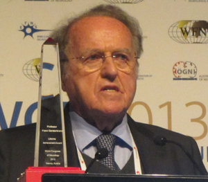
The organizers of the World Congress in Vienna have decided to award a special “one off” Lifetime Achievement Award to a most deserving person, who has truly spent his life in the service of neurology in his native Austria, the Danube region, in Europe and globally.  It is quite astonishing to realize that he was the secretary of the World Congress of Neurology here in Vienna as long ago as 1965.

Franz Gerstenbrand qualified from Vienna in 1950. Following training, he became associate professor in 1967, head of the department; Rosenhügel, Vienna, and Innsbruck in 1975.

His work in his native Austria is impressive. He was head of the Institute for Restorative Neurology and Neuromodulation, Ludwig Boltzmann Society from 1995-2006, and head of the Scientific Institute for Neurorehabilitation and Space Neurology; Karl Landsteiner Scientific Society up until 2009.

He holds many honorary doctorates; Charles University Prague, Danube University Krems , Aristotele University of Thessaloniki.

His regional achievements are most impressive. The most prominent is the bringing together the Danube and Eastern European neurologists at a time of political difficulties. He was a founding member and now is honorary president of the Danube Symposium for Neurological Sciences, and later the International Danube Neurology Association of Central and East Europe.

At the European level, a crowning achievement is that of being the founding president of EFNS. He is also the founding member of the European Society of Neuropharmacology  and World Federation for Neurorehabilitation

His involvement with the WFN started in 1965 and has continued ever since. He is chairman of the WFN Research Groups on Neuroethics and of Space and Underwater Neurology.  He is also a member of several other research groups.

He holds honorary memberships of several neurological societies:  Russia, Poland, CzechRepublic, Hungary and Myanmar. His work in Myanmar exemplifies his personality and diligence to help neurologists in all parts of the world, especially those living under difficult political regimes. He is the author of hundreds of papers and 12 books.

As far as I am aware, he is the only neurologist who is a holder of the Valeriy Gagarin Medal of the Russian Space Organization.

László Vécsei Secretary General, International Danube Neurology Association of Central and East Europe

Aspects of Neurohistory

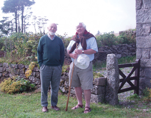
Alan Emery (left of the picture) with the author on Dartmoor in Southwest England.

Alan Emery (left) with the author on Dartmoor in Southwest England.

By Christopher Gardner-Thorpe,  MD, FRCP, FACP

I think I could turn and live with animals … Not one kneels to another, nor to his kind that lived thousands of years ago …

Walt Whitman, Song of Myself, Verse 32, 1881

This neurohistory column in World Neurology bears witness to the fact that there is increasing interest in medical history as a specialty. Many medical journals, and some textbooks, including Bailey and Love’s A Short Practice of Surgery, carry references to the history of the specialty that has always provided an intriguing glimpse of another world. Medical books and journals often discuss the history of their subjects and over recent years dedicated medical history publications have appeared and explore the subject in depth. The Journal of the History of the Neurosciences is a specialist journal edited by Peter Koehler and others and, coupled with book publications and some series of specialist history volumes, now provides a good platform for aficionados of neurohistory.

Why Medical History?

The teaching of medical history may rely upon three principal aims. The first is that it is necessary to look backward in order to understand how we have arrived where we are now and, ideally, to avoid treading the same pathway in the future where that pathway may be problematic. In other words, we learn from experience, or at least we should do so. The second is that it should be an academic discipline bringing, to the rigor of thinking, a proper academic approach to the gathering and sifting of information, and to making judgments and leading thereby to new pathways forward; a sort of dialectic where the thesis, antithesis and synthesis form a triad, each synthesis becoming a new thesis. The third is that it should be fun because things that are fun and interesting tend to capture the imagination and spur us to move forward. Perhaps this column will help achieve these aims and foster further interest in what we do and what we should do and also encourage others to join a happy band of new-age explorers.

Stanley Graveson

Stanley Graveson

The basis of any specialty is the people involved. Thus biography needs to come first. There are arguments for exploring the life history of those who contribute to a topic and biographies of neuroscientists abound. Some have achieved great things in a speciality, some have achieved great things in parallel fields including writing and some have become failed eponymists, those who have discovered things but whose name has not been attached to the discovery.

In the first group are many whose names we know from the neurology of long ago (Willis, Broca, Duchenne, Charcot, Gowers, Hughlings Jackson, Head, Critchley, Brain and others) and we can write of them. Fascinating books and papers abound, and there are many lessons to be learned from these earlier days of neurology. Thus biography grips us all, and we like to learn of the lives and experiences of our forbears. In the second group, we can be fascinated by the writings of Oliver Wendell Holmes, Arthur Conan Doyle, Somerset Maugham and in our own time Oliver Sacks, among others. Some have become medical truants. Some have written poetry (Dannie Abse) and some of the writing leans toward the neurological. In the third group, we can include Edward Meryon who described the dystrophy attributed subsequently to Duchenne, now often styled Meryon’s disease.

Many neurohistorians are collectors of books. A great worry nowadays is that since so much published on the Internet will not reach permanent paper form, the availability of publications has become greater but the ability to browse has become harder and the long-term storage of important information may fail in content and in recall. The wealth of electronic publication flying through the ether, often not peer viewed and published indiscriminately, complicates matters. Dusty old books give a pretty clear indication of the thinking of our forbears. Eponyms are often much easier to recall than disorders classified by number.

Neurohistory Collections

Collections of books in a sense comprise a museum. Specific neurological museums are uncommon, but many museums are dedicated to medical history, including the Berlin Medical Historical Museum and Virchow’s collection at the Charité, and the marvellous wax-work anatomical museums at La Specola in Florence and at the Josephinium in Vienna, the work of Joseph Towne (1806-1879) at Guys Hospital, the wax embryos at the Whipple Museum of the History of Science in Cambridge, and elsewhere. Those who state that dissection of real anatomical material is no longer necessary in the medical course are justified by some extent by the beautiful wax models that provide a three- dimensional view of the brain and the nervous system. Plasticized cadaveric material and plastic models can be quite lifelike but old timers believe dissection of real tissue is still needed. Nevertheless, today’s emerging doctors seem none the worse for having grown up without classical dissection.

Today’s junk forms tomorrow’s museum exhibits, and it is important to acquire and collect. Many personal collections of ephemera, those items that otherwise are discarded as no longer of any use — papers, equipment, drugs, collectible items that are used for only a short time — will provide to those who come after us a window on the neurological world that we experienced. X-ray films have all but disappeared in favor of CD-ROMs. Old isotope scans from rectilinear scanners and gamma cameras, carotid and vertebral angiograms on film, air encephalograms and ventriculograms, are things of the past and show the progress made eventually by CT, MRI and PET. Many of the basic ideas behind these new techniques seem so simple that they should have been conceived earlier. The truth is they depended on special technology, computing power in particular, and it is interesting to muse upon forthcoming developments and to say, for example, to those who feel that the technology behind CT is obvious, “Well, what then do you think in the future will appear obvious but which we have not yet invented?”

But it is still people who matter and who shape the future. So far we are not ruled (wholly) by computers and robots so we can make decisions and determine, or so we think, our own destiny. Neurologists might question this, believing that studies of the contingent negative variation indicate that something we think we have thought up, conjured from our own minds, is original whereas perhaps we are just made aware of some primitive activity that is programmed and not initiated voluntarily. Maybe we do not have an original thought in our heads. Maybe Jung’s collective unconscious in some way controls what we do, a sort of science fiction that has become science fact.

Our Teachers

Who has had a great influence upon each of us? Influential teachers for each of us, and bosses and colleagues who have influenced us individually. Neurohistory takes so many forms. What has caused each of us to become interested in the nervous system, and why did we enter a particular subspecialty? The evolution of clinical neuroscience into physician-  and surgeon-based, with the subdivisions by disease, by imaging and microscopy and so on, evokes many stories. Each is based upon the work of one or more individuals who were at the right place at the right time. Just as gliomas may originate from many foci seemingly simultaneously, so do ideas become reality depending upon the availability of new techniques, most obviously computers in the present era and based upon technology that we can not dream of in the future but which will become commonplace to the extent that future colleagues will say “Why was this not developed before?”

Just as we do in the investigation of malpractice claims, we must judge, if judge we must, on the basis of what was happening at the time of relevant prior events. In the future, others will ask whether in the 21st century we really did administer poisonous chemicals by mouth and by injection, whether we really did open up the body with a scalpel or the skull using a drilling machine, why we did even approach an organ (brain, heart, bile duct) by the natural tube (artery, vein, gut) that led to that organ in order to probe, enlarge, coagulate and coil, instead of heaven knows what alternative might become available instead.

We can write of those teachers who are no longer with us. For each of us, those teachers who are living now know who they are and what good influence they have had on our practice and for which we thank them. Stanley Graveson described himself as a jobbing neurologist, and this meant helping vast numbers of patients over a wide geographical area in the region of Wessex in the South of England. The stories told by colleagues and teachers regarding their experiences and research continue the stream of knowledge of what went before and of why we are where we have arrived today.

To arrive toward the end of a career in neuroscience with such a long and hard-working track record, with the ability to believe so many patients have been helped, is the aim of all, the best legacy topped by study and discovery, reading and writing, with continued learning and teaching each in our own neurological field.

Gardner-Thorpe is a consultant neurologist and editor of the Journal of Medical Biography.

New Officers and Site for WCN 2017 Announced

WFN COUNCIL OF DELEGATES REPORT

Almost 80 Delegates and representatives assembled in Vienna on Sept. 22 for the Annual General Meeting of the Council of Delegates. They arrived for registration to be greeted by a colorful oriental display from the three cities bidding to host the 2017 World Congress of Neurology — Hong Kong, Kyoto and Seoul. The importance of the occasion was evident to everyone, even more so because, in addition to the selection of the WCN 2017 venue, Delegates chose three new officers and one new elected trustee.

As well as presentations from the three member societies, Delegates heard an assessment from the Federation’s Professional Congress Organizer (PCO) and received reports from members of the site visit team to help them make up their minds. It was a difficult choice but eventually Kyoto won the day.

Just as keenly contested were the elections for officers and trustee, where 11 highly qualified candidates from across the globe stood for president, first vice president, secretary-treasurer general and elected trustee.  All of them addressed the assembly to present their vision and goals if elected.  Ballot papers were collected and counted outside the meeting by Keith Newton, WFN executive director, with assistance from Tanja Weinhart, Austrian Society executive secretary,  under the close supervision of Professor Johan Aarli, WFN past president and Professor Marianne de Visser, EFNS vice president. At the conclusion of the meeting, the results were announced.

  • WFN President: Raad Shakir (UK)
  • WFN First Vice President: William Carroll (Australia)
  • WFN Secretary-Treasurer General: Wolfgang Grisold (Austria)
  • WFN Elected Trustee: Amadou Gallo Diop (Senegal)

Delegates also received reports from officers and committee chairs, including chairs and co-chairs of the Education and Applied Research Committees.  The former gave a PowerPoint presentation of the activities of the Education Committee, including the development of standard operating procedures for committee activities, such as the monitoring of educational grants; departmental visit programs; and teaching center accreditation. Donna Bergen, chair of the Applied Research Committee, reported that new Applied Research Groups have been established on coma, neuro-oncology and neuro-infectious diseases.

The Membership Committee proposed a category of Pending Membership to speed up the process of assimilating new societies into the Federation.  Only voting rights will be temporarily withheld until all formalities are completed.  This year, three new societies joined the WFN — Oman, Tanzania and Uzbekistan — bringing the total number of neurological associations in the organization to 117.

Regional initiatives in Africa, Asia and Latin America have already begun to lay the foundations for the future and look set to build on them under the next administration now that Shakir has promised “global involvement through regional empowerment.”

By common consent, the Vienna Congress was a resounding success. Planning for Chile 2015 has already begun; Kyoto is our destination in 2017; and members are now urged to think ahead to 2019, when we return to the region of Africa or the Middle East for our biennial World Congress of Neurology.

Training Child Neurologists In Africa

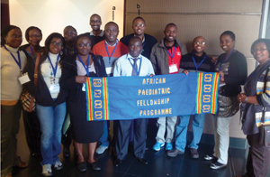
caption: The 2013 intake of doctors training under the APFP program. Roland Ibekwe from Nigeria (a) and Edward Kija from Tanzania (b) are the current child neurology trainees. Other members of the group are training in diverse paediatric specialities from gastroenteritis to paediatric intensive care from many different countries in Africa including Uganda, Kenya, Zambia and Malawi.

The 2013 intake of doctors training under the APFP program. Roland Ibekwe from Nigeria (a) and Edward Kija from Tanzania (b) are the current child neurology trainees. Other members of the group are training in diverse paediatric specialities from gastroenteritis to paediatric intensive care from many different countries in Africa including Uganda, Kenya, Zambia and Malawi.

By Jo Wilmshurst

The World Health Organization recommends a minimum of one adult neurologist per 100,000 of the population. The same group also reports that 10 percent of the pediatric population suffers from some form of neurodisability.

In Africa, the number of adult neurologists is significantly below this figure, and child neurologists are an even more scarce resource, with either no trained specialists, or at most one to service an entire country.1

The African Pediatric Fellowship Program is a project developed by the University of Cape Town to build skills capacity of health practitioners from Africa. Tertiary centers across Africa are assisted to develop strategic plans based on their countries’ key health care needs. Collaborating centers recruit teams to build skills and resources. Structured training occurs at the pediatric units through the University of Cape Town.

More than 55 specialists have completed the training program in diverse pediatric areas with a 98-percent retention rate of returning to work in their home countries since 2008. The program is evolving with training arms that support nursing and ancillary services.

Referring centers are encouraged to develop their own training modules as their trainees return, as such creating further training nodes across the continent. The trainee becomes the trainer in his or her home center, and a key opinion leader equipped to lobby for changes to health care policy.

The WFN provided a grant to support the program in 2011 over two years. Focused training has or is being provided to six general pediatricians from different centers in Nigeria, who manage large case loads of children with neurodisability and often epilepsy.

A key area of need identified by these doctors is the challenge of providing good care for children with epilepsy — centers lack the ability to perform and interpret EEG effectively and are not skilled in the clinical management of children with epilepsy.2 The University of Cape Town is in the process of developing a post-graduate diploma in “Pediatric Electrophysiology Interpretation and the Management of Children with Epilepsy.” This requires one-on-one training with a focus on areas relevant to the African context. The course should be available early in 2015.

Three doctors have been identified from Nigeria, Tanzania and Ghana, who at the request of their tertiary centers, are commencing formal training to be accredited child neurologists. Currently, Nigeria has 17 doctors with an interest in child neurology. (They are general pediatricians managing large numbers of children with neurological disorders.) Tanzania has no accredited child neurologists, and Ghana has two. Another trainee from Sierra Leone, which lacks both pediatricians (approximately eight in the entire country) and child neurologists (none), has been identified to train initially in general pediatrics and then to follow with developing relevant child neurology skills. This doctor started his pediatric training initially in Malawi in March 2013 at the Queen Elizabeth’s Children’s Hospital in Blantyre — a center with an exceptional general pediatric curriculum. After two years, he will transfer to complete his training in South Africa in our center. This system of developing other training nodes across the continent is the logical course to follow as it will build capacity to train more specialists. A goal of prevention and early intervention is one of the major aims for this project.

As such, the training is being developed to ensure that trainees return equipped with the concept of addressing health needs across all levels of health care. (They complete a “Transition and Translation of Knowledge module.”) They are expected to look at protocols, approaches and capacity to deliver health care from the primary health care clinics through to tertiary. While one person cannot address all of these needs, they can assist in the development of teaching programs whereby they train health care workers in safe and effective practice.

Building teams is essential. The program has a separate arm that focuses on nursing training, and the latest direction (the “third arm”) will be to develop ancillary services (namely physiotherapy, occupational therapy, speech therapy and so on). The benefits from these collaborations cannot be underestimated — there is much to learn from the approaches many innovative African centers undertake to cope with the challenges of scarce resources. The clinical skills of these doctors are exceptional as this and history taking are often all they have access to.

References

1.         Wilmshurst JM, Badoe E, Wammanda RD, Mallewa M, Kakooza-Mwesige A, Venter A, Newton CR. Child neurology services in Africa. J Child Neurol. 2011 Dec;26(12):1555-63.

2.         Wilmshurst JM, Cross JH, Newton C, Kakooza AM, Wammanda RD, Mallewa M, Samia P, Venter A, Hirtz D, Chugani H. Children With Epilepsy in Africa: Recommendations From the International Child Neurology Association/African Child Neurology Association Workshop. J Child Neurol. 2013;28 633-644

An American Physician’s Post-Graduate Medical Education in Vienna

boes

Christopher J. Boes, MD

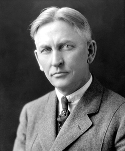
Walter DeWitt Shelden was born Feb. 2, 1870, near Windom, Minn., He grew up in Reedsburg, Wis., and graduated from the University of Wisconsin in 1891.  He attended RushMedicalCollege in Chicago, spent two years as an intern at CookCountyHospital in Chicago and worked as a general practitioner in Reedsburg for four years.  He then received internal medicine training at the University of Vienna for 1.5 years, and for the next 10 years had a lucrative private internal medicine practice in Minneapolis and taught at the University of Minnesota Medical School.  Shelden was asked by William J. Mayo to start the neurology section at the Mayo Clinic in 1913.   He was head of the neurology section at the Mayo Clinic until 1930, and died on Feb. 13, 1946.

Shelden enrolled in courses at the Viennese Medical Faculty as a so-called frequentant (student) during winter term 1901/02, summer term 1902 and winter term 1902/03, staying in Vienna from Sept. 1, 1901, through Feb. 1,1903 (University of Vienna Archives).  Approximately 10,000 Americans took some kind of formal

Walter DeWitt Shelden, MD. Photo courtesy of the Mayo Foundation for Medical Education and Research.

Walter DeWitt Shelden, MD. Photo courtesy of the Mayo Foundation for Medical Education and Research.

medical study at Vienna between 1870 and 1914.   Almost the entire post-graduate work in Vienna was given in the Allgemeines Krankenhaus (2,250 beds in 1912).

It was written that “here one could do more eye operations, deliver more babies, treat more patients and conduct more post-mortems in a week than would be possible in a year in many parts of the United States (Bonner TN.  American Doctors and GermanUniversities: A Chapter in Intellectual Relations, 1870-1914.  Lincoln:

University of Nebraska Press, 1963).”  Viennese clinicians were quick to encourage and exploit the interest of foreign doctors.  Short practical courses of four to eight weeks were offered in the clinical branches of medicine, each limited to 10 or 12 students.  In addition, adjunct professors and instructors arranged private courses. The internal medicine course given by Kovacs was controlled by Americans through a self-perpetuating membership.

Shelden worked with Edmund von Neusser (internal medicine), Friedrich Kovacs (internal medicine), Lothar von Frankl-Hochwart (neurology), Ernest Finger (syphilology  and venereology) and Alois Monti (pediatrics). Frankl-Hochwart (1862-1914) was in charge of the outpatient unit for neurology and electrotherapy in the medical department at the University of Vienna, and was professor of pathology of the nervous system (although primarily a clinician) (JAMA 1915;LXIV:353; JNMD 1917;45:189-190).  His 1891 monograph Die Tetanie contained the first summary of this disease’s clinical symptoms, and he also wrote important treatises on Meniè
re’s disease and neurogenic bladder (Lesky E.  The ViennaMedicalSchool of the 19th Century.  Baltimore: The JohnsHopkinsUniversity Press, 1976).

According to one of his students, Shelden “spent two years in Vienna, attending presentations of pathologic material and following von Neusser, whom he considered the greatest clinician he had ever known, in the wards of the Allgemeines Krankenhaus.  [Shelden] said von Neusser ‘couldn’t write worth sour grapes,’ that to learn from him one had to observe him examining patients (Mayo Clinic Archives).”

Edmund von Neusser, MD. Photo courtesy  of the National Library of Medicine.

Edmund von Neusser, MD. Photo courtesy of the National Library of Medicine.

Von Neusser (1852-1912), professor of internal medicine at the University of Vienna, was a master of diagnosis and differential diagnosis. Rumor had it that Neusser sent 140 patients to the dead-house without an error in diagnosis (Herrick JB.  Memories of Eighty Years.  Chicago: University of Chicago Press, 1949). Von Neusser had an active interest in diseases of the nervous system, and studied neurology in France, but did not become a specialist in neurology.  He was “master of the whole of internal medicine” (Lesky, 1976).

These trips to Vienna by Americans were criticized by some, but most of this criticism was directed at those without prior internship and practice experience who had short stays, partied too much, did not speak the native language, and/or came after World War I.  None of these things applied to Shelden.  Interestingly, most courses for Americans in Vienna were in English, but Shelden learned German there.  Learning the language was one of the reasons his mother encouraged him to go to Vienna.   Shelden translated German articles for an American neurologic journal later in his career.

Shelden’s training in Vienna honed his clinical skills, exposed him to scientific medicine at the highest level, and had a significant influence on his subsequent medical career.   His student Henry Woltman stated that “when the consultants gathered ‘round the autopsy table in the deadly game of turning face-down on the table a card bearing their name and diagnosis, Dr. Shelden was almost always right,” reminiscent of rumors of von Neusser’s diagnostic accuracy.  Shelden exemplified the benefit of post-graduate training in Vienna before World War I for the well-prepared American physician.

Boes is an associate professor of neurology at the Mayo Clinic in Rochester, Minn.

Stroke Definition in the ICD-11 at the WHO

strokedef_table1By Bo Norrving, MD, Didier Leys,  Michael Brainin and Steve Davis

Health classifications are a core responsibility of the World Health Organization (WHO), assigned by international treaty with 193 member countries. The International Classification of Diseases (ICD) is the oldest and historically most important. Member countries are required to report health statistics to WHO according to ICD, and ICD categories also are used as the basis for eligibility and payment for health care, social and disability benefits and services. ICD should have broad global utility, not only for specialists or neurologists, but for all physicians and health workers. All global regions are represented on WHO advisory groups, with a good representation of low- and middle-income countries.

ICD-10 was completed in 1990; the interval to ICD-11 is the longest time without revision in history of the ICD. The period has seen major advances in our understanding of cerebrovascular diseases and their treatment. The ICD-11 is mandated by the World Health Assembly and is expected to be officially approved by the 2015 World Health Assembly. A novel feature of ICD-11 is the inclusion of definitions. At WHO, the Mental Health and Substance Abuse Department is responsible for the revision of Diseases of the Nervous System. The Neurology Topic Advisory Group is chaired by Raad Shakir, and has seven individual members and more than 10 representatives from neurology associations and federations.

The World Stroke Organization (WSO) has been involved in the ICD-11 revision at WHO since 2010 and has been invited in this function as the NGO in official relations with WHO regarding stroke. The Cerebrovascular Disease ICD-11 advisory group is chaired by Bo Norrving, Sweden, with members Valery Feigin, New Zealand; Padma Gunarathne, Sri Lanka; Vladimir Hachinski, Canada; Michael Hennerici, Germany; Ming Liu, China; Peter Rothwell, UK; and Jeffrey Saver, US.

In the ICD-11, all cerebrovascular diagnoses will for the first time form one single block within Diseases of the Nervous System, which represents a major change in the classification. The work of the ICD-11 cerebrovascular working group has been reviewed by the board of the WSO and has been openly available to public comments. The document has been submitted to WHO, and the next steps include international scientific peer review of the whole ICD-11 and field trials. The aim is that the ICD-11 in its complete form is submitted to the 2015 World Health Assembly, for subsequent implementation in member countries.

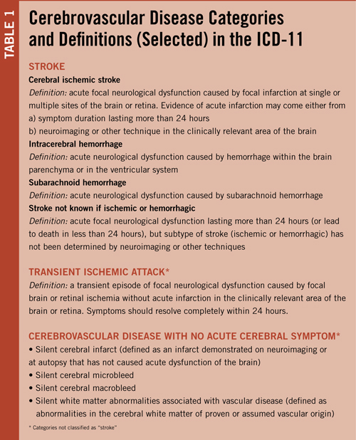
Selected key categories of the ICD-11 and their definitions are summarized in Table 1. The term “stroke” requires the presence of acute neurological dysfunction, which is in line with the definitions generally used in previous epidemiological studies, official statistics and the Global Burden of Disease project. This requirement was felt to be of particular importance in low- and middle-income countries in which a large proportion of cases will be diagnosed based only on clinical features without the addition of neuroimaging. Keeping the requirement of acute neurological dysfunction for stroke will allow comparison of data between regions and between time periods for the study of trends, which are extremely important for monitoring of global burden of diseases.

The ICD-11 cerebrovascular section includes a new cause category: “Cerebrovascular disease with no acute cerebral symptoms” which includes silent cerebral infarcts, silent cerebral microbleeds and silent white matter abnormalities associated with vascular disease.

“Silent cerebral infarct” is defined as an infarct demonstrated on neuroimaging or at autopsy that has not caused acute dysfunction of the brain. There is substantial scientific support that silent cerebral infarcts carry important consequences on brain function (cognition, gait, balance function) and prognosis. Whereas effects of specific therapies have not been demonstrated yet, risk factor assessment and control should usually be applied for preventive purposes. However, the risk of disease stigmatization also was carefully considered, and it is specifically stated that these entities do not represent a “stroke,” a distinction that may have unwanted consequences from legal or insurance perspectives.

More than 90 percent of all cerebrovascular lesions in the brain are not associated with acute neurological dysfunction; in the general elderly population the prevalence of silent cerebral infarcts and microbleeds range from one-fifth to one-half with increasing age. Use of different stroke definitions that do, or do not, include silent cerebral infarcts and microbleeds, carry a potential risk of miscoding that may seriously distort official statistics and of causing confusion within the health sector and to the general public.

As the current WSO president, Norrving was invited to the AHA/ASA working group, but when it became apparent that the group would arrive at a definition of stroke that was importantly different from the one in ICD-11 (basically the inclusion of silent cerebral infarction and silent cerebral hemorrhage within the lexicon of stroke), the issue was discussed within the organization and the decision was taken that WSO needed to withdraw.

WSO cannot officially approve another definition of stroke than the one developed within the governmental framework of the ICD-11 at WHO. Similarly, Didier Leys, as the current ESO president, was also invited to the AHA/ASA working group; ESO took the decision also to withdraw as the organization supported the definition of stroke as defined in the ICD-11, and also argued that definitions on cerebrovascular disease should be taken on a world, rather than a regional, level.

For the future, it is essential that transparent definitions are used that facilitate reporting and comparisons on a global scale. Stroke is one of the prioritized non-communicable diseases within the WHO Global Action Plan for NCDs 2013 to 2020, and the prevention and management of stroke requires the full support of all actors involved, including the stroke and neurological societies.

Norrving is immediate past president WSO and chair of the ICD-11 Cerebrovascular Advisory Group. Leys is immediate past president ESO. Brainin is ESO president. Davis is WSO president.

Implications of the AHA/ASA Updated Definition of Stroke for the 21st Century

ahaasastroke_table1By Scott E. Kasner, MD,  and Ralph L. Sacco, MD

Note: The views expressed by the authors  are their own and do not represent an official statement by the American Heart Association/American Stroke Association.

Stroke was defined by the World Health Organization (WHO) more than 40 years ago as “rapidly developing clinical signs of focal (or global) disturbance of cerebral function, lasting more than 24 hours or leading to death, with no apparent cause other than that of vascular origin.”1 This was a working definition created for a study assessing the prevalence and natural history of stroke, and it served its purpose at the time.

The ensuing decades have witnessed major advances in basic science, pathophysiology and neuroimaging that have dramatically improved our understanding of ischemia, infarction and hemorrhage in the central nervous system (CNS). There is little doubt that permanent injury occurs well before the 24-hour threshold, and therefore purely time-based definitions are inaccurate and obsolete. Further, neuroimaging has demonstrated that clinically transient symptoms are often associated with evidence of acute cerebral infarction and that infarction may occur without overt symptoms.

In 2009, the American Heart Association/American Stroke Association (AHA/ASA) published a scientific statement redefining transient ischemic attack (TIA) as, “a transient episode of neurological dysfunction caused by focal brain, spinal cord or retinal ischemia without acute infarction.”2 This statement formally addressed only one side of the proverbial coin, but clearly implied that objective evidence of infarction should be considered as a defining feature of stroke.

In the spring of 2013, the AHA/ASA published an expert consensus document with a new definition of stroke to reflect these advances.3 Authors with expertise in the fields of neurology, neurosurgery, neuroradiology, neuropathology, clinical research methods, epidemiology, biomarkers, policy and global public health were invited from within the AHA/ASA, as well as the American Academy of Neurology, the American Association of Neurological Surgeons and Congress of Neurological Surgeons, U.S. Centers for Disease Control and Prevention, the National Institute of Neurological Disorders and Stroke, the European Stroke Organization (ESO), the World Stroke Organization (WSO) and others to establish a universal definition of stroke.

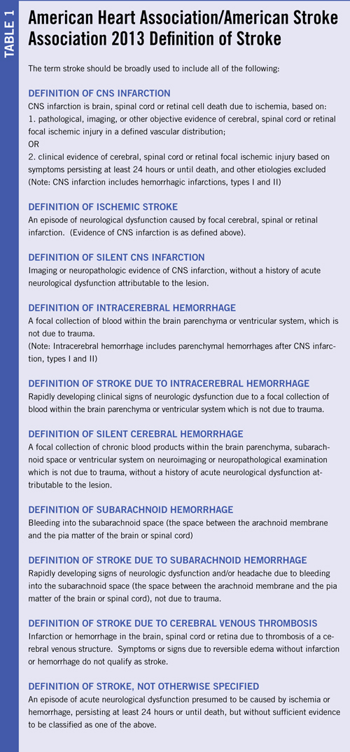
The key components of the new AHA/ASA definition are summarized in Table 1. The major fundamental change compared with older definitions is that the new broader definition of stroke includes any objective evidence of permanent brain, spinal cord or retinal cell death due to a vascular cause based upon pathological or imaging evidence with or without the presence of clinical symptoms. The new definition harmonizes with our understanding of the pathophysiology of infarction and with the recent redefinition of TIA, but also necessitates the inclusion of silent infarction and silent hemorrhage within the broad definition of stroke. Ultimately, the leaders of the ESO and WSO withdrew from participation and declined to endorse the statement because they disagreed about the inclusion of silent cerebral infarction and silent cerebral hemorrhage within the lexicon of stroke. (See “Stroke Definition in the ICD-11 at the WHO.“)

Objective Evidence of CNS Infarction: Imaging or Persistent Symptoms

The AHA/ASA defined CNS infarction based on pathological, imaging or other objective evidence of infarction. In the absence of this evidence, the persistence of symptoms of at least 24 hours or until death remained a method to define stroke. Objective evidence of infarction is generally currently available in the form of neuroimaging or less commonly neuropathological examination, but other methods such as highly sensitive and specific biomarkers may emerge in the future.

At present, imaging is not always available and also is not perfect. CT is well known to have limited sensitivity to acute infarction, especially in the first few hours, and also misses small infarctions even at later time points. MRI is far more sensitive, potentially within minutes of onset, but still fails for small infarctions, especially in the brainstem. In much of the developing world and in rural parts of more developed regions, neither of these tools may be available in the acute setting, if at all, which limits the global applicability of an imaging-based definition of stroke.

Persistence of clinical symptoms also can be taken as objective evidence of cerebral infarction. Several studies suggest that the majority of transient stroke symptoms resolve in less than 24 hours,4,5 and that persistence beyond 24 hours is almost always associated with MRI evidence of infarction. These findings support the classic threshold as a means to infer infarction when there is no confirmatory method readily available.

Definitions and Implications of Silent CNS Infarctions and Hemorrhages

CNS infarction included ischemic stroke, as well as silent CNS infarction (carefully worded to deliberately not use the term “silent stroke”). Ischemic stroke was defined as an episode of neurological dysfunction (clinical symptoms) caused by focal cerebral, spinal, or retinal infarction, while silent CNS infarction was defined as imaging or neuropathologic evidence of CNS infarction, without a history of acute neurological dysfunction attributable to the lesion. Similar definitions were crafted for cerebral hemorrhage. This major departure from past definitions is based on the observation of brain injury, either by imaging or pathological assessment, in patients without a history of well-defined neurologic symptoms.

Silent lesions have been recognized pathologically as infarctions and hemorrhages since the 1960s but were deemed of uncertain importance. However, they may not be entirely asymptomatic, as patients may have subtle cognitive, gait or other functional impairments in the absence of a typical acute presentation. To some extent, the “silence” of an infarction or hemorrhage depends on the eye of the beholder. Patients may not be aware of their symptoms due to neglect, denial or simply may attribute them to another cause and not seek a medical opinion. Physicians and other health care providers may vary in their ability to detect mild neurologic abnormalities, or they, too, may ascribe them to an alternative cause.

Silent CNS infarcts are well-recognized to be associated with impaired mobility, physical decline, depression, cognitive dysfunction, dementia and clinical stroke. Silent brain infarcts increase the risk of ischemic stroke by 2-4 times6,7 independent of other vascular and stroke risk factors. A recent review of MRI diagnostic criteria for silent brain infarcts found a threshold size of ≥3 mm to be a reliable indicator of these lesions. Silent infarcts are approximately 5 times more prevalent than ischemic strokes, found in 8 to 28 percent of patients, and are increasingly prevalent with age and in women.8-13 Chronic small parenchymal hemorrhages, or “microbleeds,” are found in up to 6 percent of healthy elderly individuals.14 These lesions, typically observed on gradient echo sequences on MRI, are the detritus of prior hemorrhages in the form of hemosiderin, typically adjacent to small blood vessels. Microbleeds appear to share the same underlying pathophysiology as larger hemorrhages, and are most commonly associated with cerebral amyloid angiopathy (CAA) and/or chronic hypertension. Since hemorrhage in the brain is always abnormal, there is no size threshold for microbleeds, unlike small infarctions. These microbleeds may not be associated with a clinical event but are associated with cognitive decline”‘5,16 as well as a high rate of subsequent ICH and ischemic stroke.17 As with silent CNS infarctions, the clinical impact may depend on the sensitivity of the observer.

The AHA/ASA included silent CNS infarctions and hemorrhages within the broadest definition of stroke for multiple reasons. First and foremost, since silent lesions have the same pathophysiology as clinically apparent ischemic and hemorrhagic strokes, it seems consistent that they should be united within the same broad disease category. Similarly, the multi-organization Universal Definition of Myocardial Infarction (MI),18 considered any pathologically defined cardiac infarction as an MI, regardless of the presence or absence of any symptoms or signs. Further, inclusion of silent CNS infarction and hemorrhage raises awareness of the potential for cognitive and functional decline that must be assessed and addressed from the perspectives of treatment and prevention. It seems clear that CNS infarctions and hemorrhages occur over a spectrum ranging from severe symptoms to very mild or even clinical silence, and the opinion of the AHA/ASA was that all must be included within the new and broader definition of stroke.

Implications for World Neurology

The new tissue-based definition of CNS infarction depends on either early objective (currently neuroimaging) evidence of infarction or persistence of symptoms for at least 24 hours. If early imaging is not available, then clinicians are left with a diagnostic dilemma in those first 24 hours since the event cannot be clearly classified as stroke. For patients with acute myocardial ischemia, the term “acute coronary syndrome” (ACS) is used before it can be determined if there is infarction or not, as assessed by electrocardiography or biomarkers. Similarly with stroke, the term “acute cerebrovascular syndrome” (ACVS) would suggest the potential diagnoses of cerebral infarction, TIA, and hemorrhage in patients presenting within the first 24 hours from onset and prior to the completion of imaging studies. Ultimately, diagnostic techniques and/or time will help define infarct or hemorrhage based on objective imaging, or TIA in the absence of positive imaging and resolution of symptoms within 24 hours from onset. A major challenge for the future will be the achievement of access to diagnostic and treatment tools in the developing world, where a substantial portion of the global burden of stroke occurs.

The inclusion of silent infarcts and microhemorrhages within the AHA/ASA definition of stroke opens many questions for clinicians. In regions with little or no access to neuroimaging, this change in definition may prove irrelevant for many years to come. However, for those with such access, silent lesions are likely to be detected as a result of the widespread use of MRI for non-cerebrovascular symptoms such as headache or dizziness. Further, clinicians and patients should be aware of the relationship between silent infarcts and hemorrhages with dementia and other impairments. The clinician should consider such patients as having evidence of cerebrovascular disease and should evaluate and treat any potential stroke risk factors. However, guidelines for secondary stroke prevention19 have been generated from clinical trials that have only included patients with symptomatic cerebrovascular disease and have not included silent infarcts. No studies have yet addressed the safety and efficacy of secondary prevention measures in patients who only have silent infarction. Future guidelines must address the available evidence for treatment in this population.

Updating the definition of disease can have prominent effects on disease surveillance and assessments of public health.  In the case of adding a large number of silent infarction cases to the existing number of stroke cases, this will increase the total number of stroke cases while likely decreasing the mortality rate due to the addition of a number of minor/silent cases.20 Updating the definition of stroke could result in reclassification of stroke cases for incidence, prevalence, and mortality in national and international statistics, disease classification coding systems and existing health surveys. This is particularly problematic if definitions are applied differently in each region of the globe, and this is a major concern of all stroke organizations. Therefore, the AHA/ASA recommended that symptomatic and silent infarctions and hemorrhages should be counted separately to allow for valid analyses of temporal and geographic trends in stroke. Although the WSO, ESO and WHO will not include the silent lesions within the definition of stroke, they recognize their importance and are going to start counting them within the scope of cerebrovascular disorders in the ICD-11.

Kasner is with the University of Pennsylvania and Sacco is with the University of Miami.

References:

1.         Aho K, Harmsen P, Hatano S, Marquardsen J, Smirnov VE, Strasser T. Cerebrovascular disease in the community: results of a WHO collaborative study. Bull World Health Organ. 1980;58:113-130.

2.         Easton JD, Saver JL, Albers GW, Alberts MJ, Chaturvedi S, Feldmann E, Hatsukami TS, Higashida RT, Johnston SC, Kidwell CS, Lutsep HL, Miller E, Sacco RL. Definition and evaluation of transient ischemic attack: a scientific statement for healthcare professionals from the American Heart Association/American Stroke Association Stroke Council; Council on Cardiovascular Surgery and Anesthesia; Council on Cardiovascular Radiology and Intervention; Council on Cardiovascular Nursing; and the Interdisciplinary Council on Peripheral Vascular Disease. The American Academy of Neurology affirms the value of this statement as an educational tool for neurologists. Stroke. 2009;40:2276-2293.

3.         Sacco RL, Kasner SE, Broderick JP, Caplan LR, Connors JJ, Culebras A, Elkind MS, George MG, Hamdan AD, Higashida RT, Hoh BL, Janis LS, Kase CS, Kleindorfer DO, Lee JM, Moseley ME, Peterson ED, Turan TN, Valderrama AL, Vinters HV. An updated definition of stroke for the 21st century: a statement for healthcare professionals from the American Heart Association/American Stroke Association. Stroke. 2013;44:2064-2089.

4.         Levy DE. How transient are transient ischemic attacks? Neurology. 1988;38:674-677.

5.         Shah SH, Saver JL, Kidwell CS, Albers GW, Rothwell PM, Ay H, Koroshetz WJ, Inatomi Y, Uchino M, Demchuk AM, Coutts SB, Purroy F, Alvarez-Sabin JS, Sander D, Sander K, Restrepo L, Wityk RJ, Marx JJ, Easton JD. A multicenter pooled, patient-level data analysis of diffusion-weighted MRI in TIA patients. Stroke. 2007;38:463.

6.         Bernick C, Kuller L, Dulberg C, Longstreth WT, Jr., Manolio T, Beauchamp N, Price T. Silent MRI infarcts and the risk of future stroke: the cardiovascular health study. Neurology. 2001;57:1222-1229.

7.         Vermeer SE, Hollander M, van Dijk EJ, Hofman A, Koudstaal PJ, Breteler MM. Silent brain infarcts and white matter lesions increase stroke risk in the general population: the Rotterdam Scan Study. Stroke. 2003;34:1126-1129.

8.         Howard G, Wagenknecht LE, Cai J, Cooper L, Kraut MA, Toole JF. Cigarette smoking and other risk factors for silent cerebral infarction in the general population. Stroke. 1998;29:913-917.

9.         Kohara K, Fujisawa M, Ando F, Tabara Y, Niino N, Miki T, Shimokata H. MTHFR gene polymorphism as a risk factor for silent brain infarcts and white matter lesions in the Japanese general population: The NILS-LSA Study. Stroke. 2003;34:1130-1135.

10.       Longstreth WT, Jr., Bernick C, Manolio TA, Bryan N, Jungreis CA, Price TR. Lacunar infarcts defined by magnetic resonance imaging of 3660 elderly people: the Cardiovascular Health Study. Arch Neurol. 1998;55:1217-1225.

11.       Price TR, Manolio TA, Kronmal RA, Kittner SJ, Yue NC, Robbins J, Anton-Culver H, O’Leary DH. Silent brain infarction on magnetic resonance imaging and neurological abnormalities in community-dwelling older adults. The Cardiovascular Health Study. CHS Collaborative Research Group. Stroke. 1997;28:1158-1164.

12.       Vermeer SE, Koudstaal PJ, Oudkerk M,  Hofman A, Breteler MM. Prevalence and  risk factors of silent brain infarcts in the population-based Rotterdam Scan Study. Stroke. 2002;33:21-25.

13.       Vermeer SE, Longstreth WT, Jr., Koudstaal PJ. Silent brain infarcts: a systematic review. Lancet Neurol. 2007;6:611-619.

14.       Roob G, Schmidt R, Kapeller P, Lechner A, Hartung HP, Fazekas F. MRI evidence of past cerebral microbleeds in a healthy elderly population. Neurology. 1999;52:991-994.

15.       Qiu C, Cotch MF, Sigurdsson S, Jonsson PV, Jonsdottir MK, Sveinbjrnsdottir S, Eiriksdottir G, Klein R, Harris TB, van Buchem MA, Gudnason V, Launer LJ. Cerebral microbleeds, retinopathy, and dementia: the AGES-Reykjavik Study. Neurology. 2010;75:2221-2228.

16.       Werring DJ, Frazer DW, Coward LJ, Losseff NA, Watt H, Cipolotti L, Brown MM, Jager HR. Cognitive dysfunction in patients with cerebral microbleeds on T2*-weighted gradient-echo MRI. Brain. 2004;127:2265-2275.

17.       Fan YH, Zhang L, Lam WW, Mok VC, Wong KS. Cerebral microbleeds as a risk factor for subsequent intracerebral hemorrhages among patients with acute ischemic stroke. Stroke. 2003;34:2459-2462.

18.       Thygesen K, Alpert JS, Jaffe AS, Simoons ML, Chaitman BR, White HD. Third Universal Definition of Myocardial Infarction. Circulation. 2012.

19.       Furie KL, Kasner SE, Adams RJ, Albers GW, Bush RL, Fagan SC, Halperin JL, Johnston SC, Katzan I, Kernan WN, Mitchell PH, Ovbiagele B, Palesch YY, Sacco RL, Schwamm LH, Wassertheil-Smoller S, Turan TN, Wentworth D. Guidelines for the prevention of stroke in patients with stroke or transient ischemic attack: a guideline for healthcare professionals from the american heart association/american stroke association. Stroke. 2011;42:227-276.

20.       Pia Sormani M. The Will Rogers phenomenon: the effect of different diagnostic criteria. J Neurol Sci. 2009;287 Suppl 1:S46-49.