WFN Training Centers

Wolfgang Grisold

Wolfgang Grisold

By Wolfgang Grisold and Steven Lewis

A worldwide concept for regional training

The mission of the World Federation of Neurology (WFN) is to foster quality neurology and brain health worldwide by promoting global neurological education and training. The WFN Education Committee has developed a concept paper to define the standards and requirements for WFN Training Centers worldwide. The purpose is to establish worldwide standards of neurological care and at the same time improve local training in neurology.

WFN Training Centers will provide excellent training in neurology regionally in all parts of the world. To be accredited by the WFN, centers must be public, openly accessible university centers. Neurology must be a department on its own, closely connected to related fields, such as neurosurgery, internal medicine, psychiatry, radiology and neuropathology.

Departments should have a defined structure of experience in general neurology, dealing with the most common neurological diseases worldwide, and also addressing local neurological needs. Training must be performed in inpatient and outpatient services. In addition, electrophysiology, neuro-ultrasound and CSF analysis should be available in the department. The objective of the training course should be the independent, unsupervised practice of neurology by the trainees after the termination of their training.

The center needs to provide the WFN with a detailed plan of the training, the teaching staff and the availability of departmental facilities and structural facilities for the WFN trainees, including a detailed description of housing and accommodations, legal issues, working permit and insurance.

Prior to the establishment of a WFN Training Center, the center must follow the accreditation process set by the WFN. This includes the submission of a formal application and detailed report about the center, written and face-to-face interviews with center staff and a site visit by representatives of the WFN.

The WFN, as a charity registered in the U.K., will only grant accredited status to a center after a thorough examination. The WFN expects that the Training Centers will conduct their programs in a responsible and economic way, in line with the WFN’s charitable aims.

Training Centers make regular reports to the WFN about the development of the trainees and confirm the end of training or other agreed milestones. WFN Training Center accreditation is for a period of two years and can be renewed.

Positions for future trainees at WFN Training Centers will be announced on the WFN website and on social media. The selection of candidates will be made by a committee composed of local/regional representatives and members of the WFN Education Committee.

Visit the WFN website for more information about WFN Training Centers.
Neurodevelopmental Disorders in India: From Epidemiology to Public Policy

Donald H. Silberberg

Donald H. Silberberg

By Donald Silberberg, MD

Epidemiologic studies address many needs, ranging from contributing to the understanding of disease pathogenesis to stimulating the development of public policy that addresses health needs. A successful example of the potential for new information to stimulate public policy is the nationwide study of the prevalence of neurodevelopmental disorders in India, carried out by members of the International Clinical Epidemiology Network (INCLEN).

When I suggested the study 10 years ago, almost no data was available for India or for neighboring countries. After successfully obtaining funding from the U.S. National Institutes of Health (NIH), the voluntary health organization Autism Speaks and The Government of India, Dr. Narendra Arora, then the newly appointed executive director of INCLEN, hosted meetings in New Delhi in order to develop the study.

The investigators included, in addition to Dr. Arora, MKC Nair, director of the Child Development Center, Medical College Thiruvananthapuram, Kerala; Sheffali Gulati, pediatric neurology chief, All India Institute of Medical Sciences, New Delhi; Vinod Bhutani, neonatologist, Stanford University, Palo Alto, California; Maureen Durkin, anthropologist/epidemiologist, University of Wisconsin, Madison; and Jennifer Pinto-Martin, epidemiologist, School of Nursing, University of Pennsylvania. Dr. Arora wisely involved the National Trust, part of the Ministry of Social Justice and Empowerment, Government of India, a choice that undoubtedly contributed to our success in helping to develop public policy.

The main objectives of our research were:

  • To estimate the prevalence of Neurodevelopmental Disorders (NDDs) among children aged 2-9 years, among urban, rural, hilly areas and tribal communities in India
  • To gather data on risk factors for NDDs
  • To develop and disseminate screening and diagnostic methodology for India and other countries in which individuals with NDDs have been underrecognized.

The domains studied were: Attention Deficit Hyperactivity Disorder (ADHD), Autism Spectrum Disorders, Intellectual Disability, Epilepsy, Learning Disability, Neuromuscular Disorders, Cerebral Palsy, Speech and Language Disorders, Hearing, and Vision Impairment.

Increasingly robust screening questionnaires were developed, expanding on the well-known “10 Questions” screen1. More than 50 clinicians and social scientists contributed to the design and execution of the study. Analysis of the data derived from the original 39-question Neurodevelopmental Screening Tool (NDST) used in gathering data from 4,000 families from six regions of India revealed that optimal sensitivity and specificity was achieved by using only 11 questions.

The questionnaire that was finally used is described in several publications2,3,4,5. Countrywide results (excluding tribal data) revealed that from 10 percent (hilly areas), 13 percent (urban areas), to 18 percent (rural areas) of children ages 2-9 years were found to have one or more NDD. The tribal prevalence was 4.96 percent, perhaps reflecting lower infant and child survival.

The study was supported by: NIH (USA) Grant R21 HD53057, MKC Nair (PI), J. Pinto-Martin and D. Silberberg (Co-PIs), S Gulati, Network Coordinator); Autism Speaks (USA); The National Trust (Government of India) and INCLEN. Importantly, the NIH funding was obtained via the Fogarty International Center’s “Brain Disorders Across the Lifetime” program. (See Dr. Donna Bergen’s article on page 4).

On the basis of the methodology and results, The Government of India has undertaken two initiatives:

  • Questions regarding disability were included for the first time in the 2011 Census of India.
  • A national program for screening, diagnosis and treatment of NDDs was launched in 2013, the Rashtriya Bal Swasthya Karyakram (RBSK; National Child Health Program). The program is funded with more than $400 million, in order to develop 630 centers for screening and subsequent care of those found to have a neurodevelopmental disorder. Activities to achieve implementation are under way in many regions of India.

This outcome serves as a case study of epidemiology as “translational research,” a term ordinarily used to describe the process of bringing research laboratory results to the bedside. Clearly, epidemiology can be used to serve advocacy, as we work to improve neurologic health in all regions of the world.

References:

1.     Zaman, Sultana S., Khan, Naila Z., Islam, Shaheen, Banu, Sultana, Dixit, Shanta, Shrout, Patrick, and Durkin, Maureen. “Validity of the ‘10 Questions’ for Screening Serious Childhood Disability: Results from Urban Bangladesh.” International Journal of Epidemiology 19, no. 3, (1990): 613-620.

2.     Juneja, Monica, Mishra, Devendra, Russell, Paul S. S., Gulati, Sheffali, Deshmukh, Vaishali, Tudu, Poma, Sagar, Rajesh, Silberberg, Donald, Bhutani, Vinod K., Pinto, Jennifer M., Durkin, Maureen, Pandey, Ravindra M., Nair, Mkc, Arora, Narendra K., and Inclen Study Group. “INCLEN Diagnostic Tool for Autism Spectrum Disorder (INDT-ASD): Development and Validation.” Indian Pediatrics 51, no. 5 (2014): 359-365.

3.     Mukherjee, Sharmila, Aneja, Satinder, Russell, Paul S. S., Gulati, Sheffali, Deshmukh, Vaishali, Sagar, Rajesh, Silberberg, Donald, Bhutani, Vinod K., Pinto, Jennifer M., Durkin, Maureen, Pandey, Ravindra M., Nair, Mkc, Arora, Narendra K, and INCLEN Study Group. “INCLEN Diagnostic Tool for Attention Deficit Hyperactivity Disorder (INDT-ADHD): Development and Validation.” Indian Pediatrics 51, no. 6 (2014) 457-462.

4.     Konanki, Ramesh, Mishra, Devendra, Gulati, Sheffali, Aneja, Satinder, Deshmukh, Vaishali, Silberberg, Donald, Pinto, Jennifer M., Durkin, Maureen, Pandey, Ravindra M., Nair, Mkc, Arora, Narendra K., and INCLEN Study Group. “INCLEN Diagnostic Tool for Epilepsy (INDT-EPI) for Primary Care Physicians: Development and Validation.” Indian Pediatrics 51, no. 7 (2014): 539-543.

5.     Gulati, Sheffali, Aneja, Satinder, Juneja, Monica, Mukherjee, Sharmila, Deshmukh, Vaishali, Silberberg, Donald, Bhutani, Vinod K., Pinto, Jennifer M., Durkin, Maureen, Tudu, Poma, Pandey, Ravindra M., Nair, Mkc, Arora, Narendra K., and INCLEN Study Group. “INCLEN Diagnostic Tool for Neuromotor Impairments (INDT-NMI) for Primary Care Physician: Development and Validation.” Indian Pediatrics 51, no. 8 (2014): 613-619.


International Neurology Forum in Kazakhstan

By Aida Kondybayeva, MD

Aida Kondybayeva

Aida Kondybayeva

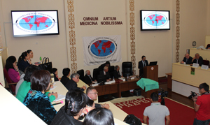
For years, the World Federation of Neurology (WFN) has reached out to Kazakhstan and its neurology community. However, language barriers made it difficult to establish a connection. After a two-year effort by Prof. Daniel Truong and Saltanat Kamenova, with the valuable assistance of Aida Kondybayeva, the International Neurology Forum for Parkinsonism and Related Disorders was held in Kazakhstan. The forum was hosted by the Asfendiyaroy Kazakh National Medical University and was attended by more than 120 Kazakh neurologists.

Prof. Aikan Akanov, rector of the university, opened the forum by thanking the organizers and emphasizing the importance of the event for Kazakhstan. Internationally known speakers included Profs. Erik Wolters from the Netherlands, Truong from the United States and Carlo Colosimo from Italy. Speakers from Kazakhstan included Guram Pichkhadze from the Virtual Institute of Neuroscience, Saltanat Kamenova, chairwoman of the department of neurology at the university and Marat Asimov, chairman of the department of medical psychology.

All of the lectures were translated in real time for the audience. The topics discussed were broad and included the diagnosis, pathology and neuropsychology of

Opening statement of the International Neurology Forum by Prof. Aikan Akanov on Sept. 23, 2014, with Prof. Guram Pichkhadze, Erik Wolters and Daniel Truong.

Opening statement of the International Neurology Forum by Prof. Aikan Akanov on Sept. 23, 2014, with Prof. Guram Pichkhadze, Erik Wolters and Daniel Truong.

Parkinson’s disease, tremor and dystonia, as well as the management of these disorders. In addition to the presentations, several workshops were offered, led jointly by Profs. Shelekov, Truong, Perlenbetov and Nurmagambetova.

The meeting was supported with a grant from the International Association for Parkinsonism and Related Disorders (IAPRD). All participants received a free textbook as a gift from the IAPRD. On the last day of the forum in her closing remarks, Prof. Kamenova noted the importance of this event for doctors and young neurologists of Kazakhstan and thanked Profs. Wolters and Truong for their noble mission in the development of the educational program.


Kondybayeva is with the Asfendiyarov Kazakh National Medical University.


A Continuing Journey: The Fight Against Stroke in India

Rohit Bhatia

Rohit Bhatia

By Rohit Bhatia, MD, DM, DNB

The stroke epidemic has arrived in India. While we were busy combating the scourge of infections and deficiency diseases, non-communicable diseases (NCDs) including stroke stealthily crept up on us.

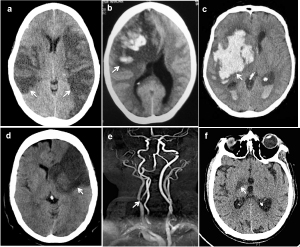
With a population of 1.2 billion today and growing, India finds itself staring at a stroke epidemic (See “The Stroke Fact Sheet in India.” on page 8.) 1,2. In addition to strokes due to conventional risk factors, cardio-embolic stroke due to rheumatic valvular heart disease, cerebral venous thrombosis, and strokes related to tuberculous meningitis still remain important causes of stroke, especially in the young Indian population. (See Figure 1.)

Types of strokes

Figure 1. Types of strokes (arrows): (a) bilateral arterial infarcts in a patient with rheumatic heart disease and atrial fibrillation (b) venous infarct in a post-partum patient with superior sagittal sinus thrombosis (c) intracerebral hemorrhage in a hypertensive patient, (d) arterial embolic infarction due to large artery athersoclerosis and carotid stenosis (e) and (f) perforator artery infarction in patient with tubercular meningitis.

The recently published Prospective Urban Rural Epidemiology (PURE) study from 18 low-, middle- and high-income countries showed that incidence of major cardiovascular disease was highest in low-income countries, despite the fact that these countries had the lowest risk-factor burden3.

Challenges in stroke care include a limited number of trained neurologists who are mostly urban, a large number of patients who are mostly rural, a lack of knowledge and awareness both about stroke risk factors and treatment in the general public and prohibitive cost of stroke care. There is a lack of uniformity and standardization of secondary and tertiary stroke care while availability of primary care in stroke is extremely unreliable. The stroke epidemic did catch us by surprise and in an unprepared state, but the situation is gradually beginning to improve and we are optimistic about the future. (See Figure 2.)

Acute stroke care has barriers, including recognition, pre-hospital delays, physician expertise, lack of ambulance services, cost of tPA and lack of critical care facilities. Although thrombolysis (using tPA) continues to be available only in urban private or academic hospitals, there has been a recent rise in the number of stroke patients getting the benefit of this treatment.

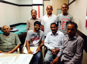
Figure 2. Recovering stroke patients at the Stroke Clinic, Neurosciences Center, AIIMS.

Figure 2. Recovering stroke patients at the Stroke Clinic, Neurosciences Center, AIIMS.

In the year 2009, 1,648 patients were thrombolysed, while in 2011, the number rose to 2,975 and a center in northwest India reported a four-fold increase in rates of thrombolysis2. About 100 centers in India currently have facilities to provide intravenous thrombolysis, and the numbers are likely to rise with awareness and experience.

In the national capital region, the cost barrier is gradually being offset for eligible patients by the provision of free tPA by the government in state-run academic hospitals, including All India Institute of Medical Sciences (AIIMS), New Delhi. The National Program on Prevention and Control of Cardiovascular Diseases, Diabetes and Stroke4 (NPCDCS) launched in 2008 by the ministry of health and family welfare (See “Major Components of NPCDCS.”) addresses NCD prevention by risk reduction, early diagnosis and appropriate management through health promotion programs for the general population and high-risk groups.

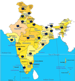
Figure 3. Map of India showing the National Program on Prevention and Control of Cardiovascular Diseases, Diabetes and Stroke (NPCDCS Program), Government of India. Red dots indicate places where it is currently implemented. Stars indicate the Indian states.

Figure 3. Map of India showing the National Program on Prevention and Control of Cardiovascular Diseases, Diabetes and Stroke (NPCDCS Program), Government of India. Red dots indicate places where it is currently implemented. Stars indicate the Indian states4.

At present, the NPCDCS program is implemented in 100 districts across 21 Indian states, and it is expected to be rolled out in 640 districts by 2017 under the 12th five-year plan. (See Figure 3.) Developing and running dedicated stroke units in the face of the extremely limited health resources is a challenge; 35-40 stroke units currently exist, mainly in bigger cities and more often in private hospitals.

Improving Access to Stroke Care

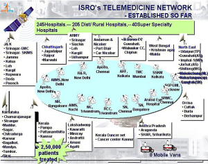
Reaching out to remotely located patients remains difficult, and telestroke is recognized as a potential solution5. Telemedicine has been successfully used by the Indian Space Research Organization (ISRO) to meet the needs of remote Indian hospitals6.

The telemedicine network implemented by ISRO in 2001 presently stretches to around 100 hospital countrywide, with 78 remote rural/district health centers connected to 22 speciality hospitals in major cities, thus providing treatment to more than 25,000 patients, including stroke patients. (See Figure 4.) A major telestroke initiative has been taken up by the state of Himachal Pradesh (HP). Telestroke Management Program has been piloted for the first time in HP in collaboration with AIIMS. Under this program, 18 primary stroke centers are being set up in HP state hospitals, which have CT scan facilities. One hundred and twenty state doctors have been trained and six patients already have been successfully treated under this program. Success of this program will pave the path for comprehensive treatment of stroke patients in more parts of the country.

Research in stroke medicine is another area that has seen improvement with increasing national and international collaborative efforts and improved funding opportunities. The Indian Council of Medical Research (ICMR), Department of Biotechnology (DBT) and Department of Science and Technology (DST) of the Government of India have increased support for basic and clinical stroke research.

The WHO stroke STEPS I version 7 was tested in the Indian Collaborative Acute Stroke Study (ICASS). During 2002-2004, 2,162 acute stroke cases were identified in the study. Analysis of results confirmed that the incidence of stroke was rising with the advance in age. Presently, there are eight stroke registries based in various states of India. Each registry has independently set up a stroke surveillance system based on the WHO STEPS guidelines7.

Figure 4. Indian Space Research Organization (ISRO) telemedicine network.

Figure 4. Indian Space Research Organization (ISRO) telemedicine network6.

The National Stroke registry of the ICMR is being run by the National Center for Disease Informatics and Research, Bangalore, where staff members have started the process of collating data on stroke patients from institutions and individual specialists who have registered with the program. The Indian Stroke Prospective Registry (INSPIRE) is a large, multicenteric prospective pilot registry run by the division of clinical trials, St. John’s Research Institute, Bangalore, with the objective of determining etiologies, clinical practice patterns and outcomes of stroke in India.

By April 2012, the study had enrolled 5,301 patients from 49 cities in 19 states. Data from these registries will provide evidence on mortality and morbidity indicators in India, which could help plan an effective stroke management program. In collaboration with Erasmus University Netherlands, AIIMS has jointly launched a large cOHORT study comprising 15,000 people above the age of 50 in rural and urban populations to prospectively examine the causes of stroke and dementia in the Indian population. The Department of Biotechnology has generously funded this endeavor with INR 340 million.

Increasing Stroke Awareness

Education programs are being carried out by hospitals and stroke support groups especially around the World Stroke Day to educate and disseminate information on stroke8. Initiatives include patient awareness programs with lectures and interactions focused on stroke symptoms, the concept of “time is brain,” the need to reach a hospital early and preventive strategies to reduce stroke occurrence; banners, advertisements and write-ups in newspapers along with talk shows on TV and radio channels are also used.

Figure 5. Educational workshop on stroke conducted by Department of Neurology AIIMS.

Figure 5. Educational workshop on stroke conducted by Department of Neurology AIIMS.

Studies have shown that lack of physician awareness delayed arrival of stroke patients to a specialized center. CMEs, physician training programs and conferences are regularly held across the country emphasizing the need for recognition and timely therapy and to appraise doctors regarding the newer developments on cerebrovascular disorders. (See Figure 5.) The Indian Stroke Association (ISA) has been organizing a stroke summer school since two years ago to train junior neurologists and physicians.

The annual meetings of ISA are well attended with invited national and international faculty and deliberations on various aspects of stroke. The national guidelines for the management of stroke in India were developed with an aim to close the gap between best and pragmatic practice. A recent study from an academic hospital in North India observed that education to the emergency staff led to an increased rate of thrombolysis and shortened door to needle time.  It is encouraging to see that students trained at academic centers are now promoting stroke awareness and timely treatment in smaller cities.

The Future of Stroke Care in India

We will never be able to treat every stroke in the country for a long time to come. So where should our emphasis lie? Preventing as many strokes as possible will probably be the best stroke care that we can provide. At present, many, if not most, strokes are a consequence of modifiable risk factors such as obesity, hypertension and smoking.

Spreading awareness on a war footing and reducing preventable strokes immediately is required. Implementation of mass screening has been recommended to reduce the burden of stroke through identification of people at high risk. Simple, practical and cost-effective measures such as identification and treatment of hypertension in the community will go a long way.  Focus also should be on effective implementation, monitoring and evaluation of present stroke programs. A stroke prevented is a much happier situation than a stroke treated.

References:

1.     http://www.sancd.org/Updated%20Stroke%20Fact%20sheet%202012.pdf. Stroke in India Factsheet (Updated 2012). Accessed September 6, 2014.

2.     Pandian JD, Sudhan P. Stroke epidemiology and stroke care services in India. Journal of Stroke. 2013;15:128-34.

3.     Yusuf S, Rangarajan S, Teo K, Islam S, Li W, Liu L, Bo J, Lou Q, Lu F, Liu T, Yu L, Zhang S, Mony P, Swaminathan S, Mohan V, Gupta R, Kumar R, Vijayakumar K, Lear S, Anand S, Wielgosz A, Diaz R, Avezum A, Lopez-Jaramillo P, Lanas F, Yusoff K, Ismail N, Iqbal R, Rahman O, Rosengren A, Yusufali A, Kelishadi R, Kruger A, Puoane T, Szuba A, Chifamba J, Oguz A, McQueen M, McKee M, Dagenais G; PURE Investigators. Cardiovascular risk and events in 17 low-, middle-, and high-income countries. N Engl J Med. 2014;28:818-27.

4.     http://health.bih.nic.in/Docs/Guidelines/Guidelines-NPCDCS.pdf. Accessed September 5, 2014.

5.     Srivastava PV, Sudhan P, Khurana D, Bhatia R, Kaul S, Sylaja PN, Moonis M, Pandian JD. Telestroke a viable option to improve stroke care in India. Int J Stroke. 2014 Jul 18. [Epub ahead of print].

6.     http://www.telemedindia.org/isro.html. Accesssed October 4, 2014.

7.     Bonita R, Beaglehole R. Stroke prevention in poor countries. Time for action. Stroke 2007;38:2871-2872.

8.     World Stroke Day celebrations: report from India. International Journal of Stroke. 2009;4:231–232.

Additional professor: Pranjal Sisodia, MSc, PhD Scholar, Department of Neurology, Neurosciences Center, All India Institute of Medical Sciences, New Delhi, India.

To correspond with the author, write to him at rohitbhatia71@yahoo.com.


Kinnier Wilson and Anglo-French Neurology in the Early 20th Century

HISTORY OF NEUROLOGY 

By Edward H. Reynolds

Edward H. Reynolds

Edward H. Reynolds

Samuel Alexander Kinnier Wilson (SAKW) (1878-1937) is distinguished throughout the neurological world for 1) the disease that bears his name, 2) his scholarly two-volume textbook which was published posthumously in 1940, and 3) his founding in 1920 of the Journal of Neurology and Psychopathology, now known as the Journal of Neurology, Neurosurgery and Psychiatry.

Born in New Jersey, to a Scottish mother and an Irish missionary Presbyterian Minister father, he returned to Scotland for his education. He graduated from the Edinburgh Medical School in 1902 and obtained a BSc with First Class Honors in Physiology in 1903. With a Carnegie Fellowship, he immediately proceeded to Paris for a year to study neurology under Pierre Marie at the Bicàªtre Hospital, followed by a few months in Leipzig. In 1904 he was appointed House Physician to the National Hospital for the Paralyzed and Epileptic in London and remained at the National Hospital for the rest of his career, as resident medical officer, registrar, pathologist, assistant physician, and finally full physician in 1921. In 1919, SAKW was also appointed junior neurologist to King’s College Hospital, one of the first of such posts in the UK to incorporate the word “neurologist.”

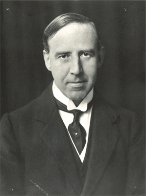
Samuel Alexander Kinnier Wilson (1878-1937).

Samuel Alexander Kinnier Wilson (1878-1937).

In 1931, Sherrington invited SAKW to participate in a symposium on muscle tone at the first International Congress of Neurology in Berne. Sherrington and SAKW were elected as president and secretary-general respectively of the second International Congress of Neurology in London in 1935, but Sherrington later had to withdraw due to ill health. In 1933 Sherrington and his joint Nobel Prize (1932) winner, Adrian, both proposed SAKW for the fellowship of the Royal Society.

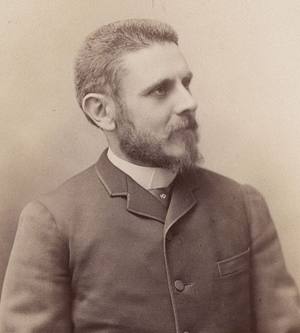
Pierre Marie (1853-1940). SAKW's first influence in Paris.

Pierre Marie (1853-1940). SAKW’s first influence in Paris.

Guided by his future father-in-law Alexander Bruce (1854-1911), an Edinburgh physician, with an interest in neurology, who also founded a neurological journal (Review of Neurology and Psychiatry 1903-1916), SAKW understood that Paris was the leading world neurological center at the turn of the 20th century. Hence, his seminal year (1904) influenced by Marie, Babinski, Dejerine and Meige among others, before proceeding to the National Hospital, Queen Square. He published his famous Edinburgh thesis on hepatolenticular degeneration (later called Wilson’s disease) in 1912, not only in Brain but also in Revue Neurologique. Furthermore he presented his work in French to the Société de Neurologie de Paris on Jan. 25, 1912, where it was very well received, but I cannot trace any record of a presentation to any UK society, such as the Section of Neurology of the Royal Society of Medicine in London. Just as Charcot had been a regular visitor to the UK in the late 19th century, so SAKW was a frequent visitor in the new century to France, where Crouzon, Guillain and Léri were particular friends and collaborators.

Kinner-Pic3

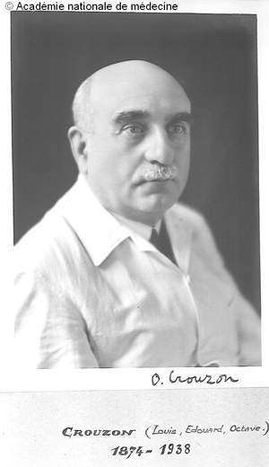
Octave Crouzon (1874-1938). SAKW’s contemporary and friend in Paris. (Courtesy of Emmanuel Broussolle).

It is interesting that even at the 17th International Medical Congress in London in 1913 the French delegation was the dominant influence in the Section of Neurology/Neuropathology, the proceedings of which were published in detail in Revue Neurologique but not in any English journal. Following that Congress SAKW and nine other British physicians were elected “Membres Correspondants Etrangers” of the Société de Neurologie de Paris, which had been founded in 1899.

In the last 25 years, I have had the privilege of working with SAKW’s son, James KW, a Cambridge-based assyriologist, on the subject of Babylonian neurology and psychiatry. Through James KW, I have learnt most about his father’s French connections. I have in my possession SAKW’s original seven-page brochure listing the members of the Société de Neurologie de Paris for 1926. It now includes 76 Paris-based neurologists, 60 French neurologists from beyond Paris and 128 international members from around the world, mainly Europe and the U.S./Canada, including 14 from the UK. Although the Neurological Society of London had been founded earlier in 1886 and had evolved in 1907 into the Section of Neurology of the Royal Society of Medicine, it had remained a small almost exclusively London-based Society. When the Association of British Neurologists (ABN) was founded in 1933, it had only 25 members.


References

Reynolds EH. Kinnier Wilson and Sherrington. J Neurol Neurosurg Psychiatry 2008;79:478-9.

Reynolds EH. Kinnier Wilson’s French connections. Rev Neurol 2014 Jun 3. pii: S0035-3787(14)00840-6. doi: 10.1016/j.neurol.2014.03.011.

Edward Reynolds is consultant neurologist and former director of the Institute of Epileptology, King’s College, London, and former president of the International League against Epilepsy. Peter J. Koehler is the editor of this history column. He is neurologist at Atrium Medical Centre, Heerlen, The Netherlands. Visit his website at http://www.neurohistory.nl


Turkey Brain Year Activities — Neurology for Public

Photo 1

Photo 1

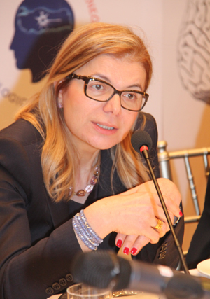
The Turkish Brain Action Group was founded from suggestions of the European Brain Council in August 2013. Group members quickly organized a “Brain Year” in Turkey in 2014. The Turkish Brain Action Group consists of neurology, neurosurgery, psychiatry, and other disciplines related to neuroscience and patient organizations. Currently, 16 different societies presents in the action group.

Photo 2

Photo 2

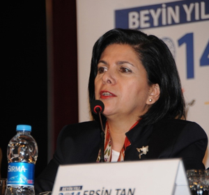
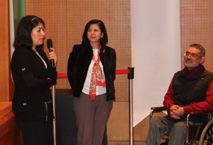
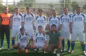
The 2014 Brain Year Turkey opening conference moderated by Prof. Rana Karabudak, under the leadership of Turkish Neurological Society, was successfully held on Feb. 18, 2014, in Ankara, Turkey. The conference aimed to announce all the projects of Brain Year in 2014 and touch on the issues regarding the brain awareness in Turkey. (See Photo 1.) The invited speakers included the representatives from the Turkish Neurological Society’s board members, experienced members, vice chancellor of Hacettepe University of Medical School and the prominent representatives of the neurological sciences specialty societies.

Photo 3

Photo 3

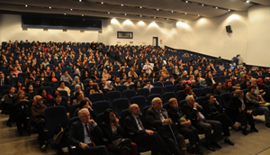
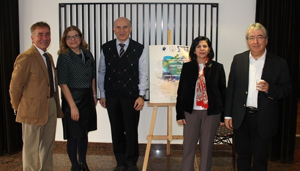
The conference brought together up to 400 participants, including Turkish press, neurologists, neurosurgeons, medical students and members of cooperated societies. (See Photo 2.)

Photo 4

Photo 4

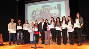
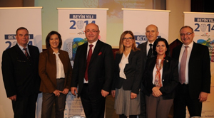
Our second meeting was held on March 12, 2014, in Ankara, Tukey. The meeting’s main topic, moderated by Prof. Serefnur Ozturk, was cerebrovascular diseases and mainly attended by medical students and delegates of the Patient Societies. (See Photo 3.) The event recorded and served from brain year web page as a continuing education. (See Photos 5, 6.)

Photo 5

Photo 5

On April 12, 2014 the Parkinson Disease Society held an event for the Brain Year. The Parkinson’s patients and the neurologists had a long walk against to Parkinson disease. This event received great attention from the press. (See Photo 7.)

Photo 6

Photo 6

An Awareness Symposium was held for the neurological diseases in May. The symposium brought together around 600 patient and patients’ relatives. They had a chance to have an interactive discussion platform to increase their knowledge. (See Photo 8.)

Photo 7

Photo 7

In addition to scientific activities, we have visited eight secondary schools and held mini conferences. We reached around 1,000 students and increased their knowledge on brain. Turkish press being so interested to all our events and this makes it more accomplished. (See Photo 9.)

Photo 8

Photo 8

In the coming days, our public service announcements will be placed on TV which will be a great opportunity to spread our word to Turkey. (See Photo 10.)

Photo 9

Photo 9

Turkish Neurological Society holding an annual congress every year with 1,500 participants. This year we are celebrating our 50th Congress and planed a special session on Brain Year in Turkey. During our national congress the contest results will be announced at the gala dinner and an outdoor event will be organized “Walking With the Neurons”.

Photo 10

Photo 10

The brain year website consists all the events and announcements and we use social media quite effectively with our Facebook and Twitter accounts.

International Parkinson and Movement Disorder Society (MDS)

International Congress in The Capital of Scandinavia

The 18th International Congress of Parkinson’s Disease and Movement Disorders was hosted this year in the beautiful city of Stockholm, Sweden on June 8-12, 2014. As the city known for hosting the Nobel Prize winners, Stockholm was the perfect location for delegates to learn about the latest research, and perspectives in the field of Movement Disorders and to network and collaborate with colleagues. Everyone’s participation made for several highlights during the week:

  • 4,500 total people registered (including press and exhibitors)
  • 86 countries represented
  • 100 travel grants awarded by MDS
  • 196 faculty members participated in the Scientific Sessions
  • 1,558 posters accepted
  • 34 late-breaking posters accepted
  • 17 MDS study group abstracts accepted
  • 4 therapeutic plenary sessions
  • 9 plenary sessions
  • 24 parallel sessions
  • 8 teaching courses
  • 24 video sessions and skills workshops
  • 16 guided poster tours
  • 28 companies exhibited
  • 23 representatives from the press attended

Under the direction of Dr. Victor Fung, the Congress Scientific Program Committee (CSPC) made every effort to ensure that the science for the 18th International Congress was diverse and educational.  The 2014 Scientific Program incorporated therapeutic plenary sessions, plenary and parallel sessions, teaching courses, video sessions, skills workshops, controversies, blue ribbon highlights, poster sessions and guided poster tours.  There were 13 sessions focused on this year’s theme of “Emerging and Experimental Therapies,” which gave participants an educational and comprehensive overview on the subject.

A unique session was introduced this year to the Scientific Program called, “Late-Breaking Clinical and Scientific Topics Relevant to Movement Disorders.” This parallel session was designed to help participants understand and appreciate the latest clinical and scientific discoveries relevant to movement disorders, as well as understand the role of new clinical discoveries for basic science and understand the role of new basic science discoveries for clinical progress.

Also of note was the Movement Disorders Grand Rounds Parallel Session. In this interactive session, volunteer patients with a known complex movement disorder were presented to one of four movement disorder “experts.” This year’s experts included Bastiaan Bloem (Netherlands); David John Burn (United Kingdom); Beom Jeon (Korea) and Claudia Trenkwalder (Germany). The patient’s history and clinical findings (including video of the movement disorder) were presented by the expert, who then reviewed the history with the patient and highlighted and demonstrated the neurological signs to the audience. The expert’s job is to generate a differential diagnosis and management plan which can be critiqued by his/her fellow experts, the audience and the chairs. The final diagnosis and learning point were then presented after the expert and audience discussion was finished. The session showed how a movement disorders expert takes a clinical history and performs a movement disorders examination of a patient to generate a diagnosis and a management plan.

The always popular video challenge was held on Wednesday night with Masters of Ceremony Anthony Lang and Kapil Sethi. A world-renowned panel of Movement Disorders experts including Victor Fung (Australia); Orlando Barsottini (Brazil); Daniel Healy (Ireland); Björn Holmberg (Sweden) and David Riley (U.S.) guided participants through unique Movement Disorder cases. The cases were presented by representatives from Movement Disorder Centers around the world and discussed by the Panel of Experts. Awards were given for the most interesting and challenging cases. The goal of this session was for attendees to learn from a series of unusual, very interesting patients and see how senior experts approach these types of challenging cases.

At the opening ceremony, President Matthew Stern distributed the following awards:

Honorary Member Awards:

  • Anthony Lang, OC, MD, FRCPC, Toronto, ON, Canada
  • William Weiner, MD, Baltimore, MD, U.S., President’s Distinguished Service Award:
  • Esther Cubo, Burgos, Spain

The International Parkinson and Movement Disorder Society (MDS) would like to extend their gratitude to faculty, supporters, exhibitors and delegates for traveling to Stockholm and for helping make the 18th International Congress of Parkinson’s Disease and Movement Disorders another successful meeting.

Mark your calendars!  MDS is already planning for the 19th International Congress in San Diego, California, June 14-18, 2015.  Please visit our website for details regarding 2015 Congress at www.mdscongress2015.org or contact the International Secretariat at congress@movementdisorders.org with any
questions.


Medical Marijuana for Epilepsy

What Do We Tell Our Patients?

164343705by Antoaneta J. Balabanov

The idea to use cannabis plants to treat seizures is not new. From ancient China, through Babylonia and Palestine, Egypt and India, cannabis plants were used to treat many different maladies — among them seizures. Throughout the centuries, cannabis plants were considered magical by some and evil by others. This is not surprising given what is now known about the psychotropic properties of these formidable plants.

The interest of cannabis and their use for treatment of seizures has come in to the limelight in the recent years. Cases of epilepsy patients who have achieved seizure freedom using marijuana have been circulating on the Internet. Social networks have contributed by organizing interest groups and chat rooms to discuss this issue. The case of Charlotte, a child with Dravet syndrome who stopped having seizures after experiencing up to 50 seizures a day, ignited the epilepsy world when presented on CNN. Charlotte had failed all antiepileptic drugs (AED) available and when finally treated with cannabidiol (CBD), a component of marijuana, the effect was miraculous.

Our patients now know that medications based on cannabinoids are already being used for treatment of nausea, spasticity and pain. As epilepsy specialists, our patients ask us daily when medical marijuana will be available for use for treatment of seizures. Frequently, the following question is asked: “Why do I have to take medications with potential long-term side effects when I can use something more natural such as marijuana? If all medications that I have used have not help me stop having seizures, why can’t I try something that may potentially help my seizures without causing significant side effects?”

The patients are absolutely right to ask all these questions. Despite having 30 AEDs to use, about 30 percent of all patients with epilepsy are medically intractable. They continue to have seizures that eventfully cause cognitive, behavioral and psychiatric problems. Patients lose their jobs, their ability to drive, and to take care of their families. Stigma associated with epilepsy still exists, SUDEP is a real tragedy. In children, the effect of continued seizures on the developing brain could be devastating. Multiple studies have shown that children with uncontrolled seizures have a lower IQ later in life. Antiepileptic drugs, even when controlling the seizures, often cause side effects that interfere with the patient’s quality of life. The long-term safety profile is not known for most of the AEDs and that alone causes additional anxiety among patients and parents of children with epilepsy.

Is cannabis the solution that we have all been waiting for?

This is what we know: Cannabis sativa and Cannabis indica are two species of the Cannabis genus plant. Both plants produce multiple compounds called cannabinoids. Among them, two are of particular interest to us: tetrahydroxycannabiol (THC) and cannabidiol (CBD). These two cannabinoids were first isolated and characterized in the 1960s. THC has gained much more interest from the research world mostly because of its psychotropic properties causing significant cost to our society. CBD accounts for up to 40 percent of the plant’s extract and has none or very little psychoactive effects. CBD, however, has shown anticonvulsive properties when tested on animal epilepsy models. It is important to note that while the CBC anticonvulsant effect appeared to be promising in the acute models of seizures, there is less evidence that the effect is the same when tested on animal models of chronic epilepsy. The effect of seizure control in humans has not been studied well enough. There were only four randomized controlled trials (RCT), which were all done between 1978 and 1990. These studies included small number of patients (total of 48) who underwent a short length of treatment. Due to these and other methodological limitations, these clinical trials failed to provide evidence about the efficacy or safety of cannabinoids in patients with epilepsy.

Cannabis and its effect on seizure control on kids with epileptic encephalopathies has been one of the most discussed topics among patients’ families. There are no studies done so far, but there was a recent survey of 19 parents that is worthwhile to note. Twelve of the parents who responded to the survey have children with Dravet syndrome, one parent was with a child with Lennox-Gastaut Syndrome (LGS). All patients were treated with CBD enriched cannabis and products containing primarily CBD. The results reported by the parents were impressive — 53 percent of the parents reported 80 percent seizure reduction and 11 percent of the children were seizure free during the three-month trial. Among the 12 patients with Dravet syndrome, 42 percent reported more than 80 percent reduction in seizures. Serious side effects were not reported, only few parents noted drowsiness and fatigue. Most parents reported increased alertness. While these results are very impressive, they should be taken with caution since they are based solely on the parents’ reporting. In addition, the exact formulation of the different cannabis extract used by the parents is unclear.

The exact mechanism of action of the cannabinoids is not completely known. TSH was found to bind to two G-protein–coupled cell membrane receptors, named type 1 (CB1) and type 2 (CB2). CB1 receptors were found primarily in the brain, but also in several peripheral tissues (GI tract, adipose tissues, pancreas, muscle). CB2 receptors are mainly found in immune and hematopoietic cells. THC is the main psychoactive agent found in cannabis. CBD does not have psychoactive properties. This is perhaps because it does not activate CB1 and CB2 receptors. CBD reduces the psychoactive effects of THC; therefore it enhances its tolerability. CBD may supplement the antispastic effect of THC. This is because when used in combination, patients can tolerate higher doses of THC. Moreover, its anti-inflamatory, antioxidant and antiexcitotoxic mechanisms also contributes to the effect. The mechanisms by which CBC works as anticonvulsant are not entirely known. Different mechanisms of action have been proposed— most of them related to the reducing of neuronal excitabilities and neuronal transmission, while some others propose an anti-inflammatory effect. CBD is potentially involved in neuroinflamation by the reduction of new astrocyte production. This summary of the mechanism of action of the cannabinoids is incomplete, leaving behind many other molecular mechanism of action identified in animal models which are not clearly associated with any anticonvulsive effects, therefore, they will not be discussed here.

So, what do I tell my patients?

I tell them that I am as interested as they are in finding new therapies for epilepsy and if cannabis is proven to be “The New Therapy” I will be very eager to use it on all my medically intractable patients. However, I tell them that the scientific evidence for cannabis is lacking. I also try to point out that the reports circulating the web should be taken with great caution. These reports are mostly biased from the selection of the patients to the reported outcomes. In addition, every case of epilepsy is different and the disease is highly variable. I ask them not to base their decision to move to a state where medical marijuana is now legal based on anecdotal reports. I remind them to consider the fact that the marijuana growers follow no regulations and safety standards and that the products that they are offering often contains different percentages of CBC and THC among the 80 or more cannabinoids that the cannabis plants produce. Interactions between cannabis and currently used AEDs have not been studied, and are therefore unknown. Adding a new substance to an already very complex mix of medications can be dangerous, especially when it is not supervised in a medical setting. The concern of the long-term side effects of the AEDs is valid, but the long term effects of cannabis products on the brain should be considered as well. There have been reports on negative effects of cannabis on the developing brain. In adults, use of cannabis has been associated with worsening of executive function over time. Viewing cannabis use as a more natural way of treatment does not mean that cannabis effects should be taken lightly. AEDs that we use today have gone through years of studies and extensive testing before being approved by the FDA. There should be no reason for the cannabis product to be treated any differently.

My conversations with patients about seizures and medical marijuana will continue. As a physician, I support the idea that CBC should be studied for its possible anti-convulsive properties. I do not support legalization of marijuana, but I also realize that current status of medical marijuana as a Federal DEA Schedule 1 controlled substance stands in the way of medical research that needs to be done, so I can give my patients more definite answers. Until this happens, I will continue with my mantra: randomized double blinded placebo-controlled studies are needed to determine the efficacy and safety of CBD and other cannabinoids as potential treatments of epilepsy.


Editor’s Update and Selected Articles From JNS

By John D. England, MD

John D. England, MD

John D. England, MD

The Journal of the Neurological Sciences (JNS) is a broad-based journal which publishes articles from a wide spectrum of disciplines, ranging from basic neuroscience to clinical cases.  In an upcoming issue of JNS, we will be acknowledging our peer reviewers.  The individuals whose names appear in the list have provided peer reviews of manuscripts which were submitted to JNS during the past year. This elite group of individuals from around the world includes some of the best and brightest minds in neuroscience and clinical neurology. They receive no remuneration for their work for the journal and take time from their busy schedules to review our articles.  All of the associate editors and I thank them for their indispensable work.  The journal would not survive without their expert advice. I express my most sincere gratitude to all of the reviewers.  They inform and enlighten all of us in the editorial office and help to make JNS a journal of the highest quality.

In our ongoing attempt to enhance accessibility of JNS articles to members of the World Federation of Neurology (WFN), we have selected two “free – access” articles, which are profiled in this issue of World Neurology.

In this issue, we feature two paired articles regarding the diagnostic accuracy of the famous Babinski sign:

  1. Despite its legendary status, controversy exists regarding the diagnostic accuracy and usefulness of the Babinski sign in determining pyramidal tract dysfunction. Much of the variability in the assessment of the diagnostic accuracy of the Babinski sign is due to studies which do not follow simple but important methodological standards.  The paper by S.P. Isaza Jaramillo, et al. is exceptional in this regard. This paper describes a simple but methodologically rigorous study to ascertain the diagnostic accuracy of the Babinski sign for identifying pyramidal tract disease. One hundred and seven (107) patients with limb weakness were assigned a random identification number for blinding purposes. Two neurologists elicited a plantar response in each patient in a standardized manner.  The examination was performed in a blind and independent manner, and each examination was filmed to quantify intra-observer variability. The reference standard for diagnosis of pyramidal tract dysfunction was provided by a senior neurologist who examined every patient independently and had available the patient’s history, complete neurological examination, laboratory and neuroimaging results. Compared to the reference standard, the Babinski sign had a very high specificity of 99 percent (CI 97.7-100), but moderately low sensitivity of 50.8 percent (CI 41.5-60.1) in identifying pyramidal tract dysfunction.  These findings indicate that a Babinski sign elicited in a standardized manner by an experienced practitioner has high diagnostic accuracy for identifying pyramidal tract disease.  However, in view of the low sensitivity, the absence of a Babinski sign does not rule out pyramidal tract dysfunction. Jaramillo SPI, Uribe CSU, Jimenez FAC, Cornejo-Ochoa W, Restrepo JFA, Roman GC.  Accuracy of the Babinski sign in the identification of pyramidal tract dysfunction.  J Neurol Sci 2014;343:66-68.
  2. In an editorial in the same issue of JNS, Austin Sumner provides an historical perspective on the Babinski sign and discusses the more recent controversy concerning its diagnostic usefulness. He concludes that the “confidence traditionally placed by clinicians on this simple test is justified” with the caveat that its absence does not rule-out pyramidal tract disease. Sumner AJ.  The Babinski sign.  J Neurol Sci 2014;343:2.
England is editor-in-chief of the Journal of the Neurological Sciences.

Breakthrough in Autism Prevention

The Importance of Measuring Thyroid Function in the Expectant Mother

By Gustavo C. Román, M.D.

Roman-EndemisCretinismEcuadorEncouraging news for the prevention of autism emerged from the results of a research study conducted by an international collaborative team conformed by Prof. Gustavo C. Román, MD, from the Methodist Neurological Institute (Houston, Texas) and researchers from the Generation R Study at Erasmus University Medical Centre in Rotterdam, The Netherlands, including Akhgar Ghassabian, MD, PhD; Jacoba J. Bongers-Schokking, MD, PhD; Vincent W.V. Jaddoe, MD, PhD; Albert Hofman, MD, PhD; Yolanda B. de Rijke, PhD; Frank C. Verhulst, MD, PhD;  and Henning Tiemeier, MD, PhD. The results point to the deficiency of maternal thyroid function during the first trimester of pregnancy as a significant factor in the causation of autism in the child.  Pregnant women with deficient thyroid hormone are nearly four times likelier to produce autistic children than women with normal thyroid function.  The study was published in the prestigious journal Annals of Neurology1.

The research was conducted in a cohort of more than 4,000 Dutch mothers and their children, and it supports the view that autism can be caused by lack of maternal thyroid hormones T4, also called thyroxine, and T3, which is crucial to the migration of fetal brain cells during embryo development, early in pregnancy. The most common cause of thyroid hormone deficiency is a lack of dietary iodine, given that T4 contains four atoms of iodine. There are also environmental contaminants and dietary factors that affect thyroid function2.

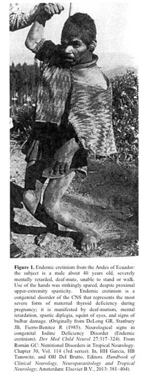
According to Prof. Román, the hypothesis linking thyroid and autism originated from his early experiences as a neurologist in the Andes of South America where the deficiency of iodine in table salt results in high prevalence of goiter and congenital problems in children including high rates of deafness, borderline mental retardation and, in the most severe cases, endemic cretinism. (See Figure 1.)  Román recently described this topic in detail3.

Iodine deficiency remains common throughout the world; including industrialized countries where it was believed to be eradicated by the use of iodinated salt.  This could explain the upward trend in the incidence of autism observed in these countries.  Another factor is the lack of determination of T4, T3 and urinary iodine levels in expectant mothers early in pregnancy, and the use of prenatal vitamin supplements lacking the extra supplementary iodine needed during pregnancy and lactation.

The complex gene network regulated by thyroid hormone during brain development is being deciphered with the use of experimental animal models4 and should provide a better understanding of the complex problem of autism spectrum disorders.

References

1. Román GC, Ghassabian A, Bongers-Shokking JJ, et al. Association of gestational maternal hypothyroxinemia and increased autism risk. Ann Neurol 2013; 74: 733–742.  doi: 10.1002/ana.23976 (E-pub 2013 Aug 13). Chang K, Shin JI. (Letter to the Editor) Association of gestational maternal hypothyroxinemia and increased autism risk: The role of Brain-Derived Neurotrophic Factor. Ann Neurol 2014;75:971–972. doi: 10.1002/ana.24143  Román GC, Ghassabian A, Bongers-Shokking JJ, et al. (Reply)  Ann Neurol 2014;75:971-972. doi: 10.1002/ana.24141

2. Román GC. Autism: transient in utero hypothyroxinemia related to maternal flavonoid ingestion during pregnancy and to other environmental antithyroid agents. J Neurol Sci 2007; 262: 15–26

3. Román GC. Nutritional Disorders in Tropical Neurology. Chapter 30, Vol. 114 (3rd series), Handbook of Clinical Neurology, Neuroparasitology and Tropical Neurology. Edited by HH Garcà­a, HB Tanowitz, and OH Del Brutto. Elsevier BV: Amsterdam, 2013: 381-404

4. Berbel P, Navarro D, Román GC:  An evo-devo approach to thyroid hormones in cerebral and cerebellar cortical development: Etiological implications for autism. Frontiers in Endocrinology 2014, ISSN: 1664-2392

Gustavo C. Román, M.D., neurologist and first director of the Nantz National Alzheimer Center at Houston Methodist Hospital, has been awarded the Doctorado Honoris Causa, the highest academic accolade given by the National University of Colombia.