Prof. Sergey Lobzin and Natalie Zinserling, North-Western State Medical University
Neurologists of Saint Petersburg and Northwestern Federal District
Saint Petersburg neurology has a great and deeply rooted history. In 1835, Prof. Shipulinsky was the first scientist who taught neurology in Saint Petersburg. Later, separately from psychiatry, neurology was developed by Prof. I. Mierzejewski (1835-1908). He worked with K. Westphal and R. Virchow in Berlin, Gudden, Leidesdorf, and Meinert in Vienna, and Broca, Ranvier, Claude Bernard, Vulpian, and Charcot in Paris.
The History of Saint Petersburg neurology was always associated with European neurological societies. Prof. Mierzejewski was a corresponding member of the Anatomical Society of Paris, an active member of the Society of Anthropology of Paris, a corresponding member of the Paris Biological Society (1876), an honorary member of Medico-Psychological Association of Great Britain, a member of American Neurology Society in New York (1882), an honorary member of the New York Academy of Anthropology (1888), and a corresponding member of the Paris Society of Medicine (1897). Prof. Mierzejewski made a significant contribution to the study of amyotrophic lateral sclerosis, and, together with V. Betz, described giant pyramidal neurons in the fifth layer of the cerebral cortex and progressive facial hemiatrophy (1883), and was a teacher of the outstanding scientist V. Bekhterev.
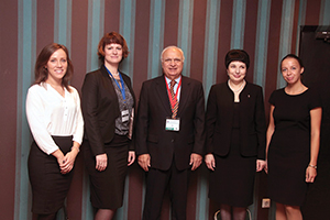
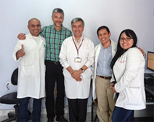
(From left) Kseniya S. Krylova, manager of the Department for International Affairs, and Natalya V. Zinserling, head of the Department for International Affairs and assistant professor in the Department of Neurology, both of North-Western State Medical University; Raad Shakir, MD,WFN president; Prof. Alla B. Guekht, Scientific and Practical Centre of Neuropsychiatry, named after Z.P. Solovyov; and Alyona D. Kubina, manager of the Department for International Affairs, North-Western State Medical University.
Prof. Vladimir Bekhterev (1857-1927) was an exceptional psychiatrist and neurologist who learned from notable scientists such as I. Mierzejewski, Paul Flechsig, and Jean M. Charcot. Prof. Bekhterev was a founder of the neurological school and author of “Pathways of the Spinal Cord and Brain” (1896). About 90 of his students became professors, and 40 of his students headed various psychoneurological institutions set up by Prof. Bekhterev. He described a specific nosological form of “spinal stiffness” known as ankylosing spondylitis (morbus Bekchterev). He and his student, L. Puusepp (the world’s first professor of neurosurgery), opened Russia’s first clinic of nervous diseases with a neurosurgical department (1897). He set up the world’s first psychoneurological institute (1907), which developed in the North-Western State Medical University, named after I.I. Mechnikov. Moreover, he created 12 scientific journals and edited them himself. Prof. Bekhterev established several departments of neurology in educational institutions and initiated the foundation of the world’s first neurosurgical institute (1918) and brain institute. He made a number of eminent discoveries in the field of neuroanatomy and neurophysiology, describing tens of neurological symptoms and syndromes. German anatomist F. Kopsch wrote, “There are only two persons who know the brain anatomy perfectly—God and Bechterev.” He brilliantly mastered hypnosis, using it in his medical practice.
Among Prof. Bekhterev’s students were two future neurologists: Michael Astvatsaturov, author of the evolutionary-biogenetic research method in neurology and founder of a nervous disease clinic (with 180-bed capacity) at the Peter the Great Hospital (now known as NWSMU, named after I.I. Mechnikov); and Alexander Triumphov, author of topical diagnosis of nervous system diseases. Prof. Alexander Panov was the first in the world who described tick-borne encephalitis (1935).
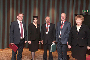
(From left) Prof. Alexey V. Silin, vice rector for Research and Innovations, North-Western
State Medical University; Prof. Alla B. Guekht, Scientific and Practical Centre of
Neuropsychiatry, named after Z.P. Solovyov; Raad Shakir, MD, WFN president; Prof. Sergey V.
Lobzin, Northwestern State Medical University; and Olga A.Kasantseva, vice governor of Saint Petersburg.
Nowadays, Saint Petersburg neurology continues to develop closely with foreign colleagues, exemplified by the annual conference Davidenkov Readings. This conference is organized by NWSMU and its Department of Neurology, named after S.N. Davidenkov. Academician Davidenkov is one of the founders of Russian neurogenetics.
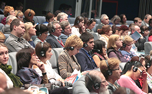
The first Davidenkov Readings were held in 1998 as a small intra-city event. However, in 2016 the number of registered attendees exceeded 1,000 participants.
On Sept. 29-30, 2016, the conference was held under the auspices of the World Federation of Neurology (WFN), and was opened by its president, Raad Shakir, MD. He delivered a speech on the challenges of global neurology education, then noted the considerable contribution of Russian scientists. Dr. Shakir highlighted the importance of further integration of Russian neurology in WFN educational programs and fruitful cooperation in clinical neurology.
Alla B. Guekht, honored doctor of Russia, delivered a commencement speech on behalf of the Russian Scientific Society. Her presentation focused on changes in the sphere of neurology education programs in Russia, as well as participation with the WFN on educational programs and collaboration prospects with European colleagues.
Within the conference was the Russian-Norwegian seminar on multiple sclerosis and migraine, nine breakout sessions on topical areas of modern neurology, and workshops on pain relief.
Prof. Lars Bø, director of the Norwegian Multiple Sclerosis Competence Centre in Norway, presented a lecture dedicated to multiple sclerosis and advances in immunomodulatory treatment, and noted the frequency of demyelinating diseases in Norway. Prof. Bø shared his experience in cell technology usage in multiple sclerosis treatment, and problems and advantages in this sphere.
Marte Helene Bjørk, MD, of the University of Bergen, Norway, gave a report on headache and cerebrovascular disease. NWSMU and the University of Bergen are partners and conduct joint educational and training programs in the field of neurology.
Summing up the results of the conference, Dr. Shakir was impressed by the conference organization and the contribution of classics of Russian neurology, and discussed topics and visited with guests from various regions of Russia and foreign countries. He expressed confidence in strengthening of relationships between the Russian Federation and the WFN.
The Neurological Society of Saint Petersburg is keen on cooperating with foreign colleagues. Please contact the University Department of International Affairs at interndept@szgmu.ru.


 On Sept. 17, 2017, at the Annual General Meeting of the World Federation of Neurology Council of Delegates during the World Congress of Neurology in Kyoto, Japan, elections will be held for the following posts: