From The Editors

By Steven L. Lewis, MD, Editor, and Walter Struhal, MD, Co-Editor

Walter Struhal

Walter Struhal

STEVEN L. LEWIS

Steven L. Lewis

We are pleased and honored to be taking on the editorship of World Neurology, the official newsletter of the World Federation of Neurology (WFN). We would like to thank President Raad Shakir and the officers and trustees of the WFN, as well as the members of WFN’s Publication Committee, for entrusting us with this responsibility. We also wish to give our sincere thanks to Dr. Donald Silberberg for his outstanding editorial leadership of World Neurology for the last three years, as well as for providing the two of us with the benefit and generosity of his ongoing guidance and knowledge as we take on this position, and for having done the work alone that is now deemed necessary for two people to perform.

We have planned a number of new initiatives for the readers of World Neurology, including contributions from authorities on breaking neurological topics that affect neurologist readers worldwide, such as the article in this issue about the Zika virus epidemic from Avi Nath, MD, and James Sejvar, MD. We also plan to develop new sections and columns over the coming issues to cover such entities as global neurological training and many other topics of interest to all neurologists worldwide.

Our plans for World Neurology include offering additional content formats (e.g., video). We will tighten the interconnection with WFN´s online footage and are currently working on implementing social media into World Neurology. This new feature will provide a convenient way to interact with other readers and discuss our articles.

We look forward to continuing to make World Neurology a trusted and sought-after resource for news and information of interest to all neurologists throughout the globe. We are also happy to field any suggestions from readers about ways to continue to make this publication evolve and be as valuable as possible for all neurologists worldwide.

Neurological Board Certification in Europe

By Jan B.M. Kuks

Jan B.M. Kuks

Jan B.M. Kuks

Young neurologists can rise to the challenge in Denmark on May 27, 2016. On that day, the 8th European Board Examination in Neurology will take place in Copenhagen.

Medical specialties in Europe are working together with the European Union of Medical Specialists (UEMS) (www.uems.eu), an organization containing 43 specialist-sections, one of these being the European Board of Neurology (EBN). Setting standards for training and practice is among the organization’s key activities. Therefore, the EBN is involved in developing harmonized models for the high-level training of the next generation of neurologists, in order to improve standards of clinical practice and, hence, patient care throughout Europe.

To achieve this, the EBN set up a core curriculum for the training of young neurologists, and — as testing drives learning — a board exam is provided as well.

Professor Wolfgang Grisold, now WFN secretary general, was the founder of this process and organized the first EBN examination in 2009. The 8th examination will take place at the site of the European Academy of Neurology (EAN) Congress. This illustrates the close cooperation between the UEMS Board of Neurology and the Academy of Neurology in Europe, an alliance without which a European training program for Neurologists would not exist.

Education in these times is not only for transferring knowledge, but is also directed toward achieving other competencies.

Successful candidates of the 7th EBN Exam in Berlin June 19, 2015 displaying their certificates

Successful candidates of the 7th EBN Exam in Berlin June 19, 2015 displaying their certificates

As in earlier days, the ability to retrieve knowledge from memory may be essential for clinical practice. But don’t we all use electronic devices in our clinics and on our ward rounds to find up-to-date knowledge as soon as possible for practicing evidence-based medicine and to offer our patients the latest achievements in our field? Is there any specialist in neurology who does not regularly want to have the opportunity for a peek inside an anatomical atlas, a handbook of neurophysiology or whatever textbook, before making a decision in clinical practice? So, today, we can’t restrict ourselves to information known by heart. We should be able to combine it with recent facts and developments. The ability to handle knowledge will become more and more important. This is the reason we offer our candidates the opportunity to take their own favorite textbooks (and in the future, electronic devices) to the examination to solve higher-order, open-book questions derived from real life, as they do in real life. Beside the great textbooks, guidelines and electronic courses from the EAN are the basis for the questions provided.

This isn’t all. Further competencies important for being a good specialist are described in several systems, such as in the CanMEDs roles (www.royalcollege.ca). In this system, a neurologist should not just be a medical expert, and the EBN exam should not be confined to testing neurological knowledge. Testing abilities in other CanMEDs roles like communicator, health advocate, professional and scholar comprise another and more essential part of the EBN examination.

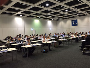
Candidates taking the written test on the 7th EBN Exam in Berlin

Candidates taking the written test on the 7th EBN Exam in Berlin

How should we test these abilities within the other competencies? Does this need just another couple of multiple choice questions? We feel that this cannot be achieved by written computer examinations. For example, public health or global health issues (being a heath advocate) have their national emphases, and ethical points of view vary in different countries. Thus, there is no absolute truth to be tested. A face-to-face discussion is more suitable than making a choice in the closed format of a multiple-choice question for testing these competencies. Therefore we invite our candidates to prepare themselves properly for a discussion by writing essays for an oral examination. Being a scholar demands the ability to make one’s own vision clear and to dive into a problem to be solved in a scientific way. Therefore we ask our candidates to make a critical appraisal of a topic of their own choice to be presented for an oral discussion.

So taking a European Board Exam for Neurology is not being dependent on having a lucky day; it can be prepared for in advance, and candidates can develop abilities over a long period of time to be successful.

CanMedsThe validity of the examination needs the input of the scientific experts at the European Academy of Neurology. The reliability of the outcome depends on the number and quality of the participating candidates. A statistical evaluation to eliminate “bad questions” only can be realized in a group of sufficient size. Establishing a passing score can be determined by specialists prior to the test. However, modification of such a score may be necessary after getting data from a sufficient number of adequate participants.

We are happy to see the number of participants grow each year. The exam becomes attractive to more candidates from inside, but also from outside Europe — many of whom want to take the exam to increase the possibility of moving between European countries or to test their abilities on a European level. In this respect, Turkey, Belgium and Italy now take a leading role by sponsoring their young neurologists to take the EBN exam, in addition to their national exit exams.

Unfortunately, by now, board exams do not yet have a legal value in Europe, and this restrains many young neurologists from taking the examination. With increasing interest in Europe and the cooperation between European countries, we are likely to establish a goal of a European exam to be taken as an exit test in order to work as a neurologist in the European continent in the near future. Striving for such a pretentious goal forces us to look at the American board exams for neurology to try to reach their high quality level, while keeping the European flavor in our own tests.

More information about the EBN Examination can be found on our website: www.uems-neuroboard.org. We would be delighted to welcome you there.

Jan B.M. Kuks is professor of clinical neurology and medical education, University Medical Centre  Groningen, Netherlands, and chair of the Examination Committee European Board of
Neurology.


Neurological Manifestations of Zika Virus Infection: What Neurologists Need to Know

By Avindra Nath, MD, and James Sejvar, MD

headshot_JJS-3

James Sejvar, MD

headshot_Avi

Avindra Nath, MD

In recent years, there has been an emergence of several major viral infections with devastating neurological consequences, including West Nile virus, dengue, chikungunya, enterovirus D68, Ebola and now Zika virus. Increased global travel and climate change, leading to changing patterns of vector distribution and behavior are among the major reasons for the emergence of these infections. Zika virus is the most recent epidemic that is having devastating effects on human populations in affected regions, and is rapidly spreading across the South American continent.

Epidemiology

Zika virus was first identified from a primate in 1947 in the Zika forest of Uganda.1 The first human cases occurred in Africa and then in Southeast Asia in the 1960s.2,3 During the intervening years, Zika virus was associated with isolated cases or small outbreaks mainly in Africa. In 2007, there was an outbreak in Yap, the Federated States of Micronesia, where nearly three-quarters of the population was infected.4, 5 This represented the largest outbreak of Zika virus infection to that point. In 2013, there was an epidemic in French Polynesia, which was associated with a reported increase in cases of the autoimmune peripheral nerve disorder Guillain-Barre syndrome, although a causal association between Zika virus and Guillain-Barre syndrome was never established.

In December 2014, Zika virus was first detected in Brazil. Although it is unknown how it was introduced into Brazil, some hypothesize that a traveler attending the 2014 football/soccer World Cup introduced the virus. The outbreak in Brazil was fast moving and large. Tens of thousands of people became ill, and likely millions of people were infected. Similar to French Polynesia, shortly after the beginning of the Zika virus outbreak, clinicians began reporting larger-than-expected numbers of Guillain-Barre syndrome. Many of these people had reported a febrile rash illness compatible with Zika in the days or weeks before their weakness onset. In addition, clinicians in Brazil noted a 20-fold increase in microcephaly in 2015, compared to previous years, with microcephalic babies born approximately eight to nine months after the first recognition of Zika virus. Some of the infants’ mothers reported a rash illness compatible with Zika virus infection while pregnant, leading to the suspicion that the microcephaly was somehow associated with Zika virus infection.

Nearly 90 percent of the cases of microcephaly occurred in the northeastern region of the country,6, 7 areas experiencing some of the heaviest burdens of Zika virus infection as well. French Polynesian health authorities reported an unusual increase in central nervous system malformations in babies born during a Zika virus outbreak on the islands from 2014 to 2015.6 The infection has now spread across most of South America and Mexico. To date, few cases have been reported in the United States among travelers returning from Zika virus-affected regions.8, 9

Virology and Pathophysiology

Zika virus is a positive-sense, single-stranded RNA virus (genome 10.7 K nucleotides) belonging to the flaviviridae family, which includes dengue, yellow fever, Japanese encephalitis, St. Louis encephalitis and West Nile virus. It has the ability to cross the placenta and cause developmental brain abnormalities in children, suggesting that the virus likely infects neural stem cells. The severity of brain malformations may be related to the stage of fetal development at the time of infection. Microcephaly would be the most common manifestation, but if infection were to occur in earlier stages of fetal development, anencephaly or lissencephaly may occur.

The pathophysiology of ascending paralysis and myelitis in adults is unknown. However, mice injected with the virus can develop paralysis, suggesting direct invasion by the virus, although an immune-mediated, post-viral syndrome is also possible. It remains unknown if once infected and recovered if an individual develops long-term immunity or not, and if recurrent infections or relapses can occur. Questions regarding long-term viral persistence in tissue reservoirs also remain unanswered.

Transmission

The virus is transmitted by the Aedes species of mosquitoes 10, in particular Aedes aegypti, the vector involved with transmission of dengue, a closely related flavivirus. Additionally, experimental evidence suggests the virus can be transmitted by Asian tiger mosquitoes (Aedes albopictus) 11, 12, which can survive in cold temperatures. Most arboviruses have an intermediary host or “reservoir.” For West Nile virus, birds, particularly corvids, serve as these reservoirs. For Venezuelan, Western and Eastern equine encephalitis viruses, horses serve this role, and for Japanese encephalitis virus, it is primarily pigs. However, the transmission of Zika virus generally occurs directly between humans and mosquitos. There is some evidence that human-to-human transmission may occur through sexual intercourse, and the virus has also been detected in saliva, so the potential for oral transmission also exists. The virus has been isolated from the amniotic fluid of pregnant women and blood and tissues of newborns, suggesting materno-fetal transmission.13 So far, an intermediary host has not been identified.

Clinical Manifestations

Zika_Photo1

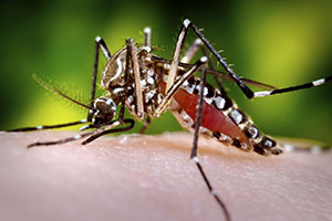
Female Aedes aegypti mosquito

The majority of Zika virus infections ­­­— 80 percent — are clinically asymptomatic.4 Among persons who develop symptoms, Zika virus infection is generally considered to be mild, causing fever, rash and body aches. Some may develop conjunctivitis. Symptoms usually last one week.

The full spectrum of neurological complications from this viral infection remains unknown. The epidemiological association between microcephaly and the infection seems strong. In Brazil, annual reported rates of microcephaly would generally be somewhere around 150 cases per year. Reportedly, between October 2015 and January 2016, more than 3,500 babies were born with the condition. CT brain scans show evidence of widespread calcification. Other malformations, such as anencephaly and lissencephaly, might also occur. It remains uncertain if other organs may be involved in addition to the brain. However, the differential diagnosis of microcephaly is broad. Hence, when presented with a patient with microcephaly, it remains important to consider other common causes, such as genetic, craniostenosis, and infections, such as toxoplasmosis, rubella, varicella zoster virus and cytomegalovirus. Intrauterine cerebral anoxia, exposure to drugs, alcohol and other toxins, malnutrition and metabolic disorders such as phenylketonuria can also cause microcephaly. Patients with microcephaly often have developmental delay, difficulty with gait and balance, mental retardation, seizures and hyperactivity.

Guillain-Barre syndrome appears to be a recurring possible complication of Zika virus infection. Following the introduction of Zika virus into French Polynesia, clinicians began reporting larger-than-expected numbers of Guillain-Barre syndrome cases on the island.14 Following the introduction of Zika virus to Brazil in December 2014, again, reports surfaced of large numbers of Guillain-Barre syndrome cases. In Brazil, few cases of Guillain-Barre syndrome had laboratory confirmation of Zika virus, but currently the primary method of diagnostic testing is through the detection of viral RNA through polymerase chain reaction. In Guillain-Barre syndrome, by the time the clinical features of limb weakness develop, it is unlikely that there would still be circulating virus, and, as such, detection of viral RNA would not be expected. Less commonly, some patients have been thought to have a myelitis or polio-like manifestations. Currently, it is unclear if these are all related or if indeed both spinal cord and peripheral nerves can be involved. Thus, in Brazil, epidemiologic evidence and the close temporo-spatial clustering of both Guillain-Barre syndrome and Zika virus cases provides intriguing circumstantial evidence for an association.

In other cases in which the virus was newly introduced, reported increases of Guillain-Barre syndrome cases have invariably appeared, including in Colombia, Venezuela and, more recently, El Salvador, which reported 46 Guillain-Barre syndrome cases in a five-week period from December 2015 to early January 2016. That is nearly three times more than the country would normally see in that timeframe. Laboratory substantiation of an association between Zika virus and Guillain-Barre syndrome has proved challenging, however. As noted, by the time of onset of weakness, the virus would be expected to be cleared from the body, and molecular techniques to identify the virus or viral RNA would not be expected to be positive. Detection of Zika virus-specific antibodies would provide evidence of current or prior infection. However, that method also has its challenges. Dengue virus is a closely related flavivirus to Zika, and invariably co-circulates in all areas currently associated with Zika virus. However, dengue virus infection has also rarely been associated with Guillain-Barre syndrome, and laboratory testing by serology is challenging due to the substantial cross-reactivity of antibodies between Zika virus and dengue virus.

Since these viruses are carried by the same mosquito vector and co-circulate at the same times of the year, it can be challenging to differentiate between infection with the two viruses.15 Development of a robust serologic assay that can reliably differentiate Zika virus from dengue and other closely related flaviviruses will be crucial in order to provide laboratory evidence of Zika-associated Guillain-Barre syndrome, as well as other late complications of Zika virus. Currently, the nature of the neuropathy is not known, as results of electrodiagnostics to determine the clinical sub-type of Guillain-Barre syndrome possibly associated with Zika virus has been rarely reported. It would be important to know if it is axonal or demyelinating and if it is immune mediated. This could affect treatment and prognosis. Recovery from demyelinating neuropathies is generally better than those due to axonal injury. Isolated reports suggest that the neuropathy may be demyelinating and may respond to treatment with intravenous immunoglobulin.14

Laboratory Diagnosis

Viremia occurs only during the first few days of the illness, but if blood samples are obtained during that time, virus can be detected by polymerase chain reaction.16 Following this phase, IgM antibodies can be demonstrated by ELISA or Western blot analysis. Previous epidemics have noted that there is cross reactivity between antibodies to Zika and other arboviruses such as dengue.5 The Centers for Disease Control and Prevention (CDC) has issued guidelines for the testing of infants born with possible Zika virus infection.17

Treatment and Prevention

Currently, there is no effective treatment or vaccine against the virus. Hence, prevention is key with control of mosquito populations and prevention of mosquito bites. Travel advisories have been issued for pregnant women not to travel to areas experiencing Zika virus outbreaks. For individuals who suffer from the neurological consequences of the infection, long-term supportive and symptomatic treatment is key. The socio-economic impact of the infection, particularly if the association between Zika virus and microcephaly holds true, will likely be huge and felt for decades. While the large number of cases of microcephaly is tragic, whatever the eventual cause turns out to be, it will result in large numbers of children with developmental disorders and begs for the need to train personnel in a wide variety of health disciplines, including neurology, rehabilitation, specialized nursing, social services, etc., to care for and treat this population. Ongoing surveillance for Zika virus in the Americas and elsewhere, to monitor its continued spread, as well as documentation of infection among travelers returning from affected areas will be critical. Development of more robust serologic assays that can differentiate Zika virus from other closely related flaviviruses will be an important tool to substantiate an association between Zika virus and devastating neurologic conditions, such as Guillain-Barre syndrome and microcephaly. Ultimately, the long-term epidemiologic pattern of Zika virus will be important to monitor.

References:

  1. Weinbren, M.P. and M.C. Williams, Zika virus: further isolations in the Zika area, and some studies on the strains isolated. Trans R Soc Trop Med Hyg, 1958. 52(3): p. 263-8.
  2. Simpson, D.I., Zika Virus Infection in Man. Trans R Soc Trop Med Hyg, 1964. 58: p. 335-8.
  3. Olson, J.G., et al., Zika virus, a cause of fever in Central Java, Indonesia. Trans R Soc Trop Med Hyg, 1981. 75(3): p. 389-93.
  4. Duffy, M.R., et al., Zika virus outbreak on Yap Island, Federated States of Micronesia. N Engl J Med, 2009. 360(24): p. 2536-43.
  5. Lanciotti, R.S., et al., Genetic and serologic properties of Zika virus associated with an epidemic, Yap State, Micronesia, 2007. Emerg Infect Dis, 2008. 14(8): p. 1232-9.
  6. Control, E.C.f.D.P.a., Rapid risk assessment: Zika virus epidemic in the Americas: potential association with microcephaly and Guillian Barre syndrome. 10 December 2015.
  7. Bogoch, II, et al., Anticipating the international spread of Zika virus from Brazil. Lancet, 2016.
  8. Control, C.f.D., CDC telebriefing: Zika virus travel alert. 15 January 2016.
  9. Hennessey, M., M. Fischer, and J.E. Staples, Zika Virus Spreads to New Areas – Region of the Americas, May 2015-January 2016. MMWR Morb Mortal Wkly Rep, 2016. 65(3): p. 55-8.
  10. Li, M.I., et al., Oral susceptibility of Singapore Aedes (Stegomyia) aegypti (Linnaeus) to Zika virus. PLoS Negl Trop Dis, 2012. 6(8): p. e1792.
  11. Grard, G., et al., Zika virus in Gabon (Central Africa)–2007: a new threat from Aedes albopictus? PLoS Negl Trop Dis, 2014. 8(2): p. e2681.
  12. Wong, P.S., et al., Aedes (Stegomyia) albopictus (Skuse): a potential vector of Zika virus in Singapore. PLoS Negl Trop Dis, 2013. 7(8): p. e2348.
  13. Pan American Health Organization and World Health Organization. Epidemiological Alert: Neurological syndrome, congenital malformations, and Zika virus infection. Implications for public health in the Americas. http://www.paho.org, 1 December, 2015.
  14. Oehler, E., et al., Zika virus infection complicated by Guillain-Barre syndrome–case report, French Polynesia, December 2013. Euro Surveill, 2014. 19(9).
  15. Carod-Artal, F.J., et al., Neurological complications of dengue virus infection. Lancet Neurol, 2013. 12(9): p. 906-19.
  16. Faye, O., et al., One-step RT-PCR for detection of Zika virus. J Clin Virol, 2008. 43(1): p. 96-101.
  17. Staples, J.E., et al., Interim Guidelines for the Evaluation and Testing of Infants with Possible Congenital Zika Virus Infection – United States, 2016. MMWR Morb Mortal Wkly Rep, 2016. 65(3): p. 63-7.
Avindra Nath, MD, is intramural clinical director and a senior investigator with the Section of Infections of the Nervous System, National Institute of Neurological Diseases and Stroke, National Institutes of Health, Bethesda, Maryland. James Sejvar, MD is a neuroepidemiologist with the division of High-Consequence Pathogens and Pathology, National Center for Emerging and Zoonotic Infectious Diseases, Centers for Disease Control and Prevention, Atlanta, Georgia. The authors have no financial relationships relevant to this article to disclose. The findings and conclusions in this article are those of the authors and do not necessarily represent the official position of the Centers for Disease Control and Prevention or National Institutes of Health.