From Cerebellum to Medulla Oblongata
By Peter J. Koehler
Nowadays, physicians know that spontaneous or (rarely) traumatic cerebellar hemorrhage may be lethal by causing hydrocephalus or brainstem compression. In the past, these observations were interpreted in a way that the cerebellum was considered an organ of vital importance. It lasted several centuries before researchers were able to distinguish cerebellar injury from pathology of the medulla oblongata.
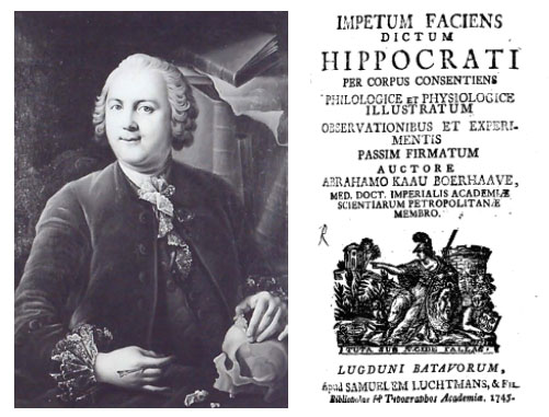
Abraham Kaau Boerhaave’s Impetum Faciens dictum Hippocrati.
Influence of Willis
In a chapter in Thomas Willis’ (1621-1675; his name is associated with the arterial circle at the base of the brain) famous Cerebri Anatome (1664) titled “De spirituum in Cerebello productorum, propter functionis involuntariae actus varia diataxi, & Exercitio diversimodo,” the author states that the cerebellum is responsible for vital and involuntary functions (breathing, heartbeat, bowel movement). Still thinking in terms of the humoral (patho)physiology of the period, he supposed that animal spirits (a hypothetic volatile substance), originating in the cerebellum, passing through the vagus nerves, are conducted to these organs (lungs, heart). So, the medulla oblongata and vagus nerves were just considered to conduct stimuli that originate in the cerebellum.
On the other hand, experiments had been done showing that the heart could continue beating after removal from the body, and the Swiss physician Johann Jacob Wepfer (1620-1695) observed continuing heartbeat for several hours after decapitation, though still believing there is an important influence from the vagus nerves on the heart that continues for a longer period.
Following William Harvey’s (1578-1657) discovery of the circulation of blood earlier in that century (1628), indeed new ideas had originated on the function of the brain and its relationship to the heart. In the past, it was thought that the brain and cerebellum had equal functions, although the ancient ventricular localization theories localized memory in the third cell (our fourth ventricle). This idea was still present in some medical minds of the 17th century [for instance, in the German physician Johan Vesling’s (1598-1649) and Dutch physician Nicolaes Tulp’s (1593-1674) work].
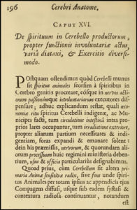
Willis’ Cerebri Anatome (1664).
However, there had already been ideas about the important vital function of this ventricle. Willis was the first to localize vital and involuntary functions in the cerebellum. Experiments seemed to confirm these ideas as injuries and removal of the cerebellum led to arrest of heart and respiration. In contrast, removal of the cerebral hemispheres was sustained much longer. These observations were considered indirect proofs of Willis’s speculations.
Experimental Cerebellar Lesions Considered Lethal
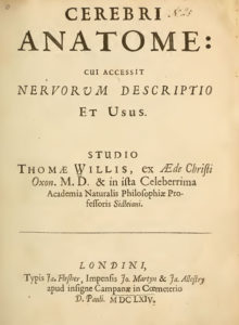
From Willis’ Cerebri Anatome.
Several 17th century experimenters reported the fatal effect of removal of the cerebellum, including the Paris physician (and architect) Claude Perrault (1613-1688), Raymond Vieussens (1641-1717; working in Montpellier), Johannes Bohn (1640-1718, who worked in Leipzig) and German physician Johann Gottfried von Berger (1659-1736).
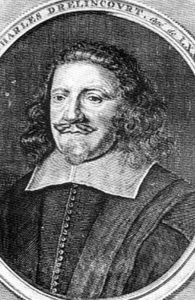
Charles Drelincourt (1633-1697), a French physician who moved to Leyden, where he preceded Herman Boerhaave (1668-1738)) stabbed a needle in the fourth ventricle, which immediately led to “epileptic cramps.” (Nowadays, we would probably call it extensor posturing and the differentiation from epileptic phenomena still may be a diagnostic clinical problem.)
“Acu in cerebelli ventriculum compulsa inter primam vertebram & os occipitis, canis, ceu epilepticus, ter quaterque concussus est universe, sed mox expiravit » (A needle was driven into the cerebellar ventricle between the occiput and first vertebra, the dog demonstrated three or four general shocks, but soon expired.) He believed to have confirmed Willis’ ideas herewith, not aware that he probably injured the medulla oblongata. However, even in this period, some observers presented results that contrasted with those mentioned before.
From Drelincourt’s
Experimenta anatomica
(1684).
French physician and anatomist Joseph-Guichard Du Verney (1648-1730) who did important work on the organ of hearing in his Traité de l’organe de l’ouïe of 1683, anticipating the work of Hermann von Helmholtz (1821-1894) for instance, was able to keep animals alive for some time after removal of the cerebral hemispheres and the cerebellum, and Pierre Chirac (1650-1732), who became personal physician of Louis XV, was able to produce survival for a while following removal of the cerebellum combined with artificial respiration (inflation of air).
“Mr. Chirac, by several experiments, he has made upon dogs, has clearly proved an animal may live some time wanting the brain and even sometimes de cerebellum.”
Experimental Cerebellar Lesions Not Lethal
During the subsequent century, several other experimenters observed that animals not always died upon removal of the cerebellum. It seemed that it depended rather on the caution with which the surgery was done. Some experimenters even pointed to the importance of the medulla oblongata.
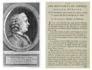
Anne-Charles Lorry. Lorry’s 1760 Memoir on movements of the brain.
François Pourfour du Petit (1664-1741) a pioneer in ophthalmology who also described dysfunction of the contralateral hemisphere in injured soldiers, looking for the possible sensory function of the cerebellum, which he could not confirm, observed survival for several days. The Italian anatomist and physician Antonio Valsalva (1666-1723), teacher of Giovanni Battista Morgagni (1682-1771), who described several of his teacher’s cases in his famous De Sedibus of 1761, removed the cerebellum of pigeons, observing that they did not immediately die. Morgagni observed that isolated injury of the cerebellum was not immediately lethal and confirmed this by pathologic-anatomical examination.
Abraham Kaau-Boerhaave (1715-1758), one of Herman Boerhaave’s nephews who moved to St. Petersburg where he became medical superintendent at the navy-hospital, disliked speculation, and after several experiments damaging the brain, the cerebellum (“stylo vel cultro deinde adacto intra cerebellum, non uno momento fuisse actum de cordis & respirationis energia” [then a pen of knife was thrust inside the cerebellum, the cardiac and respiratory function do not stop immediately) or the medulla oblongata, became one of the first to recognize the vital role of the medulla oblongata.
French physician Anne-Charles Lorry (1726-1783), one of the founders of the Société Royale de Médecine, and the German physician Justus Arnemann (1763-1806) warned against the technical deficits in many of the previous experiments on the cerebellum, resulting in massive bleeding in the subtentorial space and tried to avoid this.
Lorry, in a memoir on “Les mouvemens du cerveau” [the movements of the brain] presented at the French Académie des Sciences (1760) and experienced that young cats and pigeons were better suited to avoid these complications.
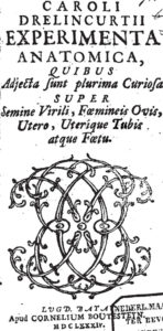
From Drelincourt’s Experimenta anatomica (1684).
With our present knowledge, the mechanism of the following experiment can be easily comprehended:
Je pressois un jour latéralement & assez fort le cervelet d’un chien gros & adulte, le hasard me fit voir tout d’un coup ce chien tomber & ronfler très-fort & très-notablement: dans mon étonnement, je lui lâchai ce viscère, il fut réveillé au même instant &: fit des efforts pour crier. Je repris le cervelet de la même manière, il se rendormit: je recommençai cette expérience plusieurs fois, jusqu’à ce que tout d’un coup une compression encore plus forte lui excita des convulsions. [One day I compressed the cerebellum of a large and adult dog from the lateral side and sufficiently strong, by chance I saw the dog falling and snoring very hard and very strikingly suddenly: in my astonishment, I released that organ, he awoke at the same moment and made efforts to scream. I again took the cerebellum by the same way, he fell asleep again: I repeated the experiment several times, until suddenly an even stronger compression excited convulsions].
No Vital Functions in the Cerebellum
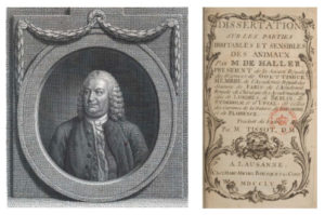
Swiss physician and pioneer experimental researcher Albrecht von Haller (1708-1777) considered the heart almost completely independent (1786), and his followers denied localization of functions in the brain, including the cerebellum. Haller, who did many experiments on all kinds of animals with improved techniques, believed Willis’ ideas were insufficiently based upon experiments and localized the soul (sensorium commune, the place in the brain, where all senses converge and result in a motor reaction) in all parts of the brain.
Charles Drelincourt.
Disorders of the cerebellum, he thought, do not lead to “certain and speedy death. For certain experiments, even of our own making, show that it has borne wounds and scirrhi, without taking away life; . . . not very rarely, wounds of the cerebellum cure” (Haller, 1786, p.217). He criticized previous physicians stating that “the source of the great error in physic has been owing to physicians … making few or no experiments, and substituting analogy instead of them” (Haller, 1755, p. 658).
Ethical Issues
Reading about these experiments and vivisection, one wonders about ethical issues at the time. Haller wrote in the introduction that he examined 190 animals since 1751, “a species of cruelty for which I felt such a reluctance, as could only be overcome by the desire of contributing to the benefit of mankind.” (Haller, 1755, p. 657) . Throughout the 19th century, the man-animal relationship, particularly with respect to cats and dogs, gradually became more emotional.
One of the possible explanations that have been presented is that pet-keeping became more common in early industrialized and urbanized countries. More information on the history of vivisection and ethical issues can be found in the work of medical historian Andreas-Holger Maehle and his pivotal article “The Ethical Discourse on Animal Experimentation, 1650-1900” (Maehle, 1993). •
Literature
Haller A. A dissertation on the sensible and irritable parts of animals. Translated from Latin by Tissot. London: Nourse, 1755/1936.
Haller A. First lines of physiology (Vol. 1). Johnson Reprint, New York, 1786. (Reprinted in 1966).
Koehler PJ. Neurology in Tulp’s Observationes medicae. J Hist Neurosci 1996;5:143-51.
Koehler PJ. Neuroscience in the work of Boerhaave and Haller. In: Whitaker H, Finger S, Smith C. Brain, mind and medicine: essays in eighteenth century neuroscience. Springer, New York, 2007, pp. 213-231.
Koehler PJ, Lameris B. The Magnus-Rademaker Scientific Film Collection: Ethical Issues on Animal Experimentation (1908-1940). J Hist Neurosci. 2016;25:102-21.
Lorry CH. Sur les mouvemens du cerveau. Second mémoire. Sur les mouvemens contre nature de ce viscère, et sur les organes qui sont le principe de son action. Mémoires de Mathématique et de Physique présentés à l’Académie Royale des Sciences. Paris, Imprimerie Royale, 1760, p.344.
Maehle AH. The ethical discourse on animal experimentation, 1650–1900. Clio Med 1993; 24: 203–251.
Neuburger M. Die historische Entwicklung der experimentellen Gehirn- und Rückenmarksphysiologie vor Flourens. Stuttgart, Enke, 1897. English edition published as The historical development of experimental brain and spinal cord physiology before Flourens. Transl. and edited, with additional material. Baltimore & London, Johns Hopkins University Press, 1981.
Voogd J, Koehler PJ. Historic notes on anatomic, physiologic, and clinical research on the cerebellum. Handb Clin Neurol. 2018;154:3-26.
Willis T. Cerebri anatome: cui accessit nervorum descriptio et usus. Londini, Ja. Flesher, 1664.