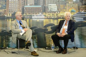
Palliative Care for Patients With Stroke

By Wolfgang Grisold, Claire J. Creutzfeldt, Gillian Mead, and Fergus N. Doubal

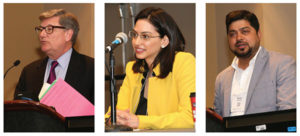
During the World Stroke Organization (WSO)-European Stroke Organization (ESO) Congress (https://eso-wso-conference.org), a teaching course on palliative care issues in patients with stroke was offered. There were four speakers: Wolfgang Grisold (Austria), Claire J. Creutzfeldt (U.S.), Gillian Mead (U.K.), and Fergus N. Doubal (U.K.). They presented four lectures.

This initiative deserves merit, as despite much progress in palliative care in many aspects of neurology, there is considerable scope for improving palliative care in stroke. Stroke is the most frequent neurological disease globally and the leading cause of disability adjusted life-years and deaths due to neurological diseases. Therefore, such an initiative is important, and, as this teaching course showed, has many facets to be discussed.

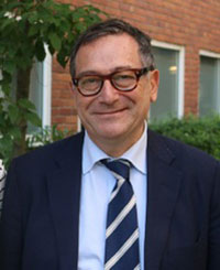
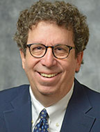
Wolfgang Grisold (Austria)
Wolfgang Grisold

W. Grisold gave an outline on the present international guidelines on palliative care in stroke. Guidelines are elaborate and define the need and the role. In addition, there is also a U.K. patient guide that is helpful. Most of the guidelines are aimed at the acute and subacute setting of stroke, and further work needs to be done for the long trajectory of stroke survivors.

Attention also needs to be given to individuals with disturbed consciousness, cognitive impairment, and speech disorders, who often can not actively participate in the process of decision-making.

From the conceptual and cultural point of view, it has to be acknowledged that the concept of palliative care is based on the patient autonomy, which is culturally perceived differently.

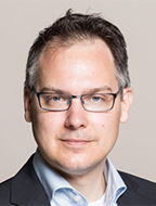
Claire J. Creutzfeld (U.S.)
Claire J. Creutzfeld

The issue of integrating palliative care and serious illness communication into high quality stroke care was addressed by C.J. Creutzfeldt. The different disease trajectories were discussed and compared with those of other illnesses often considered for palliative care, such as cancer. Building a partnership with the patient and his or her family, communicating transparently and discussing hope with both realism and compassion are key skills for stroke providers. Dr. Creutzfeldt gave a strong testimony of the need for palliative care in stroke with the goal of improving communication, decision-making, quality of life, and quality of end of life for patients with stroke and their families.

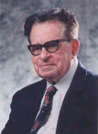
Gillian Mead (U.K.)
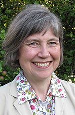
Gillian Mead

Gillian Mead emphasized the role of families and also the importance of communication. An important issue is what the individual expectancy for patients with severe stroke is, and if this would change at different time points during their disease, as survival often ensues with severe disability.

Reference to the study of Kendall et al, 2018, was made, which posed the question of outcomes, experiences, and palliative care in stroke.

One result was that palliative care still has the connotation of withdrawal, or withholding, and “dual“ narratives should be avoided. The loss of the former self of the patient is an issue for the patient and for carers. Guiding through the moral maze in discussions with the family is important for providing emotional support and dignity.

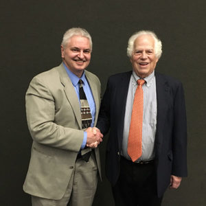
Fergus Doubal (U.K.)
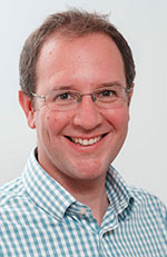
Fergus Doubal

Sudden death is common following stroke and can be due to several causes: immediate pressure effects from a large stroke causing brain edema, concurrent severe disease, often cardiac, infectious, and other medical complications, and also due to treatment withdrawal.  In young adults, stroke is the fourth most common cause of sudden death after cardiac causes, pulmonary embolism, and infection.

When stroke causes sudden death, especially in younger patients, there is a preponderance of intracerebral hemorrhages compared to ischemic stroke. When stroke causes death within 24-48 hours, the rate of intracerebral hemorrhage is higher, but as time progresses ischemic stroke become more prevalent as a cause of death.

When death is sudden (within days), this can be challenging for patients, families, and health care professionals who often need to work together to make important yet time-critical shared decisions quickly. During this process, it is important to base decisions on the patient’s values and what they would consider to be an acceptable outcome with families acting as proxies should the patient not retain capacity to participate. Often death may not be considered the worst outcome compared to survival in a highly dependent state.


References for Further Reading:

Holloway RG et al. Palliative and End-of-Life Care in Stroke. Stroke 2014; Volume 45, Issue 6,1887-1916.

https://www.stroke.org.uk/resources/national-clinical-guideline-stroke-patient-version


Claire J. Creutzfeldt, et al.  Symptomatic and Palliative Care for Stroke Survivors. J Gen Intern Med. 2012 Jul; 27(7): 853–860.


Marylin Kendall, et al. Outcomes, experiences and palliative care in major stroke: a multicentre, mixed-method, longitudinal study. CMAJ. 2018 Mar 5;190(9):E238-E246. doi: 10.1503/cmaj.170604.


Frederik Nybye Ågesen, et al. Sudden unexpected death caused by stroke: A nationwide study among children and young adults in Denmark Int J Stroke  2018 Apr;13(3):285-291. doi: 10.1177/1747493017724625. Epub 2017 Aug 1.


Wolfgand Grisold is Secretary-General of the WFN.


Claire J. Creutzfeldt is associate professor of neurology at the University of Washington – Harborview Medical Center in Seattle, Washington.


Gillian Mead is chair of Stroke and Elderly Care Medicine at the Center for Clinical Brain Sciences, University of Edinburgh.


Fergus Doubal is the Stroke Association Garfield Weston Foundation Clinical Senior Lecturer,


NHS Scotland Research Fellow at the Center for Clinical Brain Sciences, University of Edinburgh.
Simplifying the Diagnostic Criteria for ALS

New Criteria are Aimed to Facilitate Enrollment of Patients into Clinical Trials

By Matthew Kiernan, for the ALS Consensus Committee

The World Federation of Neurology recently joined forces with the International Federation of Clinical Neurophysiology and patient support groups, including the Motor Neurone Disease Association (UK) and the American ALS Association, to convene a consensus meeting to address diagnostic criteria for amyotrophic lateral sclerosis (ALS; motor neurone disease).

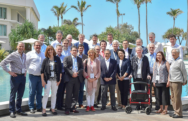
Matthew Kiernan, Chair of the ALS/MND Specialty Group, World Federation of Neurology. ALS Consensus Committee: Ammar Al-Chalabi (London, UK), Mark Baker (Newcastle upon Tyne, UK), David Burke (Sydney, Australia), Li-Ying Cui (Beijing, China), Mamede de Carvalho (Lisbon, Portugal), Andrew Eisen (Vancouver, Canada), Julian Grosskreutz (Jena, Germany), Orla Hardiman (Dublin, Ireland), Robert Henderson (Brisbane, Australia), Ryuji Kaji (Tokushima, Japan), Matthew Kiernan (Sydney, Australia), Jose Manuel Matamala (Santiago, Chile), Hiroshi Mitsumoto (New York, USA), Waulter Paulus (Gottingen, Germany), Jeremy Shefner (Phoenix,USA), Neil Simon (Sydney, Australia), Michael Swash (London, UK), Kevin Talbot (Oxford, UK), Martin Turner (Oxford, UK), Leonard van den Berg (Utrecht, The Netherlands), Renato Verdugo (Santiago, Chile), Steve Vucic (Sydney, Australia) Photo Legend: ALS Consensus Committee, Gold Coast, Australia.

The broad aim of the consensus meeting was to improve the process of diagnosis of ALS in the early stages of the disease, when clinical symptoms are minimal, thereby giving therapies the best chance of success. Previous ALS criteria, dating back to the original El Escorial and later Airlie House and Awaji criteria, used degrees of diagnostic certainty from possible to definite ALS. While such an approach makes sense, it also carries a degree of uncertainty for patients, their families, and clinicians. From a practical level, patients with a label of possible ALS may be denied entry to clinical trials, even though such patients would likely benefit from instigation of trial therapies compared with those recruited later in their disease.

Given that uncertainty around a diagnosis of ALS constitutes a potential barrier to patient enrollment in clinical trials, the consensus meeting held on the Gold Coast in Australia began by considering a broad range of data across different phenotypes, clinical presentations, and outcome measures, in addition to discussions that evaluated diagnostic technologies.

Prior to defining consensus criteria, a collective understanding of ALS was established based around key tenets: that ALS represented a progressive disorder of the motor system, that it involves dysfunction of upper and lower motor neurone compartments of the nervous system, and that there is typically a focal onset. It was accepted that while upper motor neuron signs were not always clinically evident, involvement of the lower motor neuron was more often apparent through clinical examination. In terms of diagnostic technologies, supportive evidence of lower motor neuron dysfunction has tended to be derived from electromyography and neuromuscular ultrasound, particularly the detection of fasciculations in multiple muscles. Supportive evidence of upper motor neuron dysfunction was more limited, relying on transcranial magnetic stimulation studies of the central motor nervous system, MRI, and neurofilament levels, although it was accepted that a diagnosis of ALS does not require these investigations. And finally, while ALS may include cognitive, behavioral, and psychiatric abnormalities, these features were not essential for a diagnosis.

Accepting these broad concepts of disease, combined in an integrated fashion, a consensus was reached, whereby ALS was defined by the presence of:

  • Progressive motor impairment, documented by history or repeated clinical assessment, preceded by normal motor function.
  • Upper and lower motor neurone dysfunction in at least one body region (in the same body region if only one body region was involved), or lower motor neuron dysfunction in at least two body regions.
  • Investigation findings that excluded alternative disease processes.

In adopting these simplified criteria for ALS, the previous diagnostic categories of possible, probable, and definite were abandoned. Such assessment of a likelihood of disease served to generate misinterpretation by patients, who may inadvertently have considered that such terms represented the likelihood that ALS was causing their symptoms.

In reality, the consensus group acknowledged that nearly all patients diagnosed as possible ALS continue to progress and ultimately die from ALS. Moving forward from these new consensus criteria, it is anticipated that ALS patients will be fast-tracked into clinical trials. With the advent of these new criteria, the diagnosis of ALS can be made early, and definitively. •

Reference:

Shefner JM, Al-Chalabi A, Baker MR, et al. A proposal for new diagnostic criteria for ALS. Clin Neurophysiol 2020;131:1975-1978.


COVID-19: A Neurologist’s Perspective

By Avindra Nath and B. Jeanne Billioux

The crisis we are currently facing is unprecedented in every way. Just a few months ago, we were talking about developing targeted gene therapies for a spectrum of diseases, including ultrarare diseases. Only a few weeks later, the health care system finds itself overburdened and undersupplied to the point where we are talking about rationing health care1. Maintenance care has been pushed to telemedicine clinics and elective procedures have ground to a halt. Many patients sick with respiratory symptoms are being sent home to isolate themselves, and some are dying at home. There is an acute shortage of ventilators to the point that in some hospitals one ventilator is being shared by multiple patients2.

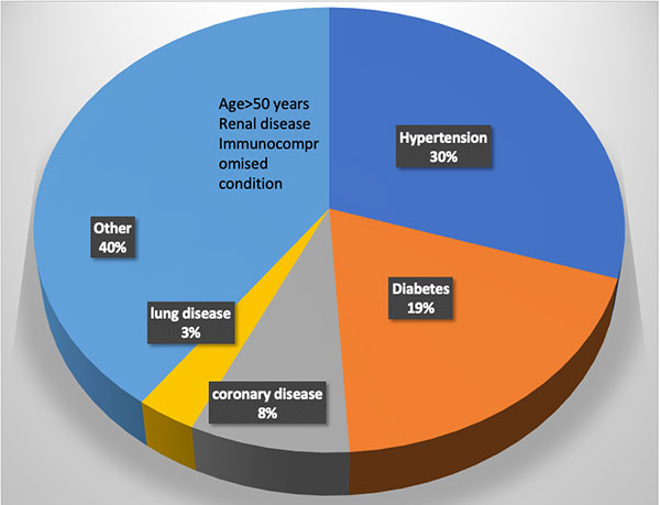
Figure 1. Distribution of comorbidities in patients requiring inpatient care due to COVID-19.

Several basic medicines are in limited supply. Although many hospitals and institutions recognized the need to stockpile personal protective equipment, several hospitals have run out of masks and gowns due to a limited supply chain. This crisis has tested community-based ingenuity, and in some hospitals, personal protective equipment is being fashioned by staff and community volunteers out of plastic visors and trash bags. Many doctors on the front line have succumbed to the infection, and many others are quarantined, a sobering reminder of these dire circumstances. The words here just a few months prior would read as a work of fiction, but this is the unfortunate reality of the crisis we face – COVID-19. Nearly every country and every major city in the world has been affected by the infection. On April 12 alone, there were over 10,000 new infections and nearly 1,000 deaths in a single day in New York. What started in Wuhan in November 2019 has become a global pandemic necessitating drastic changes in our way of life.

About COVID-19

COVID-19 is caused by the virus SARS-CoV2, a single-stranded RNA virus. Merely 60 nm in size, the virus that can only be visualized by an electron microscope has caused massive devastation. Although many pandemics have occurred in the past several decades, SARS-CoV2 has an array of features that have made it incredibly effective in spreading through the population. Perhaps the most important among these features is that asymptomatic and pre-symptomatic hosts can spread it. These asymptomatic carriers can infect large populations without knowing that they are infected with the virus. In fact, every time we speak, we release droplets into the air that can carry the virus a few feet.3 This property is the reason that distancing of at least six feet from one another and use of masks even made of cloth by the general public can be effective at lowering the degree of spread. Evidence suggests the virus can be easily inactivated by a wide variety of cleansing and disinfecting agents, including proper use of soap and water4.

Since the virus is spread through the respiratory passages, it manifests predominantly with respiratory symptoms. Most patients develop fever, dry cough, and fatigue/malaise, with many also reporting headache, myalgias, rhinorrhea, and anosmia with ageusia. Gastrointestinal symptoms occur in some patients. The symptoms may last for one to two weeks with nearly full recovery. Some symptoms such as fatigue may take longer to recover. However, nearly 20% to 30% patients may develop much more severe pulmonary symptoms. Toward the end of the first or second week, when other symptoms are improving, these patients develop dyspnea due to massive inflammation in the lungs caused by a viral pneumonia resulting in an acute respiratory distress syndrome. These patients require ventilatory support, and mortality rates are high. However, those patients who manage to survive the ordeal can recover with few residual symptoms, although the long-term consequences of the pulmonary damage are currently not known5. Patients who require hospitalization shed virus for an average of 20 days (range 8 to 37 days) from the time of symptom onset. The possibility that the virus may get reactivated has been raised. The Korean CDC is following 51 such patients who were thought to be cured but became positive again after leaving quarantine. If the virus is capable of reactivation, and whether reactivated virus is capable of infection, remains an open question. However, the findings in the Korean patients are likely related to false negative PCR testing.

Complications and Risk Factors

Multiple systemic complications may occur in patients who have severe respiratory symptoms. This may include myocarditis, which can be fatal in nearly 50% of those who develop it. A coagulopathy may occur in others resulting in both venous and/or arterial occlusions. Renal failure is a late complication of the disease.

Several risk factors have been identified for the severe manifestations of the illness. (See Figure 1). Interestingly, children only develop a mild illness and generally recover fully. Older adults have the highest risk. The complications seem to be more common in males. Hypertension and diabetes are also major risk factors which account for nearly 50% of the comorbidities in hospitalized patients6. The reasons for this are not entirely clear. One hypothesis suggests that since angiotensin converting enzyme 2 (ACE2) is the receptor for SARS-CoV-2, the use of ACE inhibitors to treat hypertension or diabetes can induce the expression of the receptor making the cells more vulnerable to infection with the virus. Clinical studies are underway to test this hypothesis. Current recommendations are to keep patients who are already on ACE inhibitors and ACE receptor blockers on their medications, as the risk of adverse events of discontinuing these medications may outweigh the minimization of risk for COVID-19.

As neurologists, we worry about our patients who have a chronic neurological illness. Can the illness itself or the medications that they are on put our patients at greater risk of severe illness? These questions are particularly important in the context of nursing homes, where neurologic comorbidities are common, and the virus has displayed rapid spread. Most certainly, patients with diseases such as Parkinson’s disease, stroke, myasthenia gravis, or other diseases that can impair mobility may also impair lung function. Patients with immune-mediated disorders such as multiple sclerosis, neuromyelitis optica, and myasthenia gravis who are on immunosuppressant drugs may be at risk for more severe complications of the illness. Various organizations such the National Multiple Sclerosis Society are collecting data on patients who develop COVID-19. These data repositories are going to be helpful in determining what medications pose greater risk of complications from the infection. In the meantime, recommendations and guidelines are emerging from various societies based on our current knowledge for the management of patients with stroke7, multiple sclerosis (nationalmssociety.org), epilepsy (ilae.org) and myasthenia gravis8.

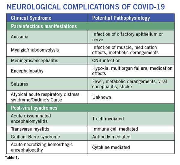
Neurological complications are rare but are being increasingly recognized9. These complications can involve the entire neuro-axis. They may occur during active viral infection and as a post-viral syndrome. (See Table 1). Some patients may present with altered mental status in the absence of respiratory or other typical COVID-19 symptoms as their sole initial presenting feature of SARS-CoV2 infection10. Anosmia is a common symptom of any upper respiratory tract infection. But anosmia with COVID-19 has received special attention. It seems to be one of the most common symptoms and often occurs in the absence of rhinorrhea. This suggests involvement of the olfactory nerve or pathway by the virus. As the majority of patients with anosmia recover their sense of smell and taste after the acute phase of the illness, the nerve endings or the cells surrounding the nerves may be affected, allowing for regeneration to occur. In a case report of a patient with sudden anosmia due to COVID-19, it was found that the olfactory clefts were inflamed, with relative sparing of the olfactory bulb11. In a mouse model of coronavirus infection, the virus can be transmitted via olfactory pathways trans-synaptically to the brain and to the brainstem12. This has raised concern about the potential long term consequences of anosmia in COVID-19. However, the mouse coronavirus uses a different receptor and hence may not replicate the human disease. Nevertheless, it is important to prospectively monitor the patients to make sure they do not develop any long-term sequelae since we know that anosmia is a recognized early symptom of neurodegenerative diseases such as Parkinson’s disease and Alzheimer’s disease.

Strokes with COVID-19

Presentation

Venous sinus thrombosis
Ischemic strokes in multiple arterial distributions
Small blood vessel occlusions
Watershed Infarcts

Pathophysiology

Coagulopathy: elevated D-dimer, PT, aPTT
Antiphospholipid antibodies
Cardioembolic
Hypoperfusion
Risk factors
Myocarditis
Known vascular risk factors
ARDS and multiorgan impairment

Table 2.

Myalgia frequently accompanies the illness. Most viral illnesses can cause body aches and pains. However, in some patients with COVID-19, the muscle aches can be quite severe. Muscle tenderness may last for several days after all other symptoms have resolved. They can involve the back muscles. A case of rhabdomyolysis13 was reported similar to what was also seen with the SARS14, although this patient was also on lopinavir/ritonavir which may have contributed to the myolysis. Since the onset of these symptoms is early in the course of the illness, it is possible that the virus invades the muscle to cause myositis, however, pathological findings have not yet been described. Importantly, these patients need proper hydration to prevent kidney damage. Also, it should be noted that potential medications used in the treatment of COVID-19 (including some protease inhibitors) may cause patients to be predisposed to muscle damage.

Meningitis and encephalitis are rare. Dull headaches are common and typically occur at the onset of the illness and resolve within a few days. They are not accompanied by any signs of meningeal irritation. However, a classical presentation of a viral meningitis has been described with COVID-19 and virus can be detected in the CSF. Encephalitis is harder to diagnose. Most patients who become comatose do so after development of ARDS and multi-organ failure, hence the CNS symptoms are attributed to hypoxia and metabolic abnormalities. Fever itself can cause delirium. However, a few cases of encephalitis where patients developed generalized seizure and coma are now being described. In one such patient from Japan, the patient had mild pleocytosis and detectable virus in the CSF. An MRI showed lesions in the temporal lobe and adjacent ventriculitis15. Few neuropathological findings have been published, but one study found low levels of SARS-CoV-2 RNA in the brain by PCR of 4 different COVID-19 patients at autopsy15a. Another case study found evidence of betacoronaviral infection of the brain with postmortem electron microscopic evaluation15b. From the earlier SARS epidemic in 2003, autopsy findings showed that the virus could be detected in the brain by multiple techniques in all patients evaluated (n=8)16. Spread of SARS-CoV-2 into the brain could involve an array of mechanisms. The virus can spread via the vasculature and enter the brain carried by infected leukocytes. Transneuronal spread has been hypothesized to also occur from the lung via the vagal nerve or from the nasal passages via the olfactory nerve.

Strokes are being increasingly recognized in this population and occur as the presenting symptom of the infection or any time during the illness. (See Table 2.) In a study from China, 5% (n=11) of 211 patients admitted with COVID-19 had acute ischemic strokes, 0.5% (n=1) had cerebral venous thrombosis, and 0.5% (n=1) had cerebral hemorrhage17. While most often these patients have underlying vascular risk factors, there are several patients where nothing other than the SARS-CoV-2 infection can be identified as a cause of the stroke. The virus is known to invade endothelial cells and can also cause a coagulopathy. Elevated D-dimer levels and increased PT and activated PTT have been described. Antiphospholipid antibodies have also been detected18. Some may develop disseminated intravascular coagulation. The virus can also cause a cardiac myositis19 which could also cause a stroke by hypoperfusion or embolism. Some patients may simultaneously develop deep vein thrombosis or vascular occlusions in other organs.

Atypical Acute Respiratory Distress Syndrome is the major cause of death in patients with COVID-19. What is atypical is that these patients have severe hypoxemia even when the lung capacity and mechanics are well preserved20. Even when the pCO2 is rising the patients are not hyperventilating and lose their respiratory drive. They develop what seems like an Ondine’s Curse. However, these patients do not have any other brainstem signs so the pathophysiology of this condition remains unclear at the present time. However, it is critical that these patients be treated with oxygen, and prone positioning also seems to help. Early ventilatory support should also be considered.

Post-viral syndromes occur when the patient is seemingly improving from the viral syndrome at about a week to three weeks after the onset of the viral prodrome. An isolated case of acute necrotizing hemorrhagic encephalopathy has been described21. This patient had bilateral thalamic lesions and other lesions in the temporal lobes which are typical of the syndrome. It is thought to be mediated by cytokine storm. A patient with transverse myelitis with quadriparesis, a sensory level and bowel and bladder involvement has been described22. However, MRI or CSF evaluation was not reported. A single case of Guillain Barre Syndrome (GBS) has been published in a patient from China23. A case series from Italy of five COVID-19 patients who developed GBS described three patients with an axonal form of GBS and two with a demyelinating process24. We recently communicated with a patient who had a sensory variant of GBS. The illness was self-limiting and did not require intervention. Acute disseminated encephalomyelitis has been recently described in an adult patient with SARS-CoV-224a; similarly, several cases have been described with the human coronavirus-OC4325 and with MERS26.

However, multiple challenges in the evaluation of patients with neurological complications exist. It is difficult to get neuroimaging when patients are acutely infected for fear of contamination of the scanners. Performing surgery or autopsies are also challenging due to the production of aerosols and lack of proper safety measures.

Therapeutic Debate

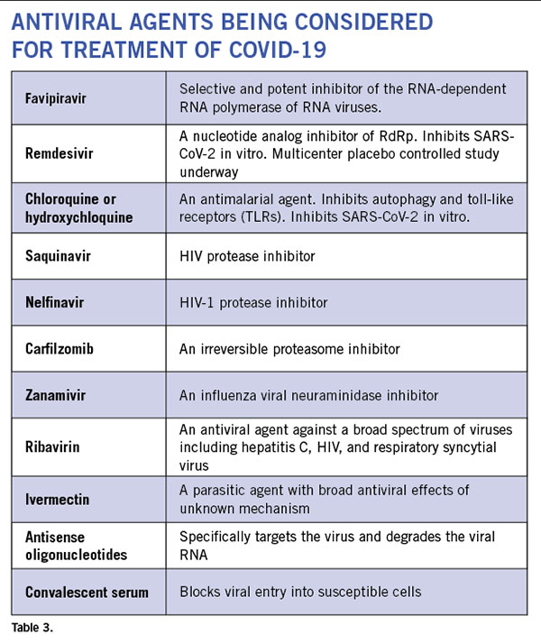
Anti-virals: Even though currently there is no proven antiviral therapy for the human coronaviruses, several drugs are being considered for clinical trials and empirical treatment of patients. (See Table 3.) There are 287 studies on coronavirus registered on www.clinicaltrials.gov. In vitro studies have shown some efficacy with chloroquine and hydroxychloroquine. These drugs cause acidification of the endosome-lysosomes and prevent viral replication. They have an anti-inflammatory effect. However, it requires pretreatment of cells prior to infection and has only a minimal effect post infection. While clinical trial results remain unpublished, these drugs have been utilized in clinic off-label in COVID-19 patients at several institutions. Well-controlled studies are necessary to know whether these drugs are efficacious against the virus. There is now a scarcity of the drug, and some countries have banned its export. Several HIV protease inhibitors have been shown to bind to the SARS-CoV-2 protease but clinical experience in small numbers of humans infected with the virus have failed to show clinical efficacy with lopinavir/ritonavir combination. Many clinical trials are currently underway that include nucleoside analogs such as remdesivir, and convalescent serum or intravenous immunoglobulin. Although the ability of most of these agents to enter the CNS is unknown, animal studies of remdesivir (GS-5734) have shown evidence of CNS penetrance, albeit at lower levels than other tissues27. Interestingly, a few drugs used to treat patients with multiple sclerosis such as teriflunomide and beta-interferons are considered to have anti-viral effects. But their effect on SARS-CoV-2 is still unknown.

Anti-inflammatory drugs: The most common cause of death is the massive immune response in the lungs leading to consolidation of the lungs with inflammatory infiltrates. Several immune suppressive medications are being used empirically. These include corticosteroids and IL-6 blockers. A case for the use of methotrexate has been made due to its broad anti-inflammatory properties and good CNS penetration.

What the Future Holds

As we continue to face the ongoing crisis, early results show reasons for optimism. In several states in the U.S., exponential growth trends have tapered. Distancing and preventive measures seem to be effective in flattening of the curve and helping institutions lower concomitant caseloads. The number of new infections and deaths are not rising as rapidly. Optimistically, we will soon face a new challenge of when and how to reopen our clinics and operating rooms. But what will this clinical environment look like? Social distancing is likely to play a role, and providers may see patients and enter any public spaces with masks on and maintain a distance of six feet from each other. Telemedicine will likely continue to play a much larger role in routine health care. A safe and effective vaccine could solve many of these issues, though development and testing of such a vaccine prior to administration to the general populace will take significant time. Another possibility is host adaptation. Most viruses are cyclical in nature. Mutations may occur that make the virus less virulent. Early signals suggest this might be the case with SARS-CoV-2. A 382 nucleotide deletion in open reading frame 8 has been identified in some circulating strains. A similar deletion also emerged in the SARS virus in 2003 that was associated with poor replication fitness28. However, until then, we will continue to see patients with COVID-19, and as neurologists we need to be vigilant for potential complications that require our attention and intervention. It is our duty to protect and advocate for the most vulnerable. •

Avindra Nath, MD, is chief of the Section of Infections of the Nervous System and Clinical Director, National Institute of Neurological Disorders and Stroke (NINDS), National Institutes of Health in Bethesda, Maryland.

B. Jeanne Billioux, MD, is staff clinician and head of the program in International Neuroinfectious Diseases within NINDS. Her research focus is on emerging infectious diseases and conducting research on the neurological consequences of infections in an International setting.

Correspondence
Avindra Nath, MD; Room 7C-103; Bldg. 10;
10 Center Drive, Bethesda, MD, 20982; U.S.
301-496-1561; e-mail: natha@ninds.nih.gov

References

1. Emanuel EJ, Persad G, Upshur R, et al. Fair Allocation of Scarce Medical Resources in the Time of Covid-19. N Engl J Med 2020.
2. Truog RD, Mitchell C, Daley GQ. The Toughest Triage – Allocating Ventilators in a Pandemic. N Engl J Med 2020.
3. Lewis D. Is the coronavirus airborne? Experts can’t agree. Nature 2020;580:175.
4. Kampf G, Todt D, Pfaender S, Steinmann E. Persistence of coronaviruses on inanimate surfaces and their inactivation with biocidal agents. J Hosp Infect 2020;104:246-251.
5. Guan WJ, Ni ZY, Hu Y, et al. Clinical Characteristics of Coronavirus Disease 2019 in China. N Engl J Med 2020.
6. Zhou F, Yu T, Du R, et al. Clinical course and risk factors for mortality of adult inpatients with COVID-19 in Wuhan, China: a retrospective cohort study. Lancet 2020;395:1054-1062.
7. Leadership AASC. Temporary Emergency Guidance to US Stroke Centers During the COVID-19 Pandemic. Stroke 2020.
8. International MGC-WG, Jacob S, Muppidi S, et al. Guidance for the management of myasthenia gravis (MG) and Lambert-Eaton myasthenic syndrome (LEMS) during the COVID-19 pandemic. J Neurol Sci 2020;412:116803.
9. Mao L, Jin H, Wang M, et al. Neurologic Manifestations of Hospitalized Patients With Coronavirus Disease 2019 in Wuhan, China. JAMA Neurol 2020.
10. Filatov A SP, Hindi F, et al. . Neurological Complications of Coronavirus Disease (COVID-19): Encephalopathy. Cureus 2020;12:e7352. doi:7310.7759/cureus.7352.
11. Eliezer M, Hautefort C, Hamel AL, et al. Sudden and Complete Olfactory Loss Function as a Possible Symptom of COVID-19. JAMA Otolaryngol Head Neck Surg 2020.
12. Dube M, Le Coupanec A, Wong AHM, Rini JM, Desforges M, Talbot PJ. Axonal Transport Enables Neuron-to-Neuron Propagation of Human Coronavirus OC43. J Virol 2018;92.
13. Jin M, Tong Q. Rhabdomyolysis as Potential Late Complication Associated with COVID-19. Emerg Infect Dis 2020; 26.
14. Tsai LK, Hsieh ST, Chang YC. Neurological manifestations in severe acute respiratory syndrome. Acta Neurol Taiwan 2005;14:113-119.
15. Moriguchi T, Harii N, Goto J, et al. A first Case of Meningitis/Encephalitis associated with SARS-Coronavirus-2. Int J Infect Dis 2020.
15a. Menter, JD; Haslbauer, R; Nienhold, S, et. al. Post-morten examination of COVID-19 patients reveals diffuse alveolar damage with severe capillary congestion and variegated findings of lungs and other organs suggesting vascular dysfunction. Histopathology. 2020 May 4.
15b. Paniz-Mondolfi A, Bryce C, Grimes Z, et al. Central Nervous System Involvement by Severe Acute Respiratory Syndrome Coronavirus -2 (SARS-CoV-2). J Med Virol. 2020 Apr 21.
16. Gu J, Gong E, Zhang B, et al. Multiple organ infection and the pathogenesis of SARS. J Exp Med 2005;202:415-424.
17. Li YW, W.; Zhou, Y.; et al., . Acute Cerebrovascular Disease Following COVID-19: A Single Center, Retrospective, Observational Study. Lancet 2020:https://ssrn.com/abstract=3550025.
18. Zhang Y, Xiao M, Zhang S, et al. Coagulopathy and Antiphospholipid Antibodies in Patients with Covid-19. N Engl J Med 2020.
19. Kim IC, Kim JY, Kim HA, Han S. COVID-19-related myocarditis in a 21-year-old female patient. Eur Heart J 2020.
20. Gattinoni L, Coppola S, Cressoni M, Busana M, Rossi S, Chiumello D. Covid-19 Does Not Lead to a “Typical” Acute Respiratory Distress Syndrome. Am J Respir Crit Care Med 2020.
21. Poyiadji N, Shahin G, Noujaim D, Stone M, Patel S, Griffith B. COVID-19-associated Acute Hemorrhagic Necrotizing Encephalopathy: CT and MRI Features. Radiology 2020:201187.
22. Zhou KH, J.; Dai, D.; Feng, Y.; Liu, L.; Nie, S. Acute myelitis after SARS-CoV-2 infection: a case report. https://wwwmedrxivorg/content/101101/2020031620035105v1fullpdf 2020.
23. Zhao H, Shen D, Zhou H, Liu J, Chen S. Guillain-Barre syndrome associated with SARS-CoV-2 infection: causality or coincidence? Lancet Neurol 2020.
24. Toscano G, Palmerini F, Ravaglia S, et al. Guillain-Barre Syndrome Associated with SARS-CoV-2. N Engl J Med 2020.
24a. Zhang T, Rodricks MB, Hirsh E. COVID-19-Associated Acute Disseminated Encephalomyelitis: A Case Report. medRxiv 2020.04.16.20068148; doi: https://doi.org/10.1101/2020.04.16.20068148
25. Yeh EA, Collins A, Cohen ME, Duffner PK, Faden H. Detection of coronavirus in the central nervous system of a child with acute disseminated encephalomyelitis. Pediatrics 2004;113:e73-76.
26. Arabi YM, Harthi A, Hussein J, et al. Severe neurologic syndrome associated with Middle East respiratory syndrome corona virus (MERS-CoV). Infection 2015;43:495-501.
27. Warren TK, Jordan R, Lo MK, et al. Therapeutic efficacy of the small molecule GS-5734 against Ebola virus in rhesus monkeys. Nature 2016;531:381-385.
28. Su YCF, Anderson, D. E., Barnaby, Y. E, et al.,. Discovery of a 382-nt deletion during the early evolution of SARS-CoV-2. Biorxiv 2020;
https://doi.org/10.1101/2020.03.11.987222.

WFN, AAN Present Invited Science Session on Infectious Disease

By John England, MD, FAAN, and Kiran Thakur

The American Academy of Neurology (AAN) held its 71st annual meeting in May in Philadelphia. During the meeting, the World Federation of Neurology (WFN) and the AAN co-sponsored a unique session on infectious disease and global health.

Left to right: John England, MD, FAAN, Kiran Thakur, MD, and Nischay Mishra, PHD. Photo courtesy of the American Academy of Neurology

The session was co-directed by John England, MD, FAAN, Richard M. Paddison professor of neurology and chair of the Department of Neurology at the Louisiana State University School of Medicine, and editor-in-chief of the Journal of the Neurological Sciences, and Kiran Thakur, the Winifred M. Pitkin assistant professor of neurology and neuroinfectious disease expert at Columbia University Irving Medical Center.

The session highlighted the joint efforts and partnership of the WFN and AAN to combat neurological diseases globally, and highlighted the work of world-renowned scientists on hot topics in infectious disease and global health. The session included presentations on the current acute encephalitis outbreaks (AES) in India, the acute flaccid myelitis (AFM) outbreaks in the United States and other global regions, the central nervous system (CNS) reservoir in HIV infection, and the newly recognized spectrum of post-infectious cases of autoimmune encephalitis.

The session’s first lecture was given by Prof. Manoj Murhekar, lead scientist and director of the National Institute of Epidemiology in Chennai, India, who spoke on acute encephalitis syndrome in Eastern Uttar Pradesh, India. His talk was followed by Dr. Nischay Mishra, a molecular biologist with expertise in advanced diagnostics and bioinformatics at the Center for Infection and Immunity at the Columbia University Irving Medical Center. He discussed molecular and serological discovery in CNS infectious diseases, highlighting his work identifying etiologies in AES outbreaks in India. Dr. Kevin Messacar and Dr. Kenneth Tyler (both at the University of Colorado School of Medicine), two major leaders of AFM investigations and members of the Center for Disease Control and Prevention (CDC) AFM taskforce, discussed features of enterovirus-associated AFM.

Audience at the invited science session. Photo courtesy of the American Academy of Neurology

Their presentations discussed human clinical features as well as an experimental mouse model, which was developed in Dr. Tyler’s laboratory. Dr. Serena Spudich, professor of neurology and division chief of infections and global neurology at Yale University School of Medicine, spoke on “Tapping into CNS reservoirs: single cell RNA sequencing of CSF in HIV.” She provided data on her recent studies using single-cell RNA sequencing, which identified a rare subset of myeloid cells that present a gene expression signature that significantly overlaps with neurodegenerative disease-associated microglia.

Prof. Josep Dalmau, director of the laboratory for the study of the pathogenesis of immune-mediated neuronal disorders at the University of Barcelona, Spain, and world authority on paraneoplastic disorders affecting the nervous system concluded the session with a lecture on viral triggers of autoimmune encephalitis. He highlighted his work in a multicenter prospective study showing that autoimmune encephalitis subsequently occurred in 27% of patients afflicted with herpes simplex encephalitis.

The session emphasized the importance of groundbreaking scientific work to combat neuroinfectious diseases globally and the importance of the WFN and AAN as organizations working together on global health initiatives. Further collaborative educational and scientific programs in infectious diseases, neuroscience, and world health will be presented at the upcoming World Congress of Neurology (WCN), which will be held Oct. 27-31 in Dubai, United Arab Emirates. •

WFN Neuroepidemiology Group Update

By Giancarlo Logroscino, MD, PhD

The Neuroepidemiology section of the World Federation of Neurology (WFN) has been particularly active in the area of teaching courses and research around the world, especially in areas where neurological research was at the starting point.

Giancarlo Logroscino, MD, PhD

The involvement of WFN members in the neurological group to the Global Burden of Disease (GBD) project has been an important asset. The GBD neurological group has been led by Valery Feigin from Auckland, New Zealand.

Two papers on the GBD of neurological diseases have been published by Lancet Neurology (Neurological Disorders Collaborator Group. Lancet Neurol. 2017 and 2019).

Lancet Neurology has launched an initiative to publish a special issue on specific neurological disorders. Disease-specific papers have been published on stroke, dementia, Parkinson disease, motor neuron diseases, and epilepsy. The activity of the GBD in the areas of the neurological disorders, even if not officially linked to WFN, have been carried out by active members of the WFN group, including Dr. Feigin, Beghi and myself.

The group has succeeded to get a small pilot grant to develop an epidemiologic course in low income countries. The first country where the WFN neuroepidemiological group decided to implement the course was Cambodia. The course with the endorsement of the WFN was held in February 2017 in Cambodia.

The overall goal was to train neurologists interested in neuroepidemiologic research and support their research activities. The chairs of the course were Giancarlo Logroscino, Italy; Pierre Marie Preux, Limoges, France; and Benoit Marin, Limoges, France.

The course was an intensive five-day course on research methods with focus on practice in low- and medium-income countries.

The course was conceived to help Cambodian neurologists to establish clinical and population-based research programs in topics of utmost interest including epilepsy, stroke, dementia, and infectious disease.

Cambodia is still experiencing a transition characterized by rapid increase both of expectation of life and incidence and prevalence of chronic diseases. In this context, neurology has a major role within medicine but has to change direction rapidly in education, clinical work, and research.

The course was held at the University of Health Sciences of Phnom Penh. It was attended by 14 out of 16 members of the Cambodian Neurological Society (eight neurologists out of nine). There was also attendance by 14 students of the School of Medicine, chosen by the faculty.

Prof. Samleng, chair of neurology in Phnom Penh, expressed the strong wish that the Cambodian Neurological Society could join the WFN, a process still ongoing.

With the same goals, we organized two courses in Albania with the endorsement of the WFN but no WFN financial support. The support was provided by a research and training grant of the University of Bari. Both courses were promoted and organized by Prof. Jera Kruja, from the University of Medicine Tirana and Prof. Giancarlo Logroscino from the University of Bari.

The first course, which was held in June 2017, focused on ALS and motor neuron diseases. The course focused on several aspects of amyotrophic lateral sclerosis: epidemiology, new classification and staging systems, biomarkers and advanced diagnostic technologies, multidisciplinary management of the disease (nutrition, pneumological assessment and supports, end of life approaches), cognition in ALS, and neuropsychological evaluation in subjects with motor impairment.

The role of hospital neurologists outside the academic centers was emphasized. The model for the care of neurodegenerative diseases patients choosing the patient home as the center of care was described. In this model, the role of the general practitioners and specialists working in the territorial unit (district) was emphasized.

Albanian and Montenegrin neurologists illustrated the available resources and the disease approach in their countries.

The second course was held in October 2017 in Albania on dementia for neuropychologists and neurologists with the participation of Thomas Bak, Edinburg, United Kingdom, chair of the aphasia and dementia group, and Prof. Raad Shakir, former WFN president. Prof. Shakir outlined the link of the public health and economic perspective in the dementias and other neurological diseases areas in the future.

The implementation of clinical care models for dementia in countries with limited resources was discussed. The changing epidemiology of dementia in the world with the general aging population has been debated with special focus on countries of the Balcanic area.

These courses were the initial framework for research for rare neurodegenerative diseases in Albania, pursuing the application of a model already established in Puglia region in Southeast Italy. A network of collaborators for the project has been established, including Albania, Kosovo, and Montenegro. An initial collection of data was started in Albania after the course.

Similar courses were held in Serbia (January 2019) and in South America, in Uruguay and Panama (August 2016 and April 2018). Both courses were primarly organized by Dr. Carlos Ketzoian from the University of Montevideo and Walter Rocca from Mayo Clinic.

I hope that the neuroepidemiological group will pursue further actions along these lines established in the next few years. It has been an honor and exceptional experience to serve for an extraordinary organization as the WFN, and the specialty group committee with interactions with recognized leaders in neurology as Prof. Shakir and Prof. Grisold.

In this perspective, Dr. Carlos Ketzoian, the new chair, has the background and the abilities to pursue the expansion of neuroepidemiology as areas of training and research all around the world with a specific emphasis in low- and medium-income countries. I will continue to serve the section with the same enthusiasm under the new leadership. •

Giancarlo Logroscino works in the Center for Neurodegenerative Diseases and the Aging Brain, Department of Basic Medicine, Center for Neuroscience and Sense Organs Department of Clinical Research in Neurology of the University of Bari at Pia Fondazione Card G. Panico Hospital Tricase (Le) University Aldo Moro Bari.


Neurology, Environment and World Brain Day

By Wolfgang Grisold, Mohammad Wasay, and Jacques Reis

The World Brain Day (WBD) was established to commemorate the foundation of the WFN, at the WCN 2013, in Vienna. The idea is to gather all member societies for a yearly event, to promote the interest of neurology.

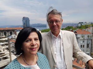
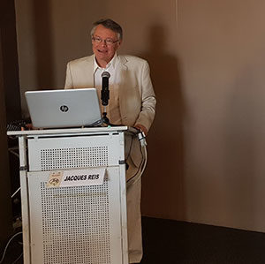
Jacques Reis, along with Serfnur Ozturk from the Turkish Neurologic Society, during World Brain Day 2018

Several topics such as stroke, dementia, and epilepsy were used in previous WBDs in cooperation with other societies.

After WBDs with topics involving epilepsy and stroke, this year’s topic was the environment. The title “Clean Air for a Healthy Brain” was chosen to signify that air pollution is a major problem relating to brain health. So far, we are aware of stroke and pollution, and there is substantial evidence that worldwide many more persons are compromised by the effects of pollution.

This WBD has brought the important message to all policy- and decision-makers around the world: Take care of our environment, notably air quality; healthy air is mandatory for our brain’s health!

The second message is for neurologists: The tremendous impact of environmental factors in the pathogenicity of many neurological diseases should not be neglected.

Presently, neurologists are not trained to manage such environmental issues. Prevention and mitigation of these environmental consequences will be important.

An important issue is prevention. For example, indoor air quality issues, such as “home fire” for cooking is still prevalent in many countries, due to lack of resources.

The great lesson, which comes from the stroke Global Burden of Disease study, shows that prevention is no longer only a personal concern but a societal challenge. Neurologists must act and advocate for a better environment.

Yet environment for many neurologists is still not as tangible as conventional neurological problems such as symptoms and diseases, and for some societies these seemed a remote topic and of less evidence than we are usually trained to think of. This may be wrong, as evidence shows that environmental factors are increasingly important for the generation of a number of diseases, including in neurology. An example is the Turkish Neurological Society, which has brought the debate to the societal level.

It has been the privilege of the WFN Applied Research Group on the environment to promote their topic, by yearly meetings, publications, and this year their effort was recognized as the topic of the WBD.

The World Brain Day team consisted of Jacques Reis, M. Wasay, Walter Struhal, and Wolfgang Grisold, with great help from Jade Levy and Laura Druce at the WFN offices in London, and we used press and promotional advice from Birgit Kofler.

This year the American Academy of Neurology (AAN) actively participated, which is a significant acknowledgement of the WBD activities, and also a sign of fruitful cooperation! We thank the AAN for their involvement.

In the past years, we have aimed to make virtual press conferences. Last year, we had a webinar to receive statements on the topic. This year, we collected a number of videos from specialists worldwide on the topic, and we hope that these interviews will be a source of ongoing information on environment and neurology.

The best immediate results of WBD are press and media echoes, as well as reports from our members on their celebration of WBD. All of this material will be collected, and will be subsequently displayed on the website to give an overall impression of this year’s WBD.

Please use our material, report on your WBD, and join us again for the next World Brain Day in 2019. The theme for 2019 World Brain Day is related to headache disorders.

RISE and the ENRG are pleased to invite you to the third meeting dedicated to the environmental impact on brain, which will be held in Strasbourg, Council of Europe Nov. 28-30, 2018. Visit asso-rise.com for more information. •


World Brain Day in Albania

By Jera Kruja

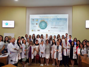
Albania, acknowledging the importance of air pollution as a risk factor for neurological disorders, organized a special event during World Brain Day. The conference was titled “Dita Botërore e Trurit: Ajër i Pastër për Tru të Shëndetshëm,” (“Clean air for brain health”).

Prof. Dr. Jera Kruja, head of the neurology service at the University Hospital Center “Mother Teresa,” advocated with the faculty of medicine at the University of Medicine, Tirana, and the Albanian Institute of Public Health to join efforts toward raising awareness on air pollution and its deleterious effects exerted on the brain. As a result, all three institutions participated actively in organizing and disseminating the information to health professionals and general public.

Besides Prof. Dr. Jera Kruja, speakers included Prof. Dr. Enver Roshi, head of the department of public health (faculty of medicine at the University of Medicine), Elida Mataj, MD, PhD, head of the environmental  epidemiology and air quality department (Institute of Public Health), Aida Quka, MD, neurologist, Neurology Service (University Hospital Center “Mother Teresa”).

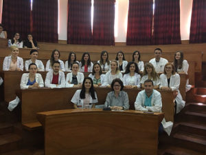
The event, organized at the faculty of medicine, was attended by health professionals and the general public, which recognized the importance of air pollution and its consequences highlighting the seriousness, gravity, and urgency of public health preventive interventions tackling environmental pollution in general and air pollution in particular.

Moreover, the topic relevance and the speakers’ excellence were appraised from the National Center of Continuous Medical Education by accrediting the event with two CME credits. Additionally, special acknowledgment was attained from the Albanian representatives of the European Industrially Contaminated Sites and Health Network.

Furthermore, the conference was reported by national television and press, and special interviews were given by the speakers addressing the general public in an effort to raise awareness on air pollution. •

Jera Kruja, MD, is a professor of neurology and head of the neurology service at the University of Medicine, Tirana, UHC Mother Teresa, Tirana, Albania, a member of the WFN Teaching Courses Committee, a former member of the European Academy of Neurology Scientific Committee, and Lancet commissioner for LMIC.

From the Editors

BY STEVEN L. LEWIS, MD, EDITOR,
AND WALTER STRUHAL, MD, CO-EDITOR

Welcome to the June issue of World Neurology. The issue begins with Prof. William Carroll’s President’s Column, where he updates us on several of the many current and upcoming activities of the WFN, which are only possible through the many valuable and multifaceted contributions of our many constituents. This issue also includes the exciting news that Vladimir Hachinski, a previous WFN president, has received the 2018 Killam Prize, the highest honor for research from the Canada Council of the Arts.

Steven L. Lewis, MD

Walter Struhal, MD

Prof. Wolfgang Grisold reports on the Austrian World Summit, which took place in May. The summit was dedicated to the environment and was organized by the Schwarzenegger Institute and the Austrian government. In this issue’s history article, Douglas Lanska provides the second part of his interview with Nobel Laureate Stanley Prusiner describing when he received the call from the Nobel Committee.

In education news, Prisca Bassole reports on the recent graduation of four trainees from the new neurophysiology fellowship in Dakar, one of the WFN-accredited teaching centers in Africa. In other education reports, three recent recipients of WFN’s Junior Traveling Fellowships provide their accounts of their travel to conferences to report on their research and to learn of cutting- edge developments in neurology.

Finally, this issue includes the statements from the candidates for the positions up for election at the upcoming Council of Delegates meeting in Berlin. For the WFN Trustee Elections, the candidates are (in alphabetical order) Morris Freeman, Alla Guekht, Jean-Marc Leger, and Daniel Truong, and for WFN Secretary General Election, the candidate is Wolfgang Grisold.

We hope you enjoy the contributions in this issue, and we continue to encourage future contributions from neurologists about neurology from around the world. We also remind all readers of the upcoming Day of the Brain on July 22, with the theme of Clean Air for Brain Health¸ and look forward to participation from neurologists and neurology societies from around the globe.•

Victor Soriano

The Soriano Lectures at the World Congresses of Neurology were named for his contributions and lasting presence

Victor Soriano was born on Feb. 8, 1909, in the Isle of Rhodes. At the time, the Isle of Rhodes was under the possession of Turkey, and soon after of Italy. Victor was the second of five siblings. His parents were Félix Soriano and Catalina Junio, both Sephardic Jews. They emigrated to Uruguay when Victor was 9 months old. This explains why Victor considered himself Turkish, Italian, and Uruguayan.

Victor Soriano

Victor Soriano

Once in Uruguay, the Soriano family settled in its capital city, Montevideo, in the historical district, where Victor chose to live the rest of his life. His father, an efficient and respected tailor, soon became popular among the Jewish community. His income quickly increased, and he gained great respect, especially after founding the first Sephardic synagogue in the city.

Young Victor was a hard-working student who in 1925 was admitted to the school of medicine, from which he graduated in 1934. During his period as a medical student, he paid special attention to Prof. Americo Ricaldoni’s teachings of clinical neurology at the Hospital Maciel. Prof. Ricaldoni was the first academic neurologist in Uruguay. Due to his efforts, the Instituto de Neurología of Montevideo was founded in 1926, having Prof. Ricaldoni as its first director.

After graduating, Victor Soriano was appointed neurologist of the Instituto de Neurología in 1935. This was a challenging time for the institute, because of the absence of a leader after Prof. Ricaldoni’s death in 1928, scarcely one year after the inauguration of the new institute.

Two years later, the authorities finally appointed Prof. Alejandro Schroeder as Ricaldoni’s successor in 1937. Prof. Schroeder started a period of reconstruction with the help of the few remaining neurologists, Victor being one of them. In 1939, Victor was promoted to assistant neurologist. Simultaneously, he was invited to teach neurology at different medical clinics in the Hospital Maciel and the Orthopedical and Traumatological Institute. In 1943, he was appointed associate professor of medicine.

In 1939, Vlctor married Clara Benzecry (Clarita, as she was known). She was his wife and his efficient secretary as well and permanently encouraged him in all his activities, both social and medical. After Victor founded the Uruguayan Committee of Friends of the Weizmann Institute of Israel, it was Clarita who organized its meetings, which were held at the Sorianos’ home. Every Tuesday evening, she hosted scientific, literary, and musical events.

In 1945, Victor traveled to the United States as a Rockefeller fellow, staying at Yale University with John Farquhar Fulton, Sterling professor of physiology, who appointed him instructor of physiology. Fulton inspired in Victor a deep and lasting admiration. In 1947, as visiting investigator of the Rockefeller Institute for Medical Research, he worked with Prof. Hiram Houston Merritt at the Montefiore Hospital of Columbia University. Since 1948, he attended with Clarita all of the meetings of the American Neurological Association (ANA). In 1950, he was named delegate for Uruguay to the Iberoamerican College of Neurologists. Victor and Clarita were regular attendees to the International Congresses of Neurology from the initial preparatory meeting in Lisbon in 1953 and the first official congress held in Brussels in 1957.

At the congress held in Rome in 1961, Earl Walker of Baltimore, Giuseppe Moruzzi of Pisa, and Victor of Montevideo paid tribute to the memory of John F. Fulton, who had died the previous year. At that meeting, Fulton’s friends and collaborators decided to organize the Fulton Society, and Victor was elected as permanent president. This society commended its president to organize a special symposium, every two years, simultaneously with the World Congress of Neurology and the ANA meeting, where selected leaders of neuroscience should give lectures on advances in nervous system research. The first of these symposia was held in Atlantic City in 1965 and continued for many decades, sponsored by the World Federation of Neurology (WFN) and the ANA and organized by Victor and Clarita.

In 1987, Victor and Clarita decided to sponsor a lectureship to be given at the ANA, after 40 years of consecutive attendance to its meetings. The lecture must be given by a member of the ANA. When asked in 1989 why they chose to sponsor a lectureship, the Sorianos answered: “When we first went to the United States in 1945, to stay with Prof. John F. Fulton and afterward with Prof. H. Houston Merritt, we enjoyed the most cordial and warm welcome everywhere, establishing lasting bonds of friendship with outstanding promising young doctors, who are now senior members of the American Neurological Association and prestigious professors. Thus, when I became a member of the American Neurological Association several decades ago, I returned since 1948 every year with the double reward of enjoying a very outstanding scientific level and seeing dear old friends. Little by little, we have regarded the ANA as a big family, to which we are united by warm affection. Sponsoring a lectureship to be given at the ANA Annual Meeting, we like to think that in future years the Sorianos will be linked to all of you, through a brilliant lecture delivered by an outstanding scientist.”

The WFN also has two related endowed lectures in every world congress of neurology. One is the Victor and Clara Soriano lecture and the second is the Fulton Symposium/Soriano lecture. At the most recent World Congress of Neurology in Kyoto in 2017, these lectures included “Grid Cells and the Medial-entorhinal Space Network” by Prof. E.I. Moser (Norway), “Defining the Future of Neurology – Japan, Asia and Oceania” by Prof. H. Mizusawa (Japan), and “Global Neurology Challenges and Way Forward” by Prof. and WFN President Raad Shakir (United Kingdom).

Victor was the author of more than 200 scientific publications in different medical journals, covering different aspects of medicine. In neurology, he preferred epilepsies, spinal cord compression, and peripheral nerve pathology. He gave lectures in several countries in America, Europe, and Asia. Many neurological societies accepted him as honorary member.

Apart from his chief interest in experimental and clinical neurology, he had many hobbies. Boxing and football were a part of his youth. He also engaged in sports journalism. In the attic of his home, he mounted an astronomic observatory provided with a powerful telescope, and he sought relaxation by watching the sky. On occasion, he invited schoolteachers and their pupils to watch the moon and the planets. During his last four decades, he devoted much of his time to medical journalism, publishing in the most important newspapers of Uruguay weekly columns on different subjects: sanitary education, innovations in medicine, historical aspects of medical practice, with the intention of popularizing and simplifying medical knowledge. He loved all kinds of music, instrumental and lyrical, but he was also an admirer of the typical music of Uruguay and Argentina and its poetry, being able to sing from beginning to end the most popular tangos.

Victor died in May 2005 and Clarita a few years later. In 2012, Soriano’s brother-in-law agreed with the National Academy of Medicine of Uruguay to sponsor the Victor and Clara Soriano Award for international research in medicine, endowed with (U.S.$)10,000. •

Stanley Prusiner on the Origin of the Term Prion

By Douglas J. Lanska, MD, MS, MSPH, FAAN

In an oral history interview for the American Academy of Neurology conducted April 27, 2017, at the Boston Convention Center, I spoke with Nobel laureate Stanley B. Prusiner, the only living neurologist to have won a Nobel Prize (Lanska and Klaffke, 2017, Lanska, 2017).

Prusiner was the sole recipient of the Nobel Prize in Physiology or Medicine in 1997 “for his discovery of Prions — a new biological principle of infection.” Here is an excerpt from that interview concerning how Prusiner created the term prion, which he introduced in a landmark paper in Science in 1982 (Prusiner 1982).

Prusiner: [In the fall of 1980,] I was with a friend of mine who was a professor at Harvard. This was [American chemist] Frank Westheimer, PhD (1912-2007). [He was receiving] an honorary professorship in the pharmacy school. He came to see me and [we] talked. I went over everything I was doing. He said, “Stan, this is really fantastic. You’ve discovered something really new, and you need to give it a name, and you need to give it a good name. You need to think about this name for a long time. A lot of work needs to go into this. Because, if you give it a crappy name, someone else will come along and give it another name, and they will end up with the lion’s share of the credit, but you will have done the work, and that’s not a good idea. This is what you’ve done with your life, and you need to make sure that you don’t screw it up. So you need to spend a lot of time on this.”

Stanley Prusiner

Stanley Prusiner

Lanska: Could you to talk about how you came up with the term?

Prusiner: OK. I’m happy to recite this… [I]t’s the spring of 1981, and I need to finish this paper [Prusiner 1982]. The only thing holding it up is the word, and I’m trying to figure out a word.
So I go through Latin dictionaries, because I knew — I still know — a lot of Latin. I’m not a scholar in it; that’s for sure. I don’t know any Greek. I don’t really know how to come up with a word. I want a word like exon. I thought that’s a great word. Where do I find somebody who can do that? I kept thinking of somebody at Berkeley [who] can help me, but then I didn’t even know who to talk to. I thought this is going to be just frivolous to go to Berkeley and try to find some professor of words who will help me.
Then, I said, “OK, I’ve got to come up with some rational approach to a word, just taking a bunch of letters, and where are these letters going to come from? Well, they’re going to come from words that have something to do … [with the responsible agent].” So I wrote out [the words] protein, infectious, and agent. I started with that. I got piaf out of that, because I wanted protein, and I wanted infectious, and then I wanted agent.

Quark: What’s in a Name?

Prusiner is certainly not alone in devoting considerable energy to devising a catchy word for a newly described entity in science or medicine, and indeed he developed exemplars based on prior scientific neologisms. Later, in the public interview, Prusiner recalled:

My model words [for prion] were virus and quark [pronounced correctly as qwork]. I thought Murray Gell-Mann was terrific with that, stealing that from Lewis Carroll [sic]. So I looked through Alice in Wonderland for another one, but I didn’t find one. (Lanska, 2017)

American physicist Murray Gell-Mann (1929-) received the 1969 Nobel Prize in physics “for his contributions and discoveries concerning the classification of elementary particles and their interactions.”

Gell-Mann coined the term quark (which he pronounced kwork) in 1963 to refer to the fundamental constituents of the nucleon (i.e., either a proton or a neutron, considered in its role as a component of an atomic nucleus) (Gell-Mann, 1964, 1995). Despite Prusiner’s recollection to the contrary, the term quark did not originate in the fantasy novel Alice’s Adventures in Wonderland (1865), written by English mathematician Charles Lutwidge Dodgson (1832-1898) under the pseudonym Lewis Carroll. Instead, Gell-Mann derived the non-phonetic spelling from a whimsical poem in Finnegans Wake (1939) by Irish writer James Joyce (1882-1941).

According to Gell-Man’s account (1995): In 1963, when I assigned the name “quark” to the fundamental constituents of the nucleon, I had the sound first, without the spelling, which could have been “kwork.” Then, in one of my occasional perusals of Finnegans Wake, I came across the word “quark” in the phrase, “Three quarks for Muster Mark.” Since “quark” (meaning, for one thing, the cry of the gull) was clearly intended to rhyme with “Mark,” as well as “bark” and other such words, I had to find an excuse to pronounce it as “kwork.” But the book represents the dream of a publican named Humphrey Chimpden Earwicker. Words in the text are typically drawn from several sources at once, like the “portmanteau” words in Through the Looking-Glass [and What Alice Found There (1871), the novel by Lewis Carroll]. From time to time, phrases occur in the book that are partially determined by calls for drinks at the bar. I argued, therefore, that perhaps one of the multiple sources of the cry, “Three quarks for Muster Mark” might be “Three quarts for Mister Mark,” in which case the pronunciation “kwork” would not be totally unjustified. In any case, the number three fitted perfectly the way quarks occur in nature. (Gell-Man, 1995, p. 180)

Joyce’s line struck Gell-Mann as particularly appropriate, because the hypothetical elementary particles combined in groups of three to form baryons, such as protons and neutrons.

Gell-Mann adopted Joyce’s spelling for his “quork,” even though Joyce clearly intended quark to rhyme with Mark.

An equivalent model to Gell-Mann’s quark model was independently proposed by Russian-American physicist George Zweig (1937-) in 1964 (Zweig 1964a,b), the same year as Gell-Mann’s model, but Zweig did not propose a similarly catchy term and ultimately Zweig did not share the 1969 Nobel Prize in Physics.
Both Prusiner and Gell-Mann described or proposed new entities that warranted new scientific nomenclature. Both saw the new names as important factors in establishing and cementing their ideas in the “scientific marketplace” (anonymous 2012), and consequently both devoted considerable time and energy to the effort of inventing a term that was short, catchy, and in some way clearly novel, but that still could be justified in a plausible way (Gell-Mann 1995; Prusiner, 2014; Lanska and Klaffke 2017; Lanska 2017). To emphasize the novelty of their terms, both chose idiosyncratic non-phonetic pronunciations and repeatedly emphasized the “correct” pronunciation in subsequent discourse.

If Prusiner’s and Gell-Mann’s later experiences are any guide, Westheimer’s advice to Prusiner was certainly prescient: The names of newly described scientific entities do matter in establishing and maintaining scientific turf. Although Lewis Carroll’s Alice character was not the source of either neologism, she might still have had something to say about this if she had been consulted: “Curiouser and curiouser!” •

Lanska: You just kind of threw on an extra “f” for fun?

Prusiner: For infectious.

Lanska: Protein, infectious, agent…p-i-a. You added an “f.”

Prusiner: So you just underline the “f” in infectious, right? That’s where the letters all come from.
I always liked [French cabaret singer and songwriter] Édith Piaf [1915-1963; nee Édith Giovanna Gassion; she adopted her stage name, Piaf, from her nickname, which is French slang for sparrow]. Now, “sparrow” is announcing all of this, right? [Prusiner laughs.]
I then sent it to a friend of mine named Sidney Udenfriend (1918-2001), an American biochemist and pharmacologist who was running the Roche Institute in Molecular Biology. This was a fabulous place that was created by Roche to do basic science that would create drugs. Eventually, they shut it all down, because they never got one drug out of spending hundreds of millions of dollars over 20 years. He reads the paper, and he says, “Stan, this is an American discovery, not a French discovery. You don’t need a French word. You need another word. Go find another word.” So that was the end of piaf.
Short. You need something short. You need to have two vowels. Great words are words like virus. That’s a fabulous word. And quark [pronounced kwork] is a great word. Those are words that I think are just A+.
So I then throw out agent, because I don’t need agent. That’s totally non-specific, and I’m left with protein and infectious. Whenever I would go to a lecture, I would write out infectious across the top and protein on the side, or vice versa. Then, I’d just pick letters randomly. I didn’t get anywhere until one day. I’d probably stumbled across the same word 10 times and never picking it out: p-r-i-o-n. I read it, and I say, “This is prion.” I could have pronounced it pry-on, but I pronounced it pree-on. Then, I said, “I’m going to write p-r-e-e-o-n as the pronunciation in parentheses.”
I leave this defunct faculty club that was now a sandwich shop across the street, and I walk upstairs. I look in my Webster’s Unabridged Dictionary, because there are no computers. I find a bird with a sawtooth beak.

Lanska: A whalebird.

Prusiner: Right. And I said, “Well, you know, this really doesn’t matter. Lots of words have more than one meaning. There’ll be two pronunciations. If I’m right, this will be the No.1 definition, and the bird will continue to live on in oblivion, because no one is interested in prions [pry-ons]. I’ve never heard of a prion [pry-on]. It’s a petrel that lives in the south, the Southern Ocean [Antarctic Ocean].”
It’s not worth worrying about the bird. I said, “There’ll be a little crap from some of my competitors who will say, ‘That’s a bird.’” But I said, “It doesn’t matter.” So that’s where it came from. It had ion, so it looked like it was something highly basic. An ion really gets right down to the essence. And it was short. It had two syllables. It was going to be OK.

Lanska: You wrote in your book, though, that one of the reviewers of your paper objected, that the name had unfortunate echoes of the author’s name, Prusiner ions (Prusiner 2014, pp. 90-91).

Prusiner: Yes. That was pretty clever, [but] there was so much vitriol in the rest of the review.

Lanska: Now, of course, proteinaceous infectious gets you proin rather than prion…

Prusiner: Right, which is like loin.

Lanska: Which has kind of a funny sound to it, of course, no matter how you pronounce it. So you made another, little flexible change in that, just to give it a more catchy flavor, I think. Is that fair?

Prusiner: Yes. Proin is not a good word.

Lanska: But proteinaceous infectious as an acronym would get you that, right?

Prusiner: Yes.

Lanska: A lot of people didn’t like the term.

Prusiner: Yes.

Lanska: There was a lot of backlash. Why do you think there was so much animosity to that term?

Prusiner: If you read what other people have said, they didn’t like it, because essentially I took over the field by putting my imprimatur on the field prematurely, because I didn’t have enough information to come up with a word. That really is what I should have done was call it a Gibbs or a Gajdusek, [after NIH researchers C. Joseph Gibbs, PhD (1924-2001) and D. Carleton Gajdusek, MD (1923-2008), who first demonstrated the experimental transmission of kuru and Creutzfeldt-Jakob disease to chimpanzees in the period from 1966-1968 (Gajdusek et al, 1966, 1968; Gibbs et al 1967)] or a Gajdusek and a Gibbs, or a Gibbs and a Gajdusek, or something like that, or a Dickinson [after British veterinary geneticist and scrapie researcher Alan Dickinson, PhD (1930-2017)]. Of course, this was nonsense from my point of view.
I did exactly what Frank Westheimer told me to do. The result was exactly what Westheimer wanted to have happen, which was to get a good word and not have somebody else take over. They had a lot of time. I was the new guy on the block. But they never could figure out anything about what was truly going on. They didn’t have a way of doing something, or they weren’t clever enough, or they weren’t sufficient showmen to figure out where all of this was going to go. But Westheimer saw it and called it completely.

Lanska: I think they were stuck in one little framework.

Prusiner: Yes.

Lanska: And they struggled to keep fiddling with “unconventional virus” [or] “slow virus.”

Prusiner: They had their own words, that’s true, but they were really dumb. “Unconventional virus” is as dumb as it gets.
There’s that great quote that’s in [my memoir] from [scrapie researcher] Iain Pattison [of the British Agricultural Research Council’s Compton Research Institute], in which he says:

“The fourth decade of my association with scrapie ended in 1978, with the causal agent still obscure, and virologists as adamant as ever that theirs was the only worthwhile point of view. To explain findings that did not fit with a virus hypothesis, they rechristened the causal agent an ‘unconventional virus.’ Use of this ingenious cover-up made ‘virus’ meaningless — for is not a cottage an unconventional castle?” (Pattison, 1988) •

References:

  • Anonymous. The scientific marketplace. Nature Materials 2012;11:259.
  • Anonymous. What Does ‘Quark’ Have to Do with Finnegans Wake? How a word used by James Joyce became the name for an elementary particle of matter. Merriam-Webster, 2017. https://www.merriam-webster.com/words-at-play/quark [Accessed 12-12-17].
  • Gajdusek DC, Gibbs CJ, Alpers M. Experimental transmission of a Kuru-like syndrome to chimpanzees. Nature. 1966;209(5025):794-796.
  • Gajdusek DC, Gibbs CJ Jr, Alpers M. Transmission and passage of experimental “kuru” to chimpanzees. Science. 1967;155(3759):212-214.
  • Gell-Mann M. A schematic model of baryons and mesons. Physics Letters. 1964;8(3):214-215.
  • Gell-Mann M. The Quark and the Jaguar: Adventures in the Simple and the Complex. New York: Henry Holt and Co., 1995:180
  • Gibbs CJ Jr, Gajdusek DC, Asher DM, Alpers MP, Beck E, Daniel PM, Matthews WB. Creutzfeldt-Jakob disease (spongiform encephalopathy): transmission to the chimpanzee. Science. 1968;161(3839):388-389.
  • Lanska DJ, Klaffke L. Stanley Prusiner, MD, FAAN. American Academy of Neurology Oral History Archive Private Interview. 69th Annual meeting of the American Academy of Neurology, Boston, Massachusetts; April 27, 2017.
  • Lanska DJ. Stanley Prusiner, MD, FAAN. American Academy of Neurology Oral History Archive Public Interview. 69th Annual meeting of the American Academy of Neurology, Boston, Massachusetts; April 27, 2017.
  • Pattison IH. Fifty years with scrapie: a personal reminiscence. Vet Rec 1988;123:661-666.
  • Prusiner SB. Novel proteinaceous infectious particles cause scrapie. Science 1982;216:136-144.
  • Prusiner SB. Madness and Memory: The Discovery of Prions—A New Biological Principle of Disease. Yale University Press, 2014.
  • Zweig G. An SU(3) model for strong interaction symmetry and its breaking. (CERN Report No.8182/TH.401. 1964a.
  • Zweig G. An SU(3) model for strong interaction symmetry and its breaking: II. CERN Report No.8419/TH.412. 1964b.

    • Douglas S. Lanska is associate chief of staff for education at the VA Medical Center, in Tomah, Wisconsin. He is also professor of neurology at the University of Wisconsin School of Medicine and Public Health in Madison, Wisconsin, and professor of psychiatry at the Medical College of Wisconsin in Milwaukee, Wisconsion and the chair of the History and Archives Committee of the American Academy of Neurology.