Austrian Day of the Brain Sheds Light on Neurology Issues

By Wolfgang Grisold, MD

Wolfgang Grisold, MD, (from left) WFN secretary general, Elisabeth Fertl, MD, OEGN president; and Reinhold Schmidt, MD, OEGN past president, represent the OEGN during the World Brain Day 2016 press conference.

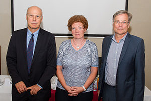
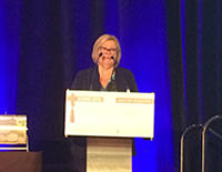
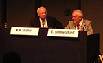
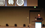
Wolfgang Grisold, MD, (from left) WFN secretary general, Elisabeth Fertl, MD, OEGN president, and Reinhold Schmidt, MD, OEGN past president, represent the OEGN during the World Brain Day 2016 press conference.

The Austrian Society of Neurology (OEGN) participated in the third Day of the Brain July 22, 2016, by discussing important topics at a press conference. The OEGN was represented by Elisabeth Fertl, MD, OEGN president, Reinhold Schmidt, MD, OEGN past president, and Wolfgang Grisold, MD, World Federation of Neurology (WFN) secretary general.

In Europe, 220 million people suffer from one or several neurological diseases. This is important because with increasing age, individuals experience disorders such as dementia, Parkinson’s disease, and stroke more frequently.

Austrian neurology, represented by the OEGN, has a strong presence with 970 neurologists, including previous neuropsychiatrists. Within Austria, there is a network of 38 acute departments with stroke units, a growing number of neurological rehabilitation centers, and other neurological centers devoted to care and rehabilitation. All stroke units are connected in a nationwide data and quality assurance system, and they have incorporated intravenous thrombolysis in their procedures. In 11 stroke units, interventional procedures can be performed for patients with acute stroke.

Dr. Schmidt gave an overview on dementia, emphasizing that efforts are needed to maintain cognitive abilities with age and in aged persons. In particular, in 75- and 80-year-old individuals, more risk for cognitive changes can be expected. The aim and strategy must be to identify the factors that positively enhance cognitive reserve. Increasingly, activity training of cognitive abilities and maintenance of social networks have been identified as positive factors.

Age is often identified as a burden (e.g., related to the burden of disease)and a contributor to increasing health care costs. This is, however, a naturally occurring phase in the life cycle with increasing needs. It is the duty of neurologists to act as advocates for our patients, to see the importance of these neurological needs for our patients, and to avoid classifying age as a burden.

Another important task in neurology is palliative care. Several neurological diseases need palliative settings of different types and time courses. For example, palliative care can be a long-term project in patients with degenerative diseases or a shorter time course in amyotrophic lateral sclerosis and brain tumors. Palliative care is not the same as end-of-life care, and it is important to include these concepts into neurology worldwide.

Worldwide, the WFN is engaged in intensive cooperation with the World Health Organization, including with ICD 11, noncommunicable diseases, and the present state of the Zika infection, where the WFN has created an international expert group.

The Austrian society had a strong echo in the media after the press conference, including several follow-up radio interviews.

BOOK REVIEW
Landmark Papers in Neurology

By Carrie Grouse, MD

Carrie Grouse, MD

Carrie Grouse, MD

In reading Landmark Papers in Neurology, the famous quote by Sir Isaac Newton, “If I have seen further, it is by standing on the shoulders of giants” comes to mind as an appropriate aphorism. The sheer scale of progress made in our understanding of neurologic pathophysiology and treatment over the last few centuries, through advancements in the fields of anatomy, molecular biology, genetics, and neuroimaging, is made evident throughout this collection. Moreover, in this book, we are shown the indefatigable efforts of the scientists and clinicians, who, by a combination of tireless effort, excellent scientific method, and at times pure serendipity, managed to make these discoveries.

Divided into 16 sections encompassing many major areas of neuroscience, the contributor or contributors for each section selected 10 important papers in each field that were particularly crucial in transforming and advancing understanding and making possible further breakthroughs. The articles themselves are not included in the volume; rather they are succinctly summarized, with the bulk of attention being paid to the scientific, social, and, at times, political context in which these papers arose, as well as the impact the papers had on their respective fields.

Landmark Papers in Neurology Book CoverAlthough it is generally understood that all medical specialties constantly are growing and evolving with new scientific and technological advancements, this collection shows just how dramatically neurologic paradigms have been transformed completely through these landmark discoveries. Given the wide breadth of immunomodulatory therapies for multiple sclerosis, it is surprising to think that the first articles investigating the possibility of the use of interferon therapy in multiple sclerosis was only in the early 1980s, and treatment with interferons was only approved in the early 1990s. It seems equally surprising that in such a prevalent disease as epilepsy, so little was known about its cause until the first article describing electroencephalography in 1929, particularly given the dramatic advancements in the field of epilepsy since then.

It was fascinating to follow the progression of knowledge in the area of Parkinson’s disease, from a humble treatise written by a relatively obscure apothecary about the “shaking palsy,” to the discovery of L-dopa as a revolutionary treatment, and more recently to the development of deep brain stimulation. Equally captivating was the recounting by Peter Goadsby, MBBS, of the circuitous path that was taken toward understanding the mechanisms behind migraine, and the substantial setback to the field that was caused by the “vascular dogma” proposed in 1938, which took 50 years to fully reverse.

Some of the most compelling aspects of this collection are the details included about the social, political, and even personal backdrops in which these discoveries were made. The seminal work performed by Alan Hodgkin and Andrew Huxley in elucidating the molecular basis of neurotransmission through their experiments on giant squid axons was almost abandoned, first due to being forced to work on radar development in World War II, then due to lack of ability to successfully catch squid for their experiments. The first description of an epileptic syndrome, West Syndrome, was in a letter to the editor of The Lancet by Dr. William James West, regarding a syndrome of unusual flexor spasms along with developmental regression that he noted in his own son. I was surprised to learn that Dr. Mary Walker, a female house physician at St. Alfege’s Hospital in London, was the first physician to treat a myasthenic patient with a cholinesterase inhibitor (physostigmine) after hearing that myasthenia gravis resembled curare poisoning, which had been found to be reversed with this agent. The political maneuvering and sabotage between competing scientists, hospitals, and laboratories, and the dramatic effect this had on what was published and how quickly new ideas were embraced, was also touched on throughout this collection, and made for fascinating reading. I often found myself regretting that each article was only afforded a few pages, such that these anecdotes were cut short.

Although a relatively minor issue, it seemed that the format of 10 articles per section was a little too strictly adhered to; some topics, such as neuroimmunology or mitochondrial diseases, could have been well covered if limited to only a few of the more seminal articles, whereas in the fields of stroke and dementia it seemed that 10 articles was impossibly limiting. Overall, however, the contributors and editors did a marvelous job with the daunting task of distilling a rich body of literature spanning multiple centuries. This book is well worth the read, as an excellent and inspiring review of the huge strides that have been made in the field of neurology in the last few centuries. It is intriguing to think of how much farther we will see in the future, thanks to the continued hard work, ingenuity, and serendipity of clinicians and scientists yet to come.

Carrie Grouse, MD, is an assistant professor of neurological sciences at Rush University Medical Center, Chicago, Illinois.

Neurology in 2050: Are You the Next Jules Verne?

The Turkish Neurological Society calls for medical student essay submissions

Serefnur Öztürk, MD

Serefnur Öztürk, MD

By Serefnur Öztürk, MD

Our century has become the Brain Century, and it is important to keep improving on this trend.

The Turkish Neurological Society has established an international essay competition for medical students to increase awareness about improvements in neurology in the Brain Century.

Medical education has various aspects, and creativity is one of the essential skills in medical education. An essay may improve oneself in various ways, including free learning, data collection, assessment of current information, improved decision-making, writing, planning, communication, and sharing.

International_Essay_Poster
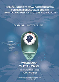
We believe we have highly talented young physicians who have the potential to become the Jules Vernes of today. We invite you to participate in our essay contest and to contribute to the future of neurology.

The subject of the contest is Neurology in Year 2050. The purpose is to raise awareness among medical students in order to attract attention to the vision of neurology in their curriculum, to raise awareness about neurology and neurological disorders in the public through the eyes of medical students, and to encourage students to study neurology as a specialty.

Our target contestants are medical students in Turkey and other countries (all World Federation of Neurology member countries).

To submit an application and access the rules, visit the World Federation of Neurology essay contest website or the Turkish Neurological Society essay contest website.

The application period is June 15 to Oct. 15, 2016. Winners will be announced Nov. 15, 2016. We look forward to receiving your creative essays. Good luck.

Serefnur Öztürk, MD, is president of Turkish Neurological Society, on the faculty of the department of neurology at Selcuk University, Konya, Turkey, and on staff at the Turkish Neurological Society.

HISTORY COLUMN
Introducing the Ophthalmoscope to British Neurologists

by Catherine E. Storey, MBBS, FRACP, MSc

Clockwise from left: May's ophthalmoscope (1935), Heine's ophthalmoscope (1995), and Morton's ophthalmoscope (1910).

Clockwise from left: May’s ophthalmoscope (1935), Heine’s ophthalmoscope (1995), and Morton’s ophthalmoscope (1910).

Hermann von Helmholtz (1821-1924), a German scientist, published the description of the first clinical ophthalmoscope in 1851. He called his invention the Augenspiegel or eye-mirror. Within a few years of his report, the ophthalmoscope became universally popular with eye specialists, many of whom attempted to make modifications and adaptations to improve the optics, illumination, and retinal image.

In Britain, one of the leading ophthalmologists of the day, Thomas Wharton Jones (1808-1891) briefly mentioned the Helmholtz ophthalmoscope in the second edition of his text, The Principles and Practice of Ophthalmic Medicine and Surgery, published in 1855. However, some 10 years later, in the third edition (1865), not only did he devote a chapter to the subject, with comprehensive illustrations to instruct on the use of the ophthalmoscope, but he hinted at the possibility of a British forerunner of the invention.

From top: Morton's ophthalmoscope (1910), May's ophthalmoscope (1935), and Heine's ophthalmoscope (1995).

From top: Morton’s ophthalmoscope (1910), May’s ophthalmoscope (1935), and Heine’s ophthalmoscope (1995).

Jones explained that he was shown a model “contrived for looking into the interior of the eye” by Charles Babbage (1791-1871), the English mathematician: “Here I ought not to omit stating that in the spring of the year 1847, Mr. Babbage showed me the model of an instrument which he had contrived for looking into the interior of the eye. The reflector was a small, plain glass mirror, with a part of the silvering rubbed off to look through.”

However, he failed to see any benefit, told Babbage that it was worthless, and, as a consequence, Babbage abandoned his prototype!

British ophthalmic surgeons embraced this new technology. A report from the Royal Ophthalmic Hospital published in The Lancet in 1855 described the contemporary position.

“It may appear at first to be a mere plaything, as did the stethoscope or microscope; but it is destined to play a very important part in the interpretation of eye cases before long. This very useful instrument continues to be more and more used and valued every year.”

If the British ophthalmologists were quick to employ the ophthalmoscope, how then was this device introduced to British (and Commonwealth) neurologists?

Three British neurologists promoted the value of the ophthalmoscope in clinical neurology:

Thomas Clifford Allbutt (1836-1925)
Thomas Clifford Allbutt (later Sir Thomas) was a well-known physician at the Leeds General Infirmary from 1861 to 1889. Although he published case reports on the value of the ophthalmoscope in medical practice from 1868, it was his text, On the Use of the Ophthalmoscope in Diseases of the Nervous System and of the Kidneys; Also in Certain Other General Disorders, published in 1871, that influenced other physicians, including William Richard Gowers. Allbutt commented in his 1868 text that “… the number of physicians, who are working with the ophthalmoscope in England may, I believe, be counted upon the fingers of one hand … it would seem that the same reproach cannot attach to our Continental neighbours … .”

John Hughlings Jackson (1835-1911)
Jackson joined the staff of the Royal London Ophthalmic Hospital (Moorfields) as early as 1859 and to the National Hospital for the Paralyzed and Epileptic, Queen Square in 1862. Jackson later reported that he learned to use the ophthalmoscope at Moorfields in 1863 and from that time urged his readers to be aware of the benefits of the instrument. He frequently claimed that it was impossible to investigate neurological disorders without using the ophthalmoscope. He drew attention to cases of optic neuritis (as papilledema was then known) when florid signs could be seen even in the presence of preserved visual acuity. He was adamant that the ophthalmic examination should never be omitted with “severe and continued headache.” Although Jackson is well known for many different facets of neurological practice, between 1877 and 1889, he devoted many of his invited lectures to medical ophthalmoscopy.

William Richard Gowers (1845-1915)
Gowers (later Sir William) was appointed to the National Hospital for the Paralyzed and Epileptic, Queen Square in 1870, and a contemporary of Jackson. He too published numerous individual case reports in which the use of the ophthalmoscope was paramount. Although best known for his Manuals, published in 1886 and 1888, Gowers’ text on medical ophthalmoscopy, A Manual and Atlas of Medical Ophthalmoscopy published in 1879, was perhaps equally influential in its time. Gowers, with his superb clinical acumen and extraordinary artistic ability, delivered a valuable resource for neurologists and other physicians, and in doing so promoted the incorporation of the ophthalmoscope into a routine neurological examination.

Commonly Used Ophthalmoscopes
Andrew Stanford Morton (1848-1927), an ophthalmologist at University College, London, and Moorfields Eye Hospital, published a small pamphlet in 1884 in which he introduced his new “improved student’s ophthalmoscope.” Although his device still relied on an external source of illumination, this small compact model now incorporated a series of 29 separate lenses propelled by a driving wheel. This mechanism provided a much improved acuity and ease of use. The Morton ophthalmoscope became one of the most popular models for the next 40 years.

Charles May (1861-1943), an ophthalmologist working in New York, is best known for the Manual of Diseases of the Eye, published in 1900. The book became a classic text and atlas of ophthalmology, going through 23 editions between 1900 and 1963. The May ophthalmoscope, with an internal illumination source, similarly went through many editions and proved to be popular in routine practice.

Catherine E. Storey, MBBS, FRACP, MSc, is a clinical associate professor at the University of Sydney School of Medicine, Australia. Peter J. Koehler, MD, PhD, FAAN, is the editor of this history column. He is neurologist at Zuyderland Medical Centre, Heerlen, the Netherlands. Visit his website at http://www.neurohistory.nl/.

Albania Celebrates the Day of the Brain

By Jera Kruja, MD

Participants gather during World Brain Day.

Participants gather during World Brain Day.

Prof. Jera Kruja gives a presentation on WFN guidelines at World Brain Day in Tirana, Albania.

Prof. Jera Kruja gives a presentation on WFN guidelines at World Brain Day in Tirana, Albania.

The Albanian Chapter of World Federation of Neurology (WFN) has celebrated World Brain Day annually since 2014. In 2016, World Brain Day was dedicated to the brain and aging and had a special meaning looking at the population figures from Albanian INSTAT.

In 2001, the population of Albania was 3,060,173, and in 2015, the population decreased to 2,889,167. However, the rate of persons over 65 years of age increased from 232,339 in 2001 to 597,420 in 2015.

We organized a meeting with the participation of neurologists from Tirana’s main public and private hospitals and outpatient clinics, delegates from the department of family medicine, and the Alzheimer Disease Association. Also, an article was published in the medical online journal, Doctor33. With these modest activities, in perfect accord to the WFN activities, we aimed to increase the attention of the society and government to the aging of the population and the associated high risk of neurological morbidity and social problems.

Jera Kruja, MD, is a professor of neurology and head of the neurology service at the University of Medicine, Tirana, UHC Mother Teresa, Tirana, Albania, a member of the WFN Teaching Courses Committee, and a member of the European Academy of Neurology Scientific Committee.

Report of the ICNMD Congress

By Wolfgang Grisold, MD

Toronto, Canada, was the backdrop for the 14th International Congress on Neuromuscular Disease in 2016.

Toronto, Canada, was the backdrop for the 14th International Congress on Neuromuscular Disease in 2016.

The 14th International Congress on Neuromuscular Disease (ICNMD), which took place July 5-9, 2016, in Toronto, Canada, brought 750 delegates.

Vera Bril, MD, organized the Congress on behalf of the World Federation of Neurology (WFN) neuromuscular research group.

This ICNMD Congress covered the broad spectrum of neuromuscular diseases and aimed to fill the gap between new developments in research, in particular genetics and immunology, and patient assessment and care. The aim was to include the spectrum of neuromuscular diseases and focus on practical issues and emerging therapies.

Prof. Vera Bril, Toronto, president of the ICNMD Congress.

Prof. Vera Bril, Toronto, president of the ICNMD Congress.

The plenary session topics focused on new genetic avenues in muscle disease, new treatment strategies in neuromuscular disease, treatment of inflammatory neuropathies, therapy in muscle disease, motor neuron disease, advances in myasthenia, and late-breaking news on the neuromuscular complications of Zika infections.

The scientific sessions were preceded by a rich selection of teaching courses, also with a wide variety of topics, such as motor neuron diseases, neuropathies, neuromuscular transmission disorders, and muscle diseases.

Prof. John England, MD, chair of the WFN neuromuscular research group, (left) and Dr. Nascimento, Brazil, at the special lecture on Zika.

Prof. John England, MD, chair of the WFN neuromuscular research group, (left) and Dr. Nascimento, Brazil, at the special lecture on Zika.

In the afternoon, a choice of workshops allowed participants to discuss diseases and other issues in small groups in more detail. The poster sessions were a site of lively discussion, and many new projects and ideas appeared.

The meeting fulfilled several important tasks, such as:

  • Giving a broad overview on the different types of neuromuscular disease
  • Adding late news and scientific knowledge
  • Having a wide educational and CME scope
  • Being a site of discussion and networking where delegates felt welcome

For the next site of the ICNMD 2018, Vienna, Austria, was elected, with Wolfgang Grisold, MD, WFN secretary general, as the Congress president. Welcome to Vienna in 2018.

Review the 2016 ICNMD Abstracts
 
View the abstracts of the 2016 International Congress on Neuromuscular Disease Congress, which were published in the Journal of Neuromuscular Diseases (Volume 3, Supplement 1, pages S3-215).

EBN, EAN Hold Joint European Board Examination in Neurology

By Wolfgang Grisold, MD

Faculty provide students with instructions for the written exam.

Faculty provide students with instructions for the written exam.

The European Union of Medical Specialists (UEMS), as the European representation of medical specialists, encourages the use of a European board examination in neurology. The exam aims to test knowledge, problem-solving, presentation abilities, and handling knowledge and aspects of ethics and public health. These European board examinations are developing well and are replacing national examinations in several disciplines and countries. The European Board of Neurology (EBN) held its Eighth European Board Examination jointly with the European Academy of Neurology (EAN) in Copenhagen, Denmark.

The UEMS/EBN/EAN board exam in neurology took place May 27, 2016, at the EAN Congress. Seventy-two candidates from several European and non-European countries participated. The exam comprised multiple-choice questions (MCQs) in open- and closed-book fashion and was followed by a presentation of a short essay on a neurology, public health, or ethics topic and a critical appraisal of a neurological topic (CAT). The language of the examination was English.

The board examination in neurology faculty gather for the UEMS/EBN/EAN board exam.

The faculty for the board examination in neurology gathers for the UEMS/EBN/EAN board exam.

Although the largest part of the examination was the MCQs (80 percent), the oral interviews for the short essays and CAT allowed more personal contact with the candidates.

The examiners took great care to listen, evaluate, and appraise the short essays and critical appraisals (right). These exam items were prepared in advance, and interesting topics relating to current problems, therapies, and procedures in neurology were discussed. Teaching materials, such as ebrain, EFNS/EAN guidelines, and textbooks were suggested for preparation.

The examiners and faculty were involved in the prior preparation of the presentations. As they were from many regions of Europe and North Africa, several authentic languages were represented, to help in addressing communication issues during the presentation.

The successful candidates and faculty join together for a final photo after the examination.

The successful candidates and faculty join together for a final photo after the examination.

The number of participants is steadily increasing, and also the number of non-European candidates is rising, which makes the UEMS/EAN board examination an attractive examination.

The next UEMS/EAN board examination will be in June 2017 during the EAN meeting in Amsterdam. The date is not fixed, but visit the UEMS/EBN website for technical details and announcements.

The WFN was invited to observe, and, as WFN secretary general, I took the opportunity to observe and make this report.

Some of the CAT Topics (Examples)

  • NMDA Receptor Antibodies in CSF and Serum
  • rTMS for Refractory Focal Epilepsy
  • Cervical Trauma and MS
  • Rituximab for Optic Neuritis
  • Tetrabenazine for the Treatment of Chorea
  • Sphenopalatine Ganglion in Cluster Headache
  • Antiviral Therapy for GBS

Some of the Essay Topics (Examples)

  • Prolonged Ventilation in ALS
  • Malnutrition in India
  • Treatment Inequalities for MS in Europe
  • Vaccination Against Meningitis During Hajj in Saudi Arabia
  • Taking Presents From industry
  • Epilepsy and Driving
  • Dealing With Medical Errors
  • A Care Driver With Epilepsy Forbidding You to Make Known His Diagnosis

EAN Breaking News Session Tackles Zika Virus

By Steven L. Lewis, MD

1

On May 29, 2016, Zika virus infections of the nervous system were the topic of a very well-attended breaking news session co-moderated by Drs. Eric Schmutzhardt and Raad Shakir (1) at the 2nd Annual European Academy of Neurology (EAN) Congress in Copenhagen, Denmark.

2

The session included talks by Dr. Shakir (2), president of the World Federation of Neurology (WFN), who spoke on behalf of Dr. John England, chair of the WFN Zika Committee, about Zika virus and its implications for world neurology.


3

Dr. John Hiscott (3), of the Istituto Pasteur-Fondazione Cenci Bolognetti in Rome, Italy, spoke about what is now known about the immunological and virological aspects of Zika virus infection.

4

Dr. Hugh Willison (4), from the University of Glasgow, Scotland, presented the newest information about the association of Zika virus and Guillain-Barré syndrome.


5

The final speaker, Dr. André Luiz Santos Pessoa (5), from the Hospital Infantil Albert Sabin in Brazil, brought the audience up to date with regard to the most critical information pertaining to Zika virus infections in newborns and children.


Record Attendance at the XII Annual Colombian Congress of Neurology

Guidelines for the Diagnosis and Treatment of Zika Virus-Associated Guillain-Barré Syndrome in Colombia

By Gustavo C. Román, MD, and Rodrigo Pardo-Turriago, MD

Gustavo Román, MD

Gustavo C. Román, MD

Organized under the direction of Yuri Takeuchi, MD, president of the Colombian Congress of Neurology and dean of the School of Health Sciences at Icesi University, Cali, Colombia, and Dr. Ignacio E. Abello, president of the Colombian Neurology Association (ACN), the XII Annual Colombian Congress of Neurology in Cali attracted more than 700 participants, a record number for the annual neurology Congress. The steady increase in the number of neurologists in this South American country and the quality of the program addressing the educational needs of Colombian neurologists explain the success of the Congress.

The scientific sessions were preceded by well-attended workshops on

  • “Intensive Care in Neurology,” conducted by guest speakers Dr. José I. Suárez, Baylor College of Medicine, Houston, Texas, and Dr. Jorge Mejà­a, Fundación Valle del Lili, Cali, Colombia
  • Rodrigo Pardo-Turriago, MD

    Rodrigo Pardo-Turriago, MD

    “Multiple Sclerosis,” presented by Jairo Quiñones, Colombia, Daniel Becker, Johns Hopkins, Baltimore, and Patricia Coyle, New York
  • “Epilepsy & EEG,” presented by Louis Wagner, The Hague, Holland, Andrew J. Cole, University of California, Los Angeles, and Ruben Kuzniecky, New York University
  • “Pediatric Neurology,” presented by J.F. Gómez, Colombia, and D. Lachhwani, Cleveland Clinic Abu Dhabi
  • “EMG & Neurophysiology,” presented by Mark Bromberg, Utah, Antonino Uncini, Italy, and Mamede de Carvalho, University of Lisbon, Portugal
  • “Neuroimaging,” presented by Ana Maria Granados and Sonia Bermudez, Colombia, and J. Romero, Harvard
  • “Abnormal Movements,” presented by Jens Volkman, Germany, Eduardo Tolosa, Spain, and Andrew Lees, London
  • “Neurooncology,” presented by Camilo Fadul, Darmouth College, Hanover, New Hampshire. There were also sessions on controversies in epilepsy, headache, neuromuscular disease, Parkinson’s disease, dementia, stroke, and multiple sclerosis. Posters and platform presentations also added to the quality of the Congress.
Official inauguration of the XII Colombian Congress of Neurology by Yuri Takeuchi, MD, Congress president.

Official inauguration of the XII Colombian Congress of Neurology by Yuri Takeuchi, MD, Congress president.

Colombia, like many other countries in South and Central America, has been affected by the Zika virus epidemic with unprecedented viral neurotropic effects manifested by Guillain-Barré syndrome (GBS) in adults and microcephaly as a result of prenatal infection of pregnant women. At the time of the Colombian Congress, a total of 31,555 cases of Zika infection had been reported in Colombia, including 25,950 confirmed clinically, 1,504 with laboratory confirmation, and 4,101 classified as suspected cases. The same vectors of dengue and chikungunya, particularly Aedes aegypti and Aedes albopictus, transmit Zika virus. Large areas of the Colombian territory are endemic or hyperendemic for dengue, suggesting that Zika infection may become widespread.

Yuri Takeuchi, MD, president of the XII Colombian Congress of Neurology, wearing the official scarf of the WFN.

Yuri Takeuchi, MD, president of the XII Colombian Congress of Neurology, wearing the official scarf of the WFN.

The guidelines for the diagnosis and comprehensive treatment of patients with GBS during the Zika epidemic were prepared for the Ministry of Health of Colombia by a panel of national and international specialists in neurology and pediatric neurology under the leadership of Dr. Rodrigo Pardo-Turriago, professor of neurology at the National University of Colombia. The group met on Feb. 29, 2016, during the Colombian Congress of Neurology in Cali, and the resulting guidelines were presented for the first time to the Colombian neurologists attending the Congress. These guidelines will be adapted and used as a model for intervention in other Latin American countries affected by the Zika virus epidemic.

Information on the clinical manifestations and epidemiological management of suspected Zika cases was prepared for the Ministry of Health of Colombia by experts in virology, public health, epidemiology, neurology, neuroimaging, pediatric neurology, genetics, maternal and fetal health, and obstetrics and gynecology.

The Colombian Ministry of Health and the Pan-American Health Organization in Washington sponsored the preparation of these guidelines.

Gustavo C. Román, MD, is the Jack S. Blanton Distinguished Endowed Chair and professor of neurology, Methodist Hospital, Houston, Texas, and Weill Cornell Medical College, New York.
Rodrigo Pardo-Turriago, MD, MSc, is an associate professor in clinical neurology and epidemiology, National University of Colombia, Bogota.


Young African Neurologists’ Message and Point of View

By Prisca-Rolande Bassolé, MD, and Yannick Fogang Fogoum, MD

PRISCA-ROLANDE BASSOLà‰, MD, BURKINA FASO

Prisca-Rolande Bassolé, MD, Burkina Faso

Africa is, in terms of population, the second most populous continent in the world, and one on which high population and economic growth forecasts are announced. For a long time, health resources in general and neurological care remained very limited. Fortunately, in the last 50 years, the number of neurologists has increased by more than 30, while the population has increased three times in sub-Saharan Africa. This has occurred simultaneously with economic growth in most countries in the region.1 This improvement in the socio-economic situation has been accompanied by increased investment in the health sector. It was marked by the opening of specialized medical training in neurology in several countries, as well as the establishment of hospitals and addition of equipment, including EEG, EMG, and neuroimaging.1 These advances are beneficial and require one organization and coordination at the regional level in order to achieve an integration of African neurology. These needs have coincided with those of the World Federation of Neurology (WFN) to assist in the establishment of Neurology Academies in different regions of the world, and, in this context, the African Academy of Neurology (AFAN) was born.

YANNICK FOGANG FOGOUM, MD, CAMEROON

Yannick Fogang Fogoum, MD, Cameroon

So, on August 29 and 30, 2015, the inauguration meeting for the creation of AFAN took place in Dakar, Senegal. Several organizations and African countries were represented: South Africa, Benin, Burkina Faso, Cameroon, Congo Brazzaville, Ivory Coast, Congo RDC, Egypt, Ethiopia, Gabon, Ghana, Guinea, Kenya, Madagascar, Mali, Morocco, Mauritania, Niger, Nigeria, Rwanda, Senegal, Sudan, Tanzania, Togo, Tunisia, Uganda, Zambia, France/PAANS, Ivory Coast/PAANS, and Burkina Faso/PAANS.

CT scan room of the neurology department at Fann Teaching Hospital, Dakar, Senegal.

CT scan room of the neurology department at Fann Teaching Hospital, Dakar, Senegal.

AFAN’s mission is to represent and unite African neurologists and provide optimal education, taking into account the advances in neuroscience.2 To meet his challenge, various opinions were sought, including those of young African neurologists.

We want to thank all our professors especially Task and Advisory Force for Africa (TAFNA) Trustee, Professor Amadou Gallo, DIOP, MD, and AFAN President Mouhamadou Mansour Ndiaye, who allowed two young African neurologists, one from Burkina Faso and the other one from Cameroon, the opportunity to identify and summarize, through a brief oral communication, the expectations of the young African neurologists’ generation to which they belong.

To start our presentation, it seemed important to us to specify major challenges, which AFAN will have to raise. It will be:

  • Improve neurology training standards.
  • Develop a core curriculum for neurology training in African countries.
  • Enhance regional and international cooperation.
  • Establish an AFAN certification Board.
  • Enhance professional development of young neurologists.
  • Encourage research and establish guidelines for a better neurological practice in our setting.
CT scan room of the neurology department at Fann Teaching Hospital, Dakar, Senegal.

CT scan room of the neurology department at Fann Teaching Hospital, Dakar, Senegal.

To achieve these objectives, AFAN has to place trust in the distal educational level of learning centers. Advantages of those centers, among others, are the existence of human resources that offer quality training and the ability to get external visiting professors during the neurology training. AFAN can also rely on facilities, although deficient in number, namely training centers (teaching hospitals) and equipment (MRI, CT, EEG, EMG, and evoked potentials). Finally, AFAN will be able to make easier the access to various training opportunities (including traineeship, congresses, and regional courses) in collaboration with other societies and organizations (WFN, the International Brain Research Organization, the Pan African Association of Neurological Sciences, the European Academy of Neurology, and the American Academy of Neurology. This will be of great help for the education of young neurologists. While welcoming the efforts already made to the efficiency of training, young African neurologists have high expectations about the creation of this AFAN.

Neurologist Yannick Fogang Fogoum, MD, Cameroon (left), and Prisca-Rolande Bassolé, MD, Burkina Faso (right), meet with WFN President Raad Shakir, MD, to discuss the African Academy of Neurology.

Neurologist Yannick Fogang Fogoum, MD, Cameroon (left), and Prisca-Rolande Bassolé, MD, Burkina Faso (right), meet with WFN President Raad Shakir, MD, to discuss the African Academy of Neurology.

These concern several points:

  • Specialized training in neurology with a unique core curriculum for all training centers in Africa (consider regional mobility of neurology trainees), the development of telemedicine and e-learning, and assistance to establish an association of trainees and young African neurologists. AFAN will also have to participate in training by allocation of scholarships, help to create a neurology textbook for Africa, as well as support the creation of sub-specialty training centers.
  • EEG room of the neurology department at Fann Teaching Hospital, Dakar, Senegal.[/caption]Continuing medical education with the creation of AFAN journals, help to obtain an accreditation and certification system for African specialists, help toward short-stay fellowships or neurology department visits, facilitate attendance to international conferences by spreading information (website, social media) and travel grant allocation, organize regular AFAN meetings, and help provide African neurology residents with subscriptions to international journals.
  • EEG room of the neurology department at Fann Teaching Hospital, Dakar, Senegal.

    EEG room of the neurology department at Fann Teaching Hospital, Dakar, Senegal.

    After training, the AFAN must facilitate young specialists’ insertion at national and international levels by creating a mentor in the various AFAN regions for young neurologists and a platform for job opportunities.
  • Concerning research, the AFAN must offer research funding, support basic research in neurosciences, and help to create regional reference centers.
  • For neurological health promotion, AFAN must help to develop standards and guidelines for:
    • Stroke units
    • Neurology emergency centers
    • Clinical neurophysiology labs
  • At last, the AFAN must encourage excellence by identifying and supporting young promising neurologists, offer clinical research fellowships, and create awards for galvanizing young researchers.

In sum, all this will be achievable only through the massive support of African neurologists in this initiative by networking. The initiation and the development of this project is vital for an integrated African neurology at the service of people and beaming worldwide.

Prisca-Rolande Bassolé, MD, and Yannick Fogang Fogoum, MD, are young African neurologists in the neurology department at FAAN Teaching Hospital, Dakar, Senegal.

References

  1. Diop AG, Gouider R. Neurology in Sub-Saharan Africa. World Neurology. 2014 Oct;29(5): 9
  2. Ndiaye MM, Charway-Felli A. The Establishment of the African Academy of Neurology. eNeurologicalSci 3 (2016) 15-16