Neurodevelopmental Disorders in India: From Epidemiology to Public Policy

By Donald Silberberg, MD

Donald H. Silberberg

Donald H. Silberberg

Two remarkable meetings took place in March in Durban, South Africa. On March 25, the National Institutes of Health (NIH) (U.S.) convened a workshop on implementation science, that is, how to use research findings, whether epidemiologic, basic science, clinical or economic studies, to influence public policy. In a previous editorial, I referred to this as “translational research,” the concept of moving from research data to utilization.

Investigators supported by the Fogarty International Center (FIC), NIH, presented the results of studies that are beginning to achieve this, in Burkina Faso, Democratic Republic of the Congo (DRC), Nigeria, South Africa, Uganda and Zambia. Discussion centered on both impediments in achieving implementation, and on what has worked in some locales. All of the presenters and discussants agreed that further efforts are needed to develop approaches that will use research findings to improve neurologic and psychiatric health in every country, and the need to provide this information to all investigators in low- and middle- income countries. Interested investigators should watch for future notices from the FIC.

The NIH workshop was followed by the 12th International Conference of the Society of Neuroscientists of Africa (SONA). In addition to basic and clinical neuroscience presentations by invited speakers from Italy, South Africa, Spain, Sweden, the U.K. and the U.S., investigators from all regions of Africa presented 157 posters and platform talks. At least half of these dealt with clinically relevant topics, ranging from studies of trypanosomiasis in the DRC, to pain following traumatic spinal cord injury in Zimbabwe. The abstracts can be viewed at www.sona2015.com. The next meeting, in 2017, will be held in Uganda. This meeting is clearly designed to include clinical neurologists as well as neuroscientists.


WFN at AAN Congress 2015

By Raad Shakir

WFN/AAN leadership meeting 67th Annual AAN Congress, Washington, DC, 2015. From left to right, Gallo Diop, Ralph Sacco, Terence Cascino, William Carroll, Tim Pedley, Catherine Rydell, Raad Shakir, Riadh Gouider, Steve Lewis and Wolfgang Grisold.

WFN/AAN leadership meeting 67th Annual AAN Congress, Washington, DC, 2015. From left to right, Gallo Diop, Ralph Sacco, Terence Cascino, William Carroll, Tim Pedley, Catherine Rydell, Raad Shakir, Riadh Gouider, Steve Lewis and Wolfgang Grisold.

The AAN is the largest single neurological association in the World Federation. The relationship goes back to the inception of the WFN. The WFN would not have existed without the generous assistance of $126,000 from the National Institutes of Health. This was only possible through the efforts of Dr. Pearce Bailey Jr., the first secretary general of the WFN and the second president of the AAN.

The AAN meeting this year was in Washington, DC, attended by 13,000 with a significant percentage of international delegates. There were 2,678 abstracts. The depth and breadth of the topics and the availability of teaching material is impressive.

Annual meetings of the leaderships of both organizations have become a tradition to discuss bilateral relations and international affairs. This year, there were several issues on the agenda, including joint support for African neurologists and full support for the burgeoning Pan American Federation of Neurological Societies bringing together Latin American neurologists. This organization is to encompass all neurological associations in Latin America and represents the Latin American region as one of six WFN regional organizations.

There was discussion on the creation of a Ted Munsat Award for Training and Education. The late Dr. Munsat was a past president of the AAN and chaired the WFN education committee for many years. There was agreement on creation of the fund, and this will be followed by more detailed discussions in the next few months.

The WFN last year created a regional liaison committee chaired by the AAN President Dr. Tim Pedley. This committee met and heard presentations from the six regions of the WFN. Closer collaboration between regions is the purpose of this committee and this is being achieved.

There is no doubt that neurological needs in Africa are paramount, and this was reflected in the leadership meetings and a special Pan African meeting, which was convened by the AAN and attended by stakeholders, including the leaderships of the two organizations and others including Dr. James Bower, Dr. Farah Mateen (chair, Global Health section) and Evelin Sipido representing the European Academy of Neurology with its special interest in annual courses for African neurologists.

Prof. Gallo Diop (Senegal) eloquently presented the needs and offered solutions with measures to ease the huge deficiency of neurologists in Africa. A tentative plan to train neurologists in Africa was discussed with the aim of opening four training centers, two in Francophone and two in Anglophone countries. Rabat and Cairo are already in operation, to be followed by Dakar and Cape Town.

The long-standing partnership between AAN and WFN continues from strength to strength. The AAN and the publisher donate the hard copies to the WFN, and the WFN distributes the course globally. It is now used in 47 countries. Dr. Steve Lewis is the editor and is a WFN co-opted trustee.

At the end of the meeting, Dr. Terence L. Cascino took over as president of the AAN, and Dr. Ralph L. Sacco was elected as president-elect.

No report on the 67th Annual Meeting of AAN could be complete without a special mention of the erudite lecture given by outgoing president Dr. Tim Pedley (link below). The title was “Moving the Academy Forward: Challenges and Opportunities,” and the talk was mesmerizing. His views on neurology as a nonprocedural specialty were spot on. Although the content understandably relates to the U.S. health care system, there are lessons for us all across the world. Indeed, our spectrum and capabilities have markedly improved but the challenges have also grown. The shortage of neurologists in the face of an aging population with increasing numbers of those affected by neurodegenerative diseases is frightening, and in a way it is now recognized by governments as the World Health Organization has emphasized in a recent meeting on dementia that this condition and its cost will cripple world economies in 30 years.

The close ties binding the WFN and the ANN will only grow stronger over the coming years.

https://www.youtube.com/watch?v=ya6mJ3rc2Gs&feature=youtube?


Myalgic Encephalomyelitis/Chronic Fatigue Syndrome: A New Name & Diagnostic Criteria

Maggie McNulty

Maggie McNulty

In March 2015, a report from the Institute of Medicine (IOM) was published in the Journal of the American Medical Association to redefine the illness known as Myalgic Encephalomyelitis/Chronic Fatigue Syndrome (ME/CFS).

Over the years, clinicians and researchers have developed different diagnostic criteria for ME and CFS; however, the two terms describe conditions with similar symptoms. In the World Health Organization’s “International Classification of Diseases,” 10th Revision, both ME and CFS are coded the same and classified as disorders of the nervous system (ICD G93.3). The term “benign myalgic encephalomyelitis” was first used in the 1950s in London when describing an outbreak in patients who experienced a variety of symptoms, including “malaise, tender lymph nodes, sore throat, pain and signs of encephalomyelitis.”

The cause was never found, but it appeared infectious in etiology, and the term “benign myalgic encephalomyelitis” was used to reflect “the absent mortality, the severe muscular pains, the evidence of parenchymal damage to the nervous system and the presumed inflammatory nature of the disorder.” Then in 1970, two psychiatrists reviewed reports of 15 of these outbreaks and concluded that the outbreaks “were psychosocial phenomena” caused by mass hysteria or altered medical perception of the community.

However, the idea that the condition was psychogenic in origin was refuted by Dr. Melvin Ramsay. In 1986, he was the first to publish diagnostic criteria for ME and, at this point, the term “benign” was dropped as the disease often was severely disabling for those patients afflicted. Around the same time, in the mid-1980s, there were two outbreaks of an illness that resembled mononucleosis characterized by “chronic or recurrent debilitating fatigue and various combinations of other symptoms, including sore throat, lymph node pain and tenderness, headache, myalgia and arthralgias.” This illness was at first linked to the Epstein-Barr virus (EBV); however, further research ruled out this as the cause, and, in 1988, the term “chronic fatigue syndrome” was coined by the Centers for Disease Control and Prevention (CDC).

ME/CFS is characterized by symptoms of profound fatigue, cognitive dysfunction, sleep abnormalities, autonomic manifestations, pain and other symptoms that are worsened by any type of exertion. The syndrome affects women more than men with an average age of onset of 33 years with a wide range of distribution ranging from 10 to 77 years old.

ME/CFS is a common disorder that is currently estimated to affect 836,000 to 2.5 million Americans. Despite the prevalence of this disorder, less than one-third of medical school curricula and only 40 percent of medical textbooks include information regarding this syndrome. This likely contributes to delays in diagnosis time for these patients (e.g., 29 percent of patients report symptoms for >5 years prior to receiving a diagnosis), and it is estimated that 84 to 91 percent of people with this condition have not yet been diagnosed.

Table1
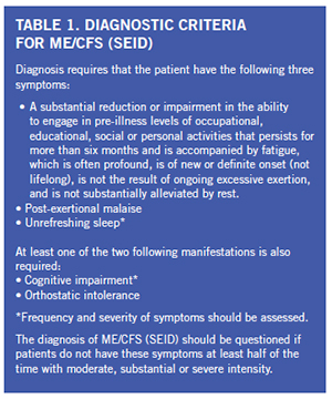
There is significant economic burden associated with this condition as one quarter of patients are bed- or house-bound at some time during their illnesses. ME/CFS patients have been found to be more functionally impaired than patients with other disabling illnesses, such as diabetes mellitus, congestive heart failure, hypertension, depression, multiple sclerosis and end-stage renal disease. Unemployment rates range from 35 to 69 percent in these patients. ME/CFS patients have loss of productivity and high medical costs that lead to an estimated economic burden of $17 billion to $24 billion yearly.

In general, ME/CFS is a condition that is poorly accepted as its pathophysiological mechanisms are poorly understood, and many contest the characteristics needed to make a diagnosis. There continue to be many misconceptions regarding ME/CFS, including that it is a psychogenic illness. Many times, patient symptoms are met with skepticism or even dismissal. After diagnosis, many people with ME/CFS report being subject to hostile attitudes from their health care providers. The cause of ME/CFS is currently unknown; however, symptoms may be triggered by different infections or other prodromal events, including “immunization, anesthetics, physical trauma, exposure to environmental pollutants, chemicals and heavy metals and, rarely, blood transfusions.”

The IOM was asked by the Department of Health & Human Services, the National Institutes of Health, the Agency for Health Care Research & Quality, the CDC, the Food & Drug Administration and the Social Security Administration to convene an expert panel to review the evidence basis for ME/CFS. This was a comprehensive committee of 15 members that convened in September 2013 and took into account data from patients, clinicians and researchers while also reviewing almost 1,000 public comments. This was followed by a comprehensive literature review to help identify new diagnostic criteria to be used by clinicians. Based upon their work, a new name was recommended to replace ME/CFS. Previous studies have shown that the term “chronic fatigue syndrome” can negatively affect patients and medical professionals’ perceptions of the illness and trivialize the seriousness of the disease. “Myalgic encephalomyelitis” is also inappropriate as there is no evidence of encephalomyelitis in these patients, and myalgia is not a core symptom of this disease. The new name that has been recommended for use is systemic exertion intolerance disease (SEID); this new name is felt to encompass the central characteristic of the disease: the fact that exertion of any kind can negatively affect patients in multiple different organ systems.

A new set of diagnostic criteria was also developed by the group with the intent to ease the process of making a diagnosis of ME/CFS (SEID) and hopefully, decrease the time to make a diagnosis for many patients. The new diagnostic criteria that were developed by the IOM committee are detailed in Table 1. The core features include: fatigue and impairment, post-exertional malaise (PEM) and unrefreshing sleep. All of these features need to be present for one to be diagnosed with ME/CFS (SEID).

Fatigue as defined in the dictionary is “weariness from bodily or mental exertion.” Sufficient evidence has been found that fatigue is profound in ME/CFS (SEID). Dramatic examples of reports of fatigue in patients with ME/CFS (SEID) include feeling “too exhausted to change clothes more than every 7-10 days” and experiencing “exhaustion to the point that speaking is not possible.” More commonly, patients report fatigue as “exhaustion, weakness, a lack of energy, feeling drained and an inability to stand for even a few minutes.” The fatigue must be associated with a significant reduction or impairment in the ability to engage in pre-illness levels of occupational, educational, social or personal activities. This degree of fatigue must also persist for more than six months.

Another core symptom, post-exertional malaise (PEM), is found consistently in patients with ME/CFS (SEID) and felt to help distinguish it from other conditions. A patient’s symptoms worsen after exposure to physical or cognitive stressors that were previously well tolerated prior to onset of the disease. Descriptions provided by patients after an exertional task include “crash,” “exhaustion,” “flare-up,” “collapse,” “debility” or “setback.” PEM may occur within 30 minutes of an exertional task or be delayed up to seven days after an exertional task with the duration of PEM lasting hours to months. In studies comparing ME/CFS (SEID) patients with healthy controls, 86 percent of patients report minimum exercise makes them tired compared to 7 percent of controls. Additionally, 85 percent of ME/CFS (SEID) patients report that they feel drained after mild activity compared to 2 percent of healthy controls.

The third and final core symptom is unrefreshing sleep for which 92 percent of ME/CFS (SEID) patients report compared to 16 percent of controls. The typical sleep-related symptoms described by ME/CFS (SEID) patients include difficulty falling asleep, frequent or sustained awakenings, early-morning awakenings and nonrestorative or unrefreshing sleep (persistent sleepiness despite adequate duration of sleep). Primary sleep disorders such as sleep-disordered breathing, restless legs syndrome and narcolepsy should be considered and evaluated for if clinically indicated as treatment of these disorders can be effective in reducing or relieving symptoms of unrefreshing sleep. However, a polysomnogram is not required to diagnose ME/CFS (SEID). Currently, there is no strong evidence to identify ME/CFS (SEID)-specific sleep pathology despite some studies revealing differences in sleep architecture in a subset of ME/CFS (SEID) patients compared to healthy controls.

In addition, there are two supportive criteria included in the new definition, of which one of two is required to be present to meet a diagnosis of ME/CFS (SEID). These include cognitive impairment and/or orthostatic intolerance. Common features of cognitive impairment that are seen in ME/CFS (SEID) include complaints of problems remembering, difficulty expressing thoughts, difficulty paying attention, slowness of thought, absent-mindedness and difficulty understanding. It is suggested that slowed information processing plays a central role in the cognitive impairment associated with ME/CFS (SEID). This deficit can lead to significant disability that results in loss of employment as well as functional capacity in social environments. Neuropsychological testing has found that patients with ME/CFS (SEID) display deficits in working memory compared with healthy controls with reduced verbal and visual memory being the most consistent finding. The second feature, orthostatic intolerance, refers to worsening of symptoms upon assuming and maintaining an upright position. Symptoms are typically improved but may not be alleviated by lying down or elevating their feet. There is sufficient evidence that indicates a high prevalence of orthostatic intolerance in patients with ME/CFS (SEID) based on bedside orthostatic vital signs, tilt table testing or by patient reported worsening of symptoms with standing in day-to-day life.

The committee also described additional frequent findings found in ME/CFS (SEID) patients, which include pain, immune impairment, infection and miscellaneous symptoms. Pain was found to be common in patients who had ME/CFS (SEID) with complaints of headaches, myalgias and arthralgias being most common. There was sufficient evidence found that supported the finding of immune dysfunction in these patients with data revealing poor NK cell cytotoxicity that correlated with illness severity in ME/CFS (SEID). This finding, however, is not specific to ME/CFS (SEID). Additionally, there was evidence that ME/CFS (SEID) can occur after infection with EBV, but there was not sufficient evidence to conclude that all cases of ME/CFS (SEID) are caused by EBV. Less frequent symptoms that were found in patients with ME/CFS (SEID) include gastrointestinal impairments, genitourinary impairments, sore throat, painful or tender axillary/cervical lymph nodes and sensitivity to external stimuli (e.g., foods, drugs, chemicals).

In summary, ME/CFS (SEID) is a serious, chronic, complex and systemic disease that often significantly limits the day-to-day activities of those affected. It is characterized by a prolonged, significant decrease in function; fatigue; post-exertional malaise; unrefreshing sleep; difficulties with information processing, especially under time pressure; and orthostatic intolerance. A thorough history, physical examination and targeted evaluation are necessary and can be sufficient to make a diagnosis. Despite the high prevalence of this condition with associated high economic burden, little research has been conducted to study the etiology, pathophysiology and effective treatment of this disease. Moving forward, it will be vital to distinguish this disease against other complex fatiguing disorders as the majority of previous research has compared ME/CFS (SEID) patients to healthy controls. Additional research into ME/CFS (SEID) is essential for further progress to be made, and the term “chronic fatigue syndrome” should no longer be used due to the associated stigma, which often precludes patients from receiving appropriate care. It is critical that we do our part to help stop the stigma associated with this condition and provide optimal care of these patients.

Maggie L. McNulty is an assistant professor at Rush University Medical Center, Department of Neurological Sciences.

Neurology Practice: The Fundamentals

Raad Shakir

Raad Shakir

While visiting various countries and looking at neurology practice, I became convinced that the fundamentals are by and large the same.  The work neurologists perform in their daily practice is duplicated across the world.  I have been privileged as WFN president to be able to attend annual congresses of neurological societies in countries as diverse as China, Macau, India, Sri Lanka, Morocco, Egypt, Sudan, Saudi Arabia, United Arab Emirates, Turkey, Albania (Figure 1), Chile and most recently Norway (Figure 2).

The diversity is clear. As the health care systems are so different, it makes one wonder if the practice is therefore affected.  There are noticeable differences. The organization of patient’s care is either through state, insurance funding, self-pay or on many occasions a combination.  As we know from the Neurology Atlas 2004, the lower income economies have a much higher probability of self-pay provision of care than in richer countries (Figure 3).  This puts a huge slant on the availability of neurological care and the difficulty in accessing specialist opinion.

Figure 1. Left to right.  Antonio Federico (Sienna Italy), Jera Kruja President Albanian Society of Neurology, Raad Shakir (WFN President), Mira Rakacolli Dean Faculty of medicine Tirana University.

Figure 1. Left to right. Antonio Federico (Sienna Italy), Jera Kruja President Albanian Society of Neurology, Raad Shakir (WFN President), Mira Rakacolli Dean Faculty of medicine Tirana University.

If we start with numbers of patients seen by a neurologist in a working day, in many instances, there are no outpatient appointment systems and patients appearing in clinics have traveled long distances.  On many occasions, this involves relatives bringing patients and expecting hospital admission.  This may well be needed on many occasions, as the neurological status is so advanced, patients need to have inpatient care.  Neurologists and neurology trainees work in crowded clinics making basic decisions, and the more detailed assessments will be carried out when a patient is admitted to a hospital bed.  This practice is the only way to cope with large outpatient loads, which would be unthinkable in other settings.  The reasons are lack of neurologists and facilities for a more time-requiring approach at outpatient clinics.  The other perhaps related reason is the centralization of neurological care in big cities. Neurologists in such settings make quick decisions on the facts as they see them.  In nearly all patients, there are no written referral letters of background information; it is quite admirable to see real coalface practice with decisions made on few available facts as they present.

Figure 2. Left to right. Anne Hege Aamodt President of Norwegian Neurological Association, Olga Bobrovnikova Renowned Pianist, MS sufferer and European Brain Council Ambassador, Raad Shakir WFN President, Hanne F Harbo Head Norwegian Brain Council. (photographer, Lise Johannessen Norwegian Medical Society).

Figure 2. Left to right. Anne Hege Aamodt President of Norwegian Neurological Association, Olga Bobrovnikova Renowned Pianist, MS sufferer and European Brain Council Ambassador, Raad Shakir WFN President, Hanne F Harbo Head Norwegian Brain Council. (photographer, Lise Johannessen Norwegian Medical Society).

In other settings, there is huge utilization of day care facilities for performing a battery of investigations within a working day.  In such settings, the care is supplemented by the availability of infusion suites, EEG/video telemetry, procedures such as lumbar punctures, muscle and nerve biopsies, detailed imaging and neurophysiological assessments, which can be performed within a day care setting. This is only possible in the presence of a well-developed administrative support and the involvement of supporting services such as radiology, laboratory and advanced nursing skills.

The relationship of neurology to general medicine is strong in many settings as the acute care at emergency departments is provided by general physicians, emergency care specialists and in other settings, neurologists are available in the emergency departments for onsite consultation and further care.  This has been made more available with the introduction of Hyper Acute Stroke Units (HASUs).  In some settings, patients with suspected stroke are brought to emergency departments where they would be assessed by neurologists for their suitability for thrombolysis.  This is a huge advance in stroke care and has the other advantage of putting neurology services at the forefront of acute medical care.  The increase in the workload requires increasing staff numbers, and this is possible in some settings but not others. The success of thrombolysis treatment in acute stroke depends on pre-hospital and in-hospital health workers. Figure 4 shows the acute stroke team at Oslo University Hospital in Norway (courtesy of Professor Espen Deitrichs).

Figure 3. Bars to the left show that 84.2% of funding of Neurological care in Low income countries is out-of-pocket.  Neurology Atlas WHO/WFN 2004.

Figure 3. Bars to the left show that 84.2% of funding of Neurological care in Low income countries is out-of-pocket. Neurology Atlas WHO/WFN 2004.

Many other neurological services are struggling with the new practice due to several factors apart from finance; logistics and the availability of a high-powered ambulance and paramedic services lead many to be behind in their ability to provide modern care.  The decision-making process of a paramedic in perhaps the two most important non-traumatic emergencies, i.e. heart attack and brain attack, lead to a major lack of highly trained individuals for this type of work.  In many countries, this shortage of staff is leading to inferior care.

The multidisciplinary approach to care with integrated multispecialty teams in acute care delivery with availability of interventional neuro-radiologists is limiting the ability for neurology to deliver.  In many instances, the availability of endovascular treatment of acute stroke is severely limited by lack of facilities.  Using telemedicine in some locations has made a great difference in acute provision of neurological care.  Acute thrombolysis is being achieved utilizing telemedicine in some locations.

Figure 4. The Pre and In hospital acute stroke team at Oslo University Hospital in Norway (courtesy of Professor Espen Deitrichs).  Photo: Fundamentals_fig5.jpg

Figure 4. The Pre and In hospital acute stroke team at Oslo University Hospital in Norway (courtesy of Professor Espen Deitrichs).
Photo: Fundamentals_fig5.jpg

The care for neurological patients with long-term needs is crucial, and this is not really available unless there is some sort of health care system, which allows regular follow-up services.  In many countries, this is possible but in others it is not.  The recent WHO executive board resolution on epilepsy on Feb. 2, 2015 (http://apps.who.int/gb/ebwha/pdf_files/EB136/B136_R8-en.pdfs) is a good example on the lack of neurological long-term care and the continued existence of a massive treatment gap.  This declaration should be translated to actual on the ground care provision.   Continued support for conditions such as epilepsy, parkinsonism, dementia, multiple cclerosis, migraine and genetically determined conditions and many others remain woeful.  The WHO can inform governments of the availability of “cheap” anti-epilepsy drugs as an example, but neurologists should aim to provide optimal up to date care for their patients. The premise that some treatment is better than nothing, although understandable and reflects current status, is not an ultimate goal to aim for.  Neurologists should endeavor to put brain health at the top of the political agenda.  However, we are delighted that the WHO executive has put brain health at the top of the agenda as it aims to reduce the treatment gap when, as an example, we know that seven out of 10 patients with epilepsy do not receive regular medication at all. I am sure that I speak of behalf of all neurologists who would not accept second- or even third-tier level of management for their patients, but would approve of the WHO executive declaration as a first step in the right direction.

Figure 5.  CME Continuum utilization in 45 countries up-to August 2014. WFN six monthly report August 2014. Helen Gallagher WFN CME coordinator.

Figure 5. CME Continuum utilization in 45 countries up-to August 2014. WFN six monthly report August 2014. Helen Gallagher WFN CME coordinator.

Returning to the fundamentals of neurology, it is my conviction that neurologists from all over the world offer their services in basically the same manner if given equivalent circumstances.  This is clear from observing training programs and the eagerness of young trainees to learn.  The knowledge base is nearly universal.  The WFN is in a position to see this when we administer the American Academy of Neurology Continuum CME program to 45 countries.  This is happening today in war-ravaged countries and in those with low income to the degree of the existence of real malnutrition.  The evaluations sent by the WFN tutors are a shining testament to the eagerness and the excellent performance of trainees from various backgrounds across continents (Figure 5).

To end on a positive note, neurology is prospering and need continuous momentum to keep brain health at the top of the health agenda of decision-makers.

Raad Shakir
London UK

Redefining Recovery from Aphasia by Dalia Cahana-Amitay and Martin Albert

BOOK REVIEW9780199811939

By Murray Grossman, MD
Cahana-Amitay D and Albert M (2015)
Redefining Recovery from Aphasia
New York: Oxford University Press
281pages, with preface and index

Language is an incredibly complex process. Yet we speak and under- stand effortlessly in order to live our lives daily. The disruption of language following a stroke is a devastating blow to an individual’s day-to-day functioning because of our extreme dependence on this modality of communication. Aphasia is extraordinarily costly to individuals and to society. Nevertheless, aphasia following a stroke is common.

Despite the high cost and common occurrence of aphasia, progress in developing successful treatments for aphasia has been slow. It has proved difficult to demonstrate that traditional speech and language therapies are better at improving communication skills than friendly social interactions. Thus, novel approaches to treatment and recovery from aphasia are desperately needed.

In this timely book, Cahana-Amitay and Albert outline an alternate approach to recovery from aphasia. Their perspective is based on the view that language is not a modular entity, but instead interacts with multiple facets of non-linguistic cognition. This includes domains such as executive functioning, visual processing, attention, memory, emotion and praxis. In turn, the authors observe that brain regions involved in language are highly interconnected with brain regions subserving these other aspects of cognition. The authors coin the phrase “neural multifunctionality” to characterize this multifaceted clinical and functional neuroanatomic approach to language.

In nine chapters, Cahana-Amitay and Albert lay out their approach to language and recovery from aphasia. The first chapter situates their volume in the context of the extensive literature describing recovery from aphasia. The second chapter outlines the authors’ multifunctionality approach to the functional neuroanatomy of language following stroke. In subsequent chapters, Cahana-Amitay and Albert discuss the relationships between language and each of the other domains of cognitive functioning that interact with language. Each chapter defines a domain of cognition, reviews cognitive aspects of the domain and its interaction with linguistic functioning, and then defines the functional neuroanatomy of the cognitive domain. Treatments for aphasia that focus on each domain of cognition are then reviewed.

In the chapter concerned with executive functioning, for example, the authors consider the ways in which language and executive functioning interact, and review disorders of executive functioning observed in aphasic patients such as perseverations, disorders of discourse, and semantic control impairments. Neural correlates of discourse and executive functions in aphasia are then reviewed. The chapter concerned with attention addresses “basic attention” concerned with vigilance and arousal, and “complex attention” selective and alternating attention. After discussing the relationship between attention and language, Cahana-Amitay and Albert examine the neural underpinnings of attention-language interactions and treatments of attention in language.

The chapter devoted to memory examines the role of working memory and other forms of memory in language processing, and the effects of working memory deficits on language functioning. The authors then consider the contribution of memory systems to aphasia treatments and recovery from aphasia as well as learning and anatomic structures implicated in aphasia therapies. In the chapter examining the role of emotion in recovery from aphasia, Cahana-Amitay and Albert examine altered emotions in aphasia, such as depression and anxiety, and interventions focusing on depression and anxiety. The chapter concerned with praxis assesses the breakdown of gesture in aphasia and the intimate connection between gestural and linguistic forms of communication in theories of apraxia. Finally, the authors consider the role of visual processing in recovery from aphasia, including the effects of visual scenes and audiovisual stimulation in language processing, and the role of visually mediated cueing in recovery from aphasia.

In the final chapter, Cahana-Amitay and Albert marshal evidence from the previous chapters to support their neural multifunctionality hypothesis. They conclude that findings from lesion, neuroimaging and electrophysiological studies support their contention that non-linguistic functions need to be incorporated into language models of the intact brain, and that recovery from aphasia must take into account the role of non-linguistic functioning.

Cahana-Amitay and Albert are experienced aphasiologists at the Harold Goodglass Aphasia Research Center of Boston University and the Boston Veterans Administration Medical Center. The authors provide a comprehensive landscape of language and the brain based on functional neuroanatomic theories that have been evolving over the past two decades. The authoritative voice of these authors compels us to reconsider classic approaches to aphasia, and develop novel forms of speech therapy that are organized around the principle of multifunctionality.

Dr. Grossman is professor of neurology, Penn Frontotemporal Degeneration Center, University of Pennsylvania

Editor’s Update and Selected Articles from the Journal of the Neurological Sciences (JNS)

By John D. England, MD

John D. England, MD

John D. England, MD

The Journal of the Neurological Sciences (JNS) is a broad-based journal that publishes articles from a wide spectrum of disciplines, ranging from basic neuroscience to clinical cases. Because JNS is the official journal of the World Federation of Neurology (WFN), the Editorial Board welcomes submissions from around the world. We also strive to publish papers with unique and original observations. In order to satisfy this latter goal, only the best manuscripts are accepted for publication in JNS.

I frequently receive correspondence asking why manuscripts are rejected. The most frequent reason for rejection is that the manuscript does not receive a high enough priority score when the scientific methodology and conclusions are assessed. In view of these criteria for acceptance, my first advice to authors is to design and perform their scientific studies in as rigorous and as thorough a manner as possible. Additionally, original and novel observations are more likely to attain higher scores by reviewers.

As a last but very important point, authors should write the manuscript as clearly and concisely as possible. The English syntax and grammar should be polished and well-edited. When we receive manuscripts that are poorly written, we always send them directly back to the authors for revision prior to more formal review. We cannot publish manuscripts that are poorly written even if the underlying scientific methodology and observations are sound.

JNSJanWe recognize that English is not the native language of many of our authors, and we will allow re-submission of manuscripts that require editing. My suggestion for authors is to have their manuscripts edited by someone who has excellent command of the English language. If you do not have ready access to such a person, please utilize one of the many excellent “English editing” services. In fact, Elsevier will provide this online service to authors for a modest fee.

In our ongoing attempt to enhance accessibility of JNS articles to members of the WFN, we have selected two more “free-access” articles, which are profiled in this issue of World Neurology.

1) Aaron Berkowitz, et al., provide a well-written and richly referenced review on the neurologic manifestations of neglected tropical diseases (NTDs). They focus the review on 17 diseases that the World Health Organization has designated neglected tropical diseases. These diseases disproportionately affect the world’s poorest populations and cause significant morbidity and mortality. In fact, at least 1 billion people around the world are affected by these diseases. Most of these diseases have significant neurologic manifestations. Importantly, these diseases can be controlled using relatively low-cost but strategic plans.
Berkowitz AL, Raibagkar P, Pritt BS, Mateer FJ. Neurologic manifestations of the neglected tropical diseases. J Neurol Sci. 2015;349:20-32.

2.) Edward Mader, et al., provide an interesting and unique case description of a young woman with biopsy-proven tumefactive multiple sclerosis and acute hepatitis C virus 2a/2c who was successfully treated with interferon beta-1a. Although this report is based upon only one case, the observations are potentially very important. The report raises the possibility that a link exists between hepatitis C infection and multiple sclerosis, and it also suggests that interferon beta-1a may be effective treatment for some patients with hepatitis C infection. One particularly reasonable suggestion is that patients with acute CNS demyelination be screened for hepatitis.
Mader EC, Richeh W, Ochoa JM, Sullivan LL, Gutierrez AN, Lovera JF. Tumefactive multiple sclerosis and hepatitis C virus 2a/2c infection: Dual benefit of long-term interferon beta-1a therapy? J Neurol Sci. 2015;349:239-242.

Dr. England is the editor-in-chief of the Journal of the Neurological Sciences.

HIV Infection Is a New Target for Stroke Prevention

Infection is an independent risk factor for both ischemic and hemorrhagic stroke

By Jerome H. Chin, MD, PhD, MPH

Jerome H. Chin, MD, PhD, MPH

Jerome H. Chin, MD, PhD, MPH

Stroke is the third-leading cause of premature death globally as measured in years of life lost1.  Demographic and epidemiologic changes, including population growth and aging, urbanization and unhealthy diets are driving a rise in the incidence of stroke in low- and middle-income countries (LMIC)2. Due to inadequate primary health care services to screen for and treat the most common stroke risk factors, particularly hypertension and diabetes, the age-standardized incidence rates of stroke in LMIC exceed those in high-income countries2. In addition, strokes due to atrial fibrillation and rheumatic heart disease contribute a significant share of the stroke burden in LMIC as a result of both diagnostic and therapeutic resource limitations for these conditions.

Figure. HIV-associated stroke: (a) middle cerebral artery infarct; 25-year-old HIV-infected male, CD4+ cell count = 42 cells/mm3; (b) bilateral basal ganglia infarcts; 25-year-old HIV-infected female, unknown CD4+ cell count.

Figure. HIV-associated stroke: (a) middle cerebral artery infarct; 25-year-old HIV-infected male, CD4+ cell count = 42 cells/mm3; (b) bilateral basal ganglia infarcts; 25-year-old HIV-infected female, unknown CD4+ cell count.

HIV infection has recently emerged as an independent risk factor for both ischemic and hemorrhagic stroke3-8. Two cohort studies conducted in the United States reported an elevated risk of ischemic stroke in HIV-infected individuals compared to non-HIV-infected individuals4,5. Lower CD4+ cell counts or higher HIV RNA levels were associated with an increased risk of stroke. Cohort studies from Canada and the United States have reported an association of HIV infection with an increased risk of intracerebral hemorrhage6,7. A community-based case-control study performed in Tanzania found HIV infection to be an independent risk factor for stroke (ischemic and hemorrhagic combined) with an adjusted odds ratio of 5·618. Only 40 percent of the 200 stroke cases had a CT scan. The pathogenic mechanisms responsible for the increased risk of stroke associated with HIV infection are multiple and may include chronic inflammation and immune activation leading to endothelial dysfunction and subsequent vasculopathy3,9. Both small-vessel (e.g. lacunar) and large-vessel strokes are observed in HIV-infected individuals (figure) in addition to intracerebral hemorrhages.

Thirty-five million people are living with HIV worldwide10. The population-attributable risk of stroke due to HIV infection will depend on the prevalence of HIV infection in a particular region or country. Given the higher prevalence of HIV infection in sub-Saharan Africa compared to other regions, a substantial number of incident strokes in sub-Saharan Africa may be the result of HIV infection. However, reliable figures for stroke incidence, mortality and comorbidities are difficult to obtain in most countries of sub-Saharan Africa due to inadequate stroke surveillance and vital registration data. The above-mentioned epidemiologic studies provide support for the inclusion of HIV antibody testing in the diagnostic evaluation of patients with acute stroke in all regions of the world. Furthermore, stroke prevention should now be considered another potential benefit of the early initiation of antiretroviral therapy in HIV-infected individuals through both a reduction in HIV-associated vasculopathy as well as through the prevention of HIV transmission to their uninfected partners.

Dr. Chin is president of the Alliance for Stroke Awareness and Prevention Project (ASAPP).

References

  1. GBD 2013 Mortality and Causes of Death Collaborators. Global, regional, and national age–sex specific all-cause and cause-specific mortality for 240 causes of death, 1990–2013: a systematic analysis for the Global Burden of Disease Study 2013. Lancet. 2015;385:117-171.
  2. Krishnamurthi RV, Feigin VL, Forouzanfar MH, et al. Global and regional burden of first-ever ischaemic and haemorrhagic stroke during 1990–2010: findings from the Global Burden of Disease Study 2010. Lancet Glob Health. 2013;1(5):e259-e281. doi:10.1016/S2214-109X(13)70089-5.
  3. Cole JW, Chin JH. HIV infection: a new risk factor for intracerebral hemorrhage? Neurology. 2014;83:1690-1.
  4. Marcus JL, Leyden WA, Chao CR, et al. HIV
    infection and incidence of ischemic stroke, AIDS. 2014;28:1911–1919.
  5. Chow FC, Regan S, Feske S, Meigs JB, Grinspoon SK, Triant VA. Comparison of ischemic stroke incidence in HIV-infected and non-HIV-infected patients in a U.S. health care system. J Acquir Immune Defic Syndr. 2012;60:351-358.
  6. Chow FC, He WS, Bacchetti P, et al. Elevated rates of intracerebral hemorrhage in individuals from a U.S. clinical care HIV cohort. Neurology. 2014;83:1705–1711.
  7. Durand M, Sheehy O, Baril JG, LeLorier J, Tremblay CL. Risk of spontaneous intracranial hemorrhage in HIV infected individuals: a population-based cohort study. J Stroke Cerebrovasc Dis. 2013;22:e34–e41.
  8. Walker RW, Jusabani A, Aris E, et al. Stroke risk factors in an incident population in urban and rural Tanzania: a prospective, community-based, case-control study. Lancet Glob Health. 2013;1:e282–e288.
  9. Benjamin LA, Bryer A, Emsley HCA, Khoo S, Solomon T, Connor MD. HIV infection and stroke: current perspectives and future directions. Lancet Neurol. 2012;11:878–90.
  10. UNAIDS, The Gap Report, available at: http://www.unaids.org/en/resources/campaigns/2014/2014gapreport/gapreport/ Accessed January 25,  2015.

Mohammad Wasay Appointed to National Research Post

Mohammad Wasay MD, FRCP, FAAN.

Mohammad Wasay MD, FRCP, FAAN.

Mohammad Wasay, MD, FRCP, FAAN, has been appointed as convener, panel of experts and member of the Advisory Committee of the Pakistan Medical Research Council. The PMRC is the premier national institute for promotion of research in Pakistan. This is the first time a person from the field of neurology has been appointed to this position. It is a symbol of recognition of the Pakistan Society of Neurology and neurological research conducted by neuroscience faculty throughout Pakistan.

Dr. Wasay is a professor in the department of neurology and chair, FHS research committee, at Agu Khan University; interim director, Clinical Trials Unit, chair, public awareness and advocacy committee, World Federation of Neurology; president, Pakistan Society of Neurology and editor, Pakistan Journal of Neurological Sciences.

News from the 1st Congress of the European Academy of Neurology

BY Jacques L. Dereuck, MD, PHD

The 1st European Academy of Neurology (EAN) congress will be held in Berlin, Germany, from June 20 to 23, 2015.

Berlin was chosen as a symbolic place, where the walls separating the city in two parts were broken down more than 20 years ago. Now the walls between the European Federation of Neurological Societies (EFNS) and the European Neurological Society (ENS) also have disappeared and a united European Academy of Neurology has been created. The congress is one of the most important achievements in Europe, showing that although there are important differences in practicing neurology in the different European countries some common goals can be achieved.

The EAN congress has been enthusiastically endorsed by most European neurologists and structured as most academies of neurology, with teaching courses, hands-on-courses, plenary symposia, interactive sessions, focused workshops, special sessions, free oral communication and poster sessions. In addition, tournaments in basic and clinical neurology for young neurologists are scheduled.

Almost 2,000 abstracts have been submitted, from which 168 will be presented as oral communications and 1,556 as poster presentations.

The creation of the EAN will reinforce the impact of world neurology on health care, showing that progression in diagnosis and treatment of neurological diseases is one of the most important issues to be reached.

Dr. De Reuck is the chairperson of the WFN Membership Committee.

European Board of Neurology Examination in Berlin in 2015

UEMS-EBN-logoThe European Board Examination in Neurology is a joint development of the UEMS Section of Neurology and the European Academy of Neurology. It is considered to be a tool for the assessment of European neurological education and to boost its European standards.

It is supervised by the examination committee of the UEMS/EBN and also observed by the EAN representing the European neurological scientific societies and the World Federation of Neurology.

The exam was held in 2009 for the first time, and since then 130 candidates have passed the exam. Beginning in 2015, the title “Fellow of the European Board of Neurology” will be conferred to European and non-European candidates.

The next UEMS/EBN examination will be organized one day prior to the 1st Congress of the European Academy of Neurology (EAN) on Friday, June 19, 2015, in Berlin, Germany. (http://www.eaneurology.org/)

UEMS-EBN-Exam-Istanbul-2014The European Board Examination in Neurology is a substantial step forward in the further harmonization and in the raising of the standards in European neurology. The cooperation with the scientific neurological societies is an important scientific input and a guarantee of continuous updates of the current knowledge of a European neurologist.

The European Examination in Neurology is a proof of excellence: Taking the examination shows the candidate’s commitment to lifelong learning. Even without legal recognition, this is known and recognized within the profession throughout Europe and the rest of the world, thus encouraging the mobility of specialists in neurology and giving an additional distinguishing mark to the individual candidate.

The deadline for application is the May 1, 2015. (http://www.uemsneuroboard.org/ebn/)

There is a reduced fee for candidates from low- and lower-middle income countries (see http://data.worldbank.org/about/country-and-lending-groups#Low_income) and for those who follow the early-bird registration procedure.

The examination consists of the following parts:

  • 80 MCQs (multiple choice questions)
  • 50 EMQs (extended matching question)
  • A short essay on a neurology-related public health or ethics-related topic to be orally discussed with the examiners.
  • A critical appraisal of a neurological topic to be discussed with the examiners.

Results of these four parts of the examination will be combined to one final mark.

We are happy to note that the number of participants taking the European Board Exam in Neurology is increasing year by year, and we aim to develop an exam that will be taken by all neurology trainees, particularly those who wish to extend their experience beyond the borders of their own country.

Any questions and comments can be sent to uems-sbn@medacad.org

Professor Dr. Jan Kuks: Chair of the examination committee: j.b.m.kuks@umcg.nl

Professor Dr. Wolfgang Grisold: UEMS/EBN past chair of the examination committee wolfgang.grisold@wienkav.at

Dr Walter Struhal: WFN website and social media, w.struhal@aesculapian.net

CONTACT address:

Mag.Gabrielle Lohner: uems-sbn@medacad.org

Section of Neurology –European Board of Neurology
c/o Vienna Medical Academy
Alser Strasse 4, 1090 Vienna
AUSTRIA
T (+43 1) 405 13 83 – 32
F (+43 1) 407 82 74
www.uems-neuroboard.org