Research Group on Neuroethics

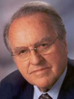
Franz Gerstenbrand

Franz Gerstenbrand

Ethics in neurology are the  basis of all practical work  as well as for research  activities. In a critical analysis of  the used ethical principles, it has to  be stated that the contemporary ethical laws are missing different religious, traditional and cultural requirements. The contemporary ethics are based on the philosophy  of Socrates, Plato and Aristotle,  with the incorporation of the  Christian philosophy of Saint  Augustinus and Thomas Aquinas  and should be called Western  Ethics. But Western Ethics must  not be acceptable worldwide. The ARG Neuroethics decided to found  a task force with the aim to harmonize the ethical principles in  neurology including transcultural considerations. As members of the task force,  representatives of the Buddhism and  Hinduism, the Islamic and  Mosaic religions and the different Christian communities are invited.

As a continuous activity of  the ARG Neuroethics, the  organization of teaching courses  and informative workshops has to  be mentioned. The organizers  of neurological congresses and  symposia are asked to include  special lectures and training  courses in the program. The  great lack of ethical knowledge  in scientific reports about new  treatment programs or research  projects is shameful fact and has to  be strictly changed. A continuing ethical education is necessary.

The ARG Neuroethics is asking for more active cooperation and  for helpful support to follow the  education for a better knowledge  in ethical thinking and moral  responsibility. The aim for a  worldwide acceptable basis for  ethical principles in modern  neurology needs great knowledge and an intensive preparatory work in philosophical, moral and ethical issues.

Gerstenbrand is chairman of the Research Group on Neuroethics. He can be reached at f.gerstenbrand@aon.at.
Organization and Delivery of Neurological Services

Members of the WFN Applied Reserach Group (ARG) from many different countries are active in their regions. In 2012, we were focused mainly on Central and Eastern European countries, and we organized several meetings.

As part of these activities, members of the ARG were active in their research, related to importance of delivery of neurological services, educational activities and spreading the knowledge to our colleagues in many parts of the world. Vida Demarin was active at the Eighth Congress of the Society for the Study of Neuroregeneration and Neuroplasticity (SSNN) in March 2012 in Dubrovnik; Management of Pain in November 2012 in Moscow; Neurological Meeting on Stroke in March 2012 in Ljubljana, Slovenia; Challenges on Stroke in April 2012 in Belgrade, Serbia; and Neurological Symposium on Headache in May 2012 in Tuzla, Bosnia and Herzegovina, to mention just a few of them.

Our gorgeous city of Dubrovnik was the host of the 23rd Summer Stroke School with international participation June 4-8. For the first time, the summer school was under the auspices of Croatian Academy of Sciences and Arts. The usual and proud auspicates are Inter-University Center Dubrovnik, Croatian Stroke Society, Medical School University of Zagreb, Central and Eastern European Stroke Society and Research Group on Delivery of Neurological Services (RGODNS) of World Federation of Neurology (WFN). The joint meeting of ARG ODNS and the 23rd Summer Stroke School was organized in Interuniversity Center in June 2012 in Dubrovnik.

The aim of the course was to support the cooperation and promote exchange of knowledge and experience among participants from different countries. This meeting shed new light on epidemiology of stroke, its primary and secondary prevention, diagnostics, therapy and rehabilitation. All participants had a unique opportunity to share their national stroke data and discuss about specific stroke problems of their country. Countries of the East and West Europe had an opportunity to work together on solving problems related to stroke, whether they were medical, economical or of some other nature. We can’t skip mentioning that all the hard work was awarded by rich social program during the evenings. We look forward to 2014 course already.

All participants had a unique opportunity to share their national stroke data and discuss about specific stroke problems of their country. Countries of the East and West Europe had an opportunity to work together on solving problems related to stroke.

The 52nd International Neuropsychiatric Pula Congress (INPC) was held June 20-23, 2012 under the auspices of the Croatian president Ivo Josipovic. The congress is traditionally held in Verudela Hotels Brioni and Histria, and organizers are: Kuratorium International Neuropsychiatric Pula Congress, Sestre Milosrnice University Hospital Center, Zagreb, and this year, the Department of Medical Sciences of the Croatian Academy of Sciences and Arts. The main sponsors of the congress were the Ministry of Science, Education and Sports of the Republic of Croatia, the city of Graz, the city of Zagreb and Istria County. This year, more than 350 international particpants attended the congress.

The main themes of the congress were recent achievements in restorative neurology, new advances in psychopathology — the interaction of biological and psychological factors. The theme of the joint meeting of the International Neuropsychiatric Pula Congress in the Section of Neuroscience Alps-Adria was discussions about different therapeutic procedures. Besides the main topic during this year’s congress, numerous symposia were also held: roundtables and workshops in collaboration with the Central and Eastern European Stroke Society (CEESS), World Federation of Neurology, Research Group on Delivery of Neurological Services (WFN RG ODNS) and one topic symposium related to dilemmas in therapies for different neurological disease (stroke, multiple sclerosis, epilepsy and Parkinson’s disease) and the Croatian branch of the Italian Cochrane Centre (CBICC) on the topic: Introduction to the Cochrane systematic review and application of evidence-based medicine in everyday practice, the meeting of the European Society of young neurologists and residents (European Association of Young Neurologists and Trainess – EAYNT), European Summer School of psychopathology in Pula, fifth Pula International Symposium on Epilepsy, neuro-otological course of vertigo, the European standards for doctoral studies in the field of neurology, a symposium on forensic psychiatry, the German Symposium on the ongoing training in neuropsychiatry (Deutschsprachiges Neuropsychiatrisches Fortbildungs Symposium), a symposium on the quality of life of psychiatric patients, a symposium on connecting providers in neurorehabilitation, a symposium on the role and experiences of nurses in the care of patients with incontinence, symposium on deep brain stimulation, interactive school about headache and symposium on palliative care.

We are proud to announce that this year’s 53rd INPC Congress in Pula June 19-22, 2013, had the endorsement of the WFN. We are grateful for all efforts related to ARG organization and its activities.

Battistin is president of the ARG OSNS, and Demarin is the secretary-general of the ARG ODNS.

Highlights of the 53rd International Neuropsychiatric Pula Congress

The 53rd International Neuropsychiatric Pula Congress (INPC) was held June 19-22, 2013, under the auspices of the Croatian President Prof. dr. Ivo Josipovic. The congress is traditionally held in Verudela Hotels Park Plaza Histria and Brioni, and organizers are: Kuratorium International Neuropsychiatric Pula Congress and the Department of Medical Sciences of the Croatian Academy of Sciences and Arts. The supporting organizations of the Congress are: World Federation of Neurology, WFN Applied Research Group on the Delivery of Neurology Services, International Interdisciplinary Medicine Association, European Psychiatric Association, Central and Eastern European Stroke Society.

This year, the congress was endorsed by WFN. The main sponsors of the congress were the Ministry of Science, Education and Sports of the Republic of Croatia, the City of Graz and the City of Zagreb. More than 350 participants attended representing Australia, Austria, Bosnia, Croatia, Czech Republic, Germany, Great Britain, Herzegovina, Hungary, India, Italy, Lithuania, Macedonia, Montenegro, New Zeeland, Poland, Romania, Russia, Serbia, Slovenia, South Korea, Spain, Sweden, Switzerland, Thailand, Turkey, Ukraine, the United States, and many more.

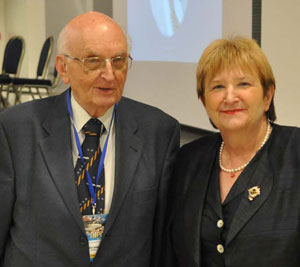
Vida Demarin, MD, honorary president of the INPC Congress, and Bosko Barac, MD.

Vida Demarin, MD, honorary president of the INPC Congress, and Bosko Barac, MD.

The main theme of this year’s congress was Depression and Pain — What Is the Link? The theme of the joint meeting of the International Neuropsychiatric Pula Congress in the Section of Neuroscience Alps-Adria offered discussions about different therapeutic approaches. Beside the main theme during this year’s congress, numerous symposiums were also held: the Second European Summer School of Psychopathology in Pula, the Sixth Pula International Symposium on Epilepsy, the Second Interface Providers in Neurorehabilitation Symposium, the First Pula Neuro-interdisciplinary School, psychiatric symposia on Law on Psychotherapy, Forensic Psychiatry, CSF Signaling and CSF Biomarkers, neurological symposia on Music and Rhythm in Restoring the Brain, Advanced Treatment of Parkinson´s Disease and Diagnosis and Treatment of Low Back Pain. On Wednesday evening, the traditional academic lecture was given by Professor Luigi Agnati, who presented the latest findings on the Volume Transmission Mode and Potential Clinical Impact.

This year’s International Neuropsychiatric Pula Congress has once again proved that there is a need and interest to organize such a congress where multidisciplinary approaches to numerous interesting topics in the field of both neurology and psychiatry is maintained, and which has during more than half a century maintained the continuity of meeting that has become known as “The Pula School of Science and Humanism,” as it was often emphasized by prominent speakers, honored guests and participants of the oldest international congress in the field of neurology, psychiatry and neuropsychiatry.

Report from the European Stroke Conference

I’m deeply grateful to the WFN for awarding me the Junior Traveling Fellowship in 2013 to visit the European Stroke Conference held May 27-31, in London.

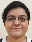
Shaily Singh

Shaily Singh

The conference was attended by more than 3,500 delegates from  around  the world. Following is the brief outline of the meeting and its structure and discussion items.

The meeting was broadly divided into different themes, with parallel symposia, teaching courses, debates and controversies, oral paper sessions, poster presentations and plenary lectures.

The various themes were acute stroke treatment both interventional and medical, intracerebral hemorrhage, translational neuroscience, arteriopathies, childhood stroke, rehabilitation, small vessel disease, imaging, neuroprotection, outcomes and quality of health services, risk factors, anticoagulation in stroke, and alternative therapies to name a few.

The plenary sessions were on the awards on stroke research and also new trial results announced, including INTERACT 2, SPS3, STITCH 2, imaging results of IST3, etc.

A huge number of research papers and posters were presented during this meeting.

There were parallel halls for oral presentations with different themes to select the area of stroke one is interested in. The timing, discipline and attendance was par excellence.

I had one abstract accepted as first author and was a co-author on one other.

Special e-poster sessions with presentations and question-and-answer sessions were undertaken for the first time for highly rated abstracts. There was a lot of interaction during my e-poster presentation and new ideas generated.

Overall, this meeting was an excellent feast of stroke academics and gave me a great opportunity to present our institutional research, to interact with other stroke researchers from around the world and to collaborate for future research.

Singh is assistant professor of Neurology at  the Institute of Human Behavior and Allied Sciences In Delhi, India.

Neurology Nursing in Kenya: The Current State and the Way Forward

Jolynne Mokaya

Jolynne Mokaya

Neurological disorders make up at least 25 percent of the global burden of disease and are responsible for an even greater proportion of persons living with disability (Silberberg and Katabira, 2006). Factors such as malnutrition, adverse perinatal conditions, malaria, human immunodeficiency virus and acquired immune deficiency syndrome (HIV/AIDS), meningitis, demographic transitions, increased road traffic accidents and persistent regional conflicts have increased the prevalence of neurological disorders in Sub-Saharan Africa (Silberberg and Katabira, 2006).

A study of Parkinson’s disease carried out in rural Tanzania showed that  prevalence rates were higher than  what has been previously reported  from Sub-Saharan Africa. The crude prevalence rates were 30/100,000 (men), 11/100,000 (women) and 20/100,000  (combined) showing only a slight difference when compared to the prevalence rates with the U.K. population. This illustrates the fact that neurological diseases are equally a problem in Africa as they are in the Western world. Many Parkinson’s disease patients and many individuals with other neurological diseases in Sub-Saharan Africa may never be diagnosed or treated, with consequent reduction in their life expectancy and quality of life (Dotchin et al., 2008)

With the current increase of neurological diseases globally, health care professionals are expected to see an increasing number of patients living with neurologic conditions including dementia, stroke, epilepsy, Alzheimer’s and Parkinson’s disease.  The World Health Organization identified a shortfall of 4.3 million trained health workers in 2006, with the biggest disparity in Sub-Saharan Africa.

With the current increase of neurological diseases globally, health care professionals are expected to see an increasing number of patients living with neurologic conditions including dementia, stroke, epilepsy, Alzheimer’s and Parkinson’s disease. Policymakers, therefore, must help ensure there are properly trained clinicians available to provide high-quality care (American Academy of Neurology Professional Association). The World Health Organization identified a shortfall of 4.3 million trained health workers in 2006, with the biggest disparity in Sub-Saharan Africa.

Kenya has an estimated population of 41 million, most of whom live in the rural areas (Index Mundi, 2012). Medical standards, such as income distribution, reveal huge differences between various segments of the population. Like many developing nations, there are too few health care providers for the growing population, and only 30 to 40 percent of all Kenyans have access to improved sanitation, clean water and decent health care (InterNations, nd).

Public (government service), private and missionary hospitals are the three major providers of health care in Kenya, with public hospitals being the major health care providers to the majority of the Kenyan population (Amayo, 2006). Kenyatta National Hospital is the largest public hospital, with 7.5 percent of all medical cases seen in this hospital being neurological illnesses (Kwasa 1992).

The HIV/AIDS pandemic has evolved to being the major clinical challenge of neurology practice in Kenya due to the rise of neurological complications associated with it. More than 60 percent of all hospital beds are occupied by HIV-related illness in public hospitals. On the other hand, non-communicable diseases with their neurological sequelae are becoming epidemic due to the changing pattern of lifestyle. Once considered rare, multiple sclerosis is now being diagnosed frequently, and age-related diseases are on the rise as the population ages offering major management challenges (Amayo, 2006).

With awareness of the massive burden associated with neurological disorders, it is recognized that neurological services and resources are disproportionately scarce especially in low income and developing countries (WHO, 2005). The situation in Kenya is worse; currently, there are only 11 neurologists with most of them in private practice and working in Nairobi. In Kenya, the majority of health care providers are nurses who receive little or no training in how to diagnose and treat the common neurologic conditions that present to them every other day. There is not a single nurse trained in neurology, nor is there a training institution of neurology for nurses in Kenya. Lack of mentorship makes it difficult for most nurses to be involved in neurology, and it is sad that the majority of nurses are not aware that they could specialize in neurology (Hooker, 2013).

Nurses bring an important voice and point of view to management and policy discussions. It is crucial to involve them not only in hospital management and patient care but also in being part of various organizations, societies and bodies promoting neurology worldwide.

Training of nurses is particularly important globally. In low income countries such as Kenya where few physicians exist, nurses are involved in making diagnostic and treatment decisions.

Previous  training of  nurses in Kenya concentrated on  the cadre of  enrolled nurses  and registered nurses. However, with the increasing development of the society, a higher level of training for nurses has become necessary to meet the challenging demand for high-quality nursing care (NNAK, 2009). The health sector reforms that are currently being introduced in Kenya not only require highly qualified nurses but also highly specialized nurses for effective and successful implementation (Muga et al, nd). It is crucial and essential for nurses to be endowed with the necessary knowledge, skills and attitudes to be able to give quality service.

Availability of funds, scholarships, training opportunities and mentorship programs should also be made available to nurses and other health care providers in low income countries and regions with few or non-specialists in neurology, to encourage and have more health care providers train and specialize in neurology. •

References

1. Amayo E., (2006).Neurological letter from Kenya. Practical Neurology; Vol 6(4):261 doi:10.1136/jnnp.2006.097519

2. American Association of Neurology  Professional Association. The critical role of neurologists in our health care system. (www.aan.com/advocacy/issues/tools/109.pdf, accessed 30 may 2013)

3. Dotchin C., Msuya O., Kissima J., Massawe J., et al. (2008). The prevalence of Parkinson’s disease in rural Tanzania. PubMed; Vol 23(11): 1567-672. doi: 10.1002/mds.21898

4. Hooker J., (2013). Nurturing Neuroscience and Neurology in Kenya and East Africa-Some reflections. Kenya Association of Physicians. (http://kapkenya.org/index.php?option=com_phocadownload&view=category&download =233:nurturing-neurology-and-neuroscience… &id=13:17th-annual-scientific-conference &Itemid=127, accessed 30 May 2013).

5. Index Mundi, (2012). Kenya’s population.  (www.indexmundi.com/kenya/population.html, accessed 30 May 2013)

6. InterNations,(nd). Health care in Kenya.(www.internations.org/kenya-expats/guide/life-in-kenya-15768/healthcare-in-kenya-2, accessed 30 May 2013)

7. Kwasa T., (1992). The pattern of neurological disease at Kenyatta National Hospital. East African Medical Journal;69(5):236-9.

8. Muga R., Kizito P., Mbaya M., Gakuru T., (nd). Overview of health systems in Kenya.(http://www.measuredhs.com/pubs/pdf/SPA8/02Chapter2.pdf, accessed 28 May 2013)

9. National Nurses Association Of Kenya, (2009). Strategic plan 2009-2014. (www.nnak.or.ke, accessed 29 may 2013)

10. Preamble to the Constitution of the World Health Organization as adopted by the International Health Conference, 1946. In: Basic documents, 45th ed. Geneva, World Health Organization, 2005

11. Silberberg D., Katabira E., (2006). Disease and Mortality in Sub-Saharan Africa: Neurological Disorders, 2nd edition, Bookshelf ID: NBK2295PMID: 21290656

12. World Health Organization. Neurological disorders: public health challenges, (2006).

Mokaya is a final year student in the bachelor’s degree program in Nursing Sciences at the University of Nairobi. The University of Nairobi is the pioneer institution of university education in Kenya, situated in Nairobi. It is committed to quality through teaching, research and creative works, fostering an intellectual culture that bridges theory with practice, and producing holistic graduates prepared for a life of purpose, service and leadership.

The Forced Migration of German-Speaking Neuroscientists

“Vienna’s culture was one of extraordinary power, and it had been created and nourished in good part by Jews. My life has been profoundly shaped by the collapse of Viennese culture in 1938. … The sense of loss is heightened by the fact that Vienna was my birthplace, my home.”

– Eric Kandel, In Search of Memory (2007)

Frank W. Stahnisch, MD, MSc (Courtesy of the University of Calgary)

Frank W. Stahnisch, MD, MSc (Courtesy of the University of Calgary)

If one looks at cultural influences on modern brain research from a historical perspective, it is useful to take into account the ground-breaking developments in 20th century clinical neuroscience that emerged through the forced migration of German-speaking neurologists, neuropathologists and neurosurgeons after the rise of Nazism and Fascism in Central Europe. Following the massive expulsion of Jewish and politically opposed neuroscientists from the German-speaking countries to North America after 1933 (a development that saw approximately 600 researchers and physicians with neurological training and scientific experience driven into exile), the various relationships between neurology, psychiatry, pathology and experimental psychology underwent gradual readjustment.

The effect this process had on the pre-existing research cultures in the U.S. and Canada was the rapid transformation of the brain research field into one of the most prolific areas of biomedical knowledge production. The founding of the National Institutes of Health in 1948 and particularly the research conducted by the National Institutes of Mental Health and the National Institute of Neurological Diseases and Blindness in Bethesda, Md., since the 1950s were landmark events that mark this transformation phase in the formation of early neuroscience. This was a period when many émigré doctors and neuroscientists became relicensed and intended to resume their work in clinical care in North American postwar neuroscience institutions.

Eric Kandel (left) at a medical conference at the NIMH in Bethesda, MD, circa 1965. (Courtesy NIH)

Eric Kandel (left) at a medical conference at the NIMH in Bethesda, MD, circa 1965. (Courtesy NIH)

Stahnisch currently conducts a historical research project … that aims to document and analyze the impact of the forced migration of German-speaking neuroscientists.

Often whole research schools were expelled under the Nazi and Fascist governments: An illustrative example of this is the group of academic disciples and coworkers of the director of the clinical department of psychiatry at the Charité in Berlin, Karl Bonhoeffer (1868-1948). Nearly one third of his longtime research associates had to seek refuge in North America. This illustrious group of psychiatrists and neurologists included Paul B. Jossmann (1891-1978) who went to the Veterans Administration Outpatient Clinic in Boston; Lothar Bruno Kalinowsky (1899-1992) who shifted his work to the Mount Sinai Hospital in New York City; Franz Joseph Kallmann (1897-1965) who led the Genetics Laboratory of the New York State Psychiatric Institute between 1938 and 1961; Fred Quadfasel (1902-1981) who worked at the Hospital of the Veterans Administration in Boston; Herta Seidemann (1900-1984) who likewise went to New York City, where she assumed the post of a staff attending physician at Montefiore Hospital in Brooklyn, and Erwin W. M. Strauss (1891-1971) who was the only physician from the former Berlin group to settle in the traditionally southern state of Kentucky.

Karl Bonhoeffer, 1938

Karl Bonhoeffer, 1938

Stahnisch currently conducts a historical research project — funded by the Canadian Institutes of Health Research (CIHR) — that aims to document and analyze the impact of the forced migration of German-speaking neuroscientists to Canada and the United States after the 1930s and 1940s, while specifically focusing on theoretical concepts and scientific applications of interdisciplinarity in 20th-century neuroscientific research. The aims of the research project are: first, to describe the general research topic; second, to show how a new model can be applied to historiography and social studies of neuroscience; third, to provide a deeper understanding of the influence of Central-European émigré-researchers on the emerging field of neuroscience after WWII.

With this article, Stahnisch seeks assistance from the international community of neurologists regarding existing archival collections, personal papers and diaries, along with personal accounts by former colleagues, pupils and family members, in order to gain information about German-speaking émigré neuroscientists in North America.

For a preliminary article on the scope and depth of the research project, see: OEZG. 2010;21:36-68 (www.univie.ac.at/oezg/OeZG103.html#A2).

Stahnisch is a medical historian at the University of Calgary, Alberta, Canada. Visit the history of medicine and health care program at homhcp.ucalgary.ca.

The Past and the Present: Parents of the Future

Vladimir Hachinski

Vladimir Hachinski

When Johan Aarli, my predecessor as president of the World Federation of Neurology (WFN), first mentioned that he was writing a history of the WFN, I realized that this was an important task. However, I did not expect that institutional history would be very exciting.

As Aarli began sharing the chapters that he was writing, I happily realized that I was totally wrong. It is a fascinating book that has gained from the author’s strategy of creating a context by describing what was occurring in the world, then what was happening in the world of the brain sciences and practice and then addressing the specific story of the WFN at different times.

One of his interesting findings is the crucially important role that the National Institute of Health played in the launching of the WFN. The book describes colorful personalities, crises and both generic and specific issues. One could easily conclude at times “same plot, different players.”

The past WFN president gives a lively account of the history of the WFN up to the end of his tenure. I offer a provisional account of the present of the WFN in the accompanying reprinted article that I wrote at the request of the Editor of the Canadian Journal of Neurological Sciences, Bryan Young. The future of the WFN will be determined in Vienna, where a new president, a new vice president, a new secretary-general and one new trustee will be elected. There are multiple candidates for all of the roles, reflecting a healthy interest of individuals in leadership positions for the WFN. Whether the new officers will build on the leadership role that the WFN has achieved with the major world brain organizations during this administration, or revert to a more traditional and limited role will be conditioned by the WFN’s past and present.

In addition to crucial elections, the World Neurology Congress in Vienna promises to be exceptional. The scientific program and teaching courses are of the highest quality, and the congress will be enhanced by the presence of another author: Eric Kandel, the Noble Prize winner, will speak about the highlights of his recent book “The Age of Insight.” Johan Aarli’s book on the history of the WFN, titled “The WFN: The First Half Century” will not yet be available, but can be ordered at the congress.

Vienna has its own attractions beyond that of the congress, and we expect a record number of attendees.

Looking forward to seeing you in Vienna!

See Also

See also Vladimir  Hachinski’s article, “World Federation of Neurology: Moving into the Future,” Can J Neurol Sci. 2013; 40: 609-611.

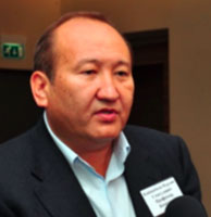
Kayshibaev Nurlan Smagulovich

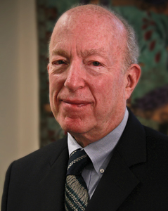
smagulovichProfessor Kayshibaev Nurlan Smagulovich passed at the age of 51 on June 17, 2013. Smagulovich was a famous neurologist of Kazakhstan. After graduating from medical school in 1984, he moved his way up from doctor-intern to the head of the Institute of Neurology.
During his career, he also headed the departments of neurology of Almaty city clinics, was the director of a health center in South Kazakhstan region, was a leading neurologist of Health Care Ministry of Republic of Kazakhstan, and was a leading neurologist of Almaty Department of Health Care Ministry.

He founded and headed the Institute of Neurology and the League of Neurologists of Kazakhstan, the member society of WFN (World Federation of Neurology).

He was a head of the Department of Neurology of the Almaty State Institute of Advanced Training of Physicians and a member of the Association of Neurologists of Kazakhstan.

He trained medical students and doctors from all over the country. His teaching career included work at the South Kazakhstan State Medical Academy, Kazakh National Medical University, Almaty State Institute of Advanced Training of Physicians and the Institute of Neurology.

He was particularly interested in cerebrovascular diseases, neurorehabilitation, degenerative and demyelinating diseases of nervous system, and diseases of the peripheral nervous system. One special area of focus was transient ischemic attacks (TIA), to which his master’s and doctoral dissertations were devoted.

He is the author of more than 78 scientific works, including a training manual for graduate students, interns and doctors, as well as methodical recommendations, including “Diagnosis and treatment of atherosclerotic transient cerebral ischemia” and “Electroneuromyographic criteria for early diagnosis and monitoring of pharmacological correction of subclinical and clinical manifestations of polyneuropathy in patients with newly diagnosed Diabetes mellitus type 2.”

Smagulovich and the members of the Institute of Neurology and the League of Neurologists of Kazakhstan in collaboration with the Kazakh National Medical University and the Almaty State Institute of Advanced Training of Physicians organized “Actual Problems of Neurology,” an annual conference in Almaty with international participation.

He was awarded the badge “High Achiever of Health Care of Kazakhstan” and certificates of honor of Kazakh National Medical University.

Smagulovich was a doctor of the highest qualification category, great clinician, gifted teacher, caring husband, father, son, brother, true friend, kind and sympathetic person.

Condolences to the family, friends and colleagues for this great loss.

Welcome to Editorial Advisory Board, Assistant Online Editor

Donald H.  Silberberg

Donald H. Silberberg

I am pleased to present the members of the Editorial Advisory Board. These individuals will help guide the continued development of World Neurology Online. Each of these distinguished neurologists has agreed to provide and solicit announcements and articles of interest from their regions and beyond.

I also am pleased to announce that Keith Newton, the World Federation of Neurology’s executive director, has agreed to serve as assistant editor of World Neurology Online. Please send your announcements, reports and articles to me silberbe@mail.med.upenn.edu and/or to Keith Newton keith@wfneurology.org.

Keith Newton

Keith Newton

As you prepare for what promises to be an exciting and informative World Congress in Vienna, keep in mind that two democratic processes will take place during the meeting.

The first is the selection of new officers for the WFN; the second is the choice of country for the 2017 World Congress. The WFN Council of Delegates votes both selections. If you wish to share your thoughts with your national delegate, and might not be sure who that is, you can obtain his or her name and email address from Keith Newton.

I look forward to seeing and meeting many of you in Vienna.

Editorial Advisory Board


Donna Bergen
Chicago, Illinois
USA


Amadou Gallo Diop
Dakar
Senegal


Geoffrey Donnan
Melbourne
Australia


Jerome Engel, Editor, World Federation of Neurology Website
Los Angeles, California
USA


John England, Chief Editor, Journal of Neurological Sciences
New Orleans, Louisiana
USA


Riadh Goudier
Tunis
Tunisia


Mark Hallett
Bethesda, Maryland
USA


Ryuji Kaji
Tokushima
Japan


Man Mohan Mehndiratta Mahndiretta
New Delhi
India


Girish Modi
Johannesburg
South Africa


Gustavo Roman
Houston, Texas
USA


Keith Newton, Executive Director of the World Federation of Neurology, Assistant Editor, World Neurology

Richard Bright Advanced Research on Apoplexy, Stroke

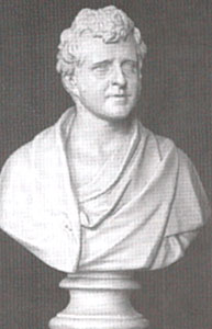
Figure 1. Marble bust depicting a young  Richard Bright (from Barry and Mackenzie 1998).

Figure 1. Marble bust depicting a young Richard Bright (from Barry and Mackenzie 1998).

Richard Bright was born in 1789 in Bristol. He received his medical training at the University of Edinburgh and at Guys Hospital, which he completed in 1812 with a doctorate on infectious erysipelas. (Berry and Mackenzie).

In 1820, Bright was elected assistant physician to Guy’s Hospital. There, he spent time in the company of a number of physicians whose names like his own became household names in medicine — James Parkinson, Thomas Addison and Thomas Hodgkin.

The eminent surgeon and scholar Sir Astley Cooper encouraged Bright in his research activities and impressed upon him the value of autopsies. After the publication of his epochal studies on diseases of the kidney in 1827, Bright recorded his observations on diseases of the brain. Volume II of his “Reports of Medical Cases” (Bright 1831) contains a wide range of neurological disorders.

The portrait of Bright that accompanies many of his biographical sketches shows him as a somewhat perplexed grandfather. It is likely however, that during his productive period, he resembled more a marble bust of him, which suggests a bright, alert and inquisitive young man. (See Figure 1.)

Bright made seminal observations on diseases of many organs, but it was his work on diseases of the kidneys that earned him undying fame and the title of “The Father of Nephrology.” This monumental accomplishment has overshadowed his important observations on diseases of the brain and other organs.

Apoplexy

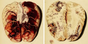
Figure 2. A: Plate V – Thrombosis of the superior sagittal sinus and feeding veins.  B: Plate VI – Petechial hemorrhages in cerebral venous thrombosis

Figure 2. A: Plate V – Thrombosis of the superior sagittal sinus and feeding veins. B: Plate VI – Petechial hemorrhages in cerebral venous thrombosis

Advances in anatomy and physiology initiated in the 16th and 17th centuries and information that accrued from autopsies of apoplectics provided facts that were not reconcilable with the humoral theory. Nevertheless, scholarly physicians at the beginning of the 19th century interested in what was then known as “apoplexy” still faced a bewildering assortment of opinions, dogma and hypotheses. “Apoplexy” was a term used by professionals and the educated laity for a disorder that “struck abruptly, causing a sudden abolition of all the activities of the mind, with the preservation, for a time, of the pulse and respiration” (Bonet).

The rest of the populace used the term “stroke” for this condition. Jean Fernel (1544) and Johann Jacob Wepfer (1658) found intracranial hemorrhage at autopsies of apoplectics. These

Figure 3. Sagittal section though the left hemisphere showing cerebral softening.

Figure 3. Sagittal section though the left hemisphere showing cerebral softening.

observations were repeatedly confirmed, and the notion took hold that apoplexy (stroke) may be a disorder of cerebral blood vessels, rather than an accumulation of phlegm or some other humor in the cerebral ventricles, as proposed by Galen on the basis of the humoral theory.

The classification of apoplexy that emerged from autopsy reports published in the 17th and 18th centuries was still accepted at the beginning of the 19th century: “sanguineous apoplexy,” caused by intracranial hemorrhage, and “serous apoplexy,” in which the effusion of serum was held responsible for the apoplectic state.

Morgagni (1769) recognized a third type of apoplexy, in which neither blood nor serum was effused.

bright_fig4

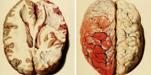
Figure 4. Images from Plates XV and XVI. A: Swelling and softening of the left cerebral hemisphere. B: Striking hyperemia of the swollen hemisphere. The labeling on Plate XV indicates that the right hemisphere was swollen, whereas in Plate XVI the hyperemia is said to be on the left side. It seems that the images are mislabeled.

On the authority of Morgagni and other “great masters,” any amount of fluid present in the ventricles and the subarachnoid space was, for a time, deemed to be abnormal by many scholars and a cause of apoplexy. The concept of “serous apoplexy” endured until the latter part of the 19th century.

Ramolissement

New thoughts on apoplexy emerged as the result of studies at the Paris Hospitals at the turn of the 19th century (Karenberg). While Rochoux (1812) defined apoplexy as cerebral hemorrhage, Rostan’s discovery, published in 1819, of the phenomenon of ramolissement (cerebral softening) in patients with the syndrome of apoplexy, led to new notions concerning this condition. Rostan suggested that ramolissement, was a “disease sui generis,” a primary disease, and that it was peculiar to the brain, while Lallemand (1820) speculated that ramolissement was an inflammatory condition (Karenberg).

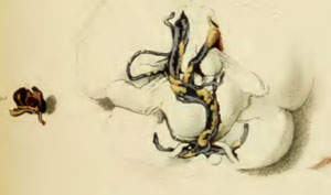
Figure 5. Images from Plate IX. Left: Partially clotted aneurysm from a patient with a subarachnoid hemorrhage. (Case CXXV) Right: Arteries of the brain with extensive cerebral atherosclerosis in a patient with intracerebral hemorrhage (case CXXXV).

Figure 5. Images from Plate IX. Left: Partially clotted aneurysm from a patient with a subarachnoid hemorrhage. (Case CXXV) Right: Arteries of the brain with extensive cerebral atherosclerosis in a patient with intracerebral hemorrhage (case CXXXV).

Bright’s Reports of Medical Cases

Bright held that the ultimate object of medical research was:”to devise remedies most likely to be beneficial in each form of disease,” which, he said, was bound to be facilitated by the understanding of their causes.

To this end, he arranged his cases of diseases of the brain into sections according to what were then understood to be “…. the more obvious sources of disease,” namely inflammation, pressure, irritation and inanition.

This article provides a review of Bright’s observations and thoughts on apoplexy or stroke, which he recorded in the sections on inflammation and pressure.

Inflammation  and Cerebral Softening

In the section on inflammation, Bright described cases illustrative of various inflammatory disorders, such as meningitis, intracranial suppuration, as well as cerebral congestion and also deficient cerebral circulation.

With the first case in this group, Bright established that cerebral softening may be caused by cerebral congestion due to cerebral venous thrombosis. (See Figure 2.) He concluded that venous thrombosis occurred in debilitated states, which in his patient he attributed to pneumonia. To illustrate “the tendency to coagulate which the blood acquires when the system is under the influence of different debilitating causes,” he added three cases with systemic venous thrombosis associated with chronic illness and one with ovarian cancer.

Ramolissement Due to  Deficient Blood Supply

In the last five cases in this section, cerebral softening was presented as the principal pathological finding. Bright stated that the symptoms generated by cerebral softening resemble those caused by “pressure,” which he believed to be the ultimate source of the cardinal symptoms of apoplexy, namely paralysis and impaired consciousness.

Case LXXXI was a 68-year-old man who suddenly became unconscious. When he regained consciousness after 10 minutes, he was hemiplegic on the right, with right hemisensory loss and aphasia. Death occurred about three weeks after the onset of his illness. Softening of the anterior part of the left cerebral hemisphere extending from the fossa Sylvii to the corpus striatum was discovered at autopsy. (See Figure 3.) (Case LXXXI, p 178). The arteries of “fossa Sylvii” “fossa Sylvii,” the carotids and of the circle of Willis were extensively diseased, the mitral valve was abnormal.

In the next two cases, the illness developed gradually, and the patients were young (28 and 30 years). Autopsy showed extensive cerebral softening. In one of these cases, there was a remarkable cortical hyperemia of the swollen and softened cerebral hemisphere. (See Figure 4.)

These cases showed severe ossification of the arteries and extensive cerebral softening of one cerebral hemisphere. Bright stated that abnormal arteries: “… certainly produced in my mind an impression that the proper supply of blood had been cut off by some change in the vessels of the pia mater, or some obstruction in their passage through the cineritious substance, and in this way death and disorganization of the brain had been produced.”

Bright stated that “ossification of the arteries might well be considered as laying the foundation for defective circulation,” and that cerebral softening corresponded to gangrene of the limbs.

These three cases constitute the basis for a concept that was novel at that time, namely that apoplexy (stroke) may be caused by arterial obstruction as well as by the extravasation of blood.

Cases Illustrating  the Effects of Pressure

The section on the effects of “pressure” includes cases illustrative of the effects of vascular turgescence, “pressure of the brain from serous effusion independent of inflammation,” cases in which some organic change had given rise to pressure, and cases in which blood had been effused from ruptured vessels.

Bright’s attempts at localization, which he believed was of importance for accurate diagnosis, represents a significant advance toward modern neurology.

Pressure Due to Effused Serum

Bright maintained that by generating pressure effused serum caused or enhanced apoplexy. He did not use the term “serous apoplexy,” but his cases “illustrating the occurrence of pressure of the brain from serous effusion independent of inflammation” show that he could not free himself of the concept of “serous apoplexy.”

Pressure Due to  Extravasation of Blood

The subsection on intracranial hemorrhage begins with five cases “in which the effusion of blood has been found upon the surface of the brain.” Here Bright gives excellent descriptions of the symptoms that characterize subarachnoid hemorrhage.

In one of these cases, a 19-year-old man “while sitting on the chamber utensil suddenly exclaimed, ‘Oh my head.'” Bright found blood on the surface of the brain and a ruptured, partially clotted middle cerebral artery aneurysm. (See Figure 5a.)

The remainder of the cases in this section showed intracranial hematomas of various ages and a subdural hematoma. The arteries showed evidence of atherosclerosis (See Figure 5b), not only in older patients, but also in young apoplectics.

Bright noted severe bradycardia in a patient with a subdural hematoma and suggested that “it is by no means improbable that the pressure experienced by the brain during the existence of the coagulum…had some share in producing the retarded circulation.”

A patient who suffered “successive paralytic attacks” and eventually reached a “state of imbecility” showed multiple small softenings of the brain at autopsy, “the brain resembling ‘Parmasan’ ” (sic) cheese.” The aortic and mitral valves were “very much thickened.” Bright declared that the size of the softenings was related to the size of the diseased vessel.

In this description, we recognize TIAs (transient ischemic attacks), RINDs (reversible ischemic neurologic deficits), vascular dementia and lacunes.

Additional symptoms preceding apoplexy recorded by Bright were: headache, giddiness, migraine-like headaches, numbness left face, two or three attacks of hemiplegia, successive slight attacks of transient hemiplegia, affecting sensation more than motion, occasional vertigo and episodic incoherence.

Bright maintained that the location of disease in the brain or the blood vessels determined the nature of the symptoms. He furthermore suggested that the awareness of this fact would enhance diagnostic accuracy, which in turn was bound to improve treatment.

Unusual Cases

Bright recorded two unusual cases: Hemiplegia ipsilateral to the cerebral lesion and “tormenting pain” in paralyzed limbs that were deprived of sensation.

He was skeptical about the genuineness of signs ipsilateral to the brain lesion: “having once or twice nearly deceived myself by the imperfect accounts of friends in such cases, I am not willing to admit them hastily.”

Treatment of Apoplexy

“It is impossible to remove a strong attack of apoplexy and not easy to remove a weak attack.” (Hippocrates)

This Hippocratic aphorism did not deter physicians from trying to cure apoplexy, both strong and weak.

Although it was well known that some apoplectics survived without any treatment. Survival was usually credited to treatment, and it was often implied that death resulted when treatment was inadequate or withheld. This helped to perpetuate the belief in the effectiveness of the diverse interventions then in fashion.

“In the treatment of apoplexy, the most important point is the employment of bleeding; the judicious use of which powerful remedy the cure greatly depends.” (Bright 1831, p 334)

Although the methods of treatment in the early 19th century were still based largely on surviving humoral concepts, Bright’s rationale for abstracting blood was “a view of checking the hemorrhage,” and the hope of reducing congestion rather than the restoration of a balance of the four humors. The supplemental maneuvers had the same rationale for Bright.

Whatever the rationale, the abstraction of blood was the recommended remedy for apoplexy and a nearly universal practice in Bright’s time. Venesection or arterial section close to exsanguination, supplemented by the application of leeches, cupping, both wet and dry, were standard procedures in the treatment of apoplexy and for many other ailments. The pulse was monitored and abstraction was stopped when the pulse became depressed. Caution was advocated in the exsanguination of feeble individuals. The energetic bleeding that was practiced at that time must have lowered blood pressure for a time, and therefore might have reduced bleeding.

Blisters and Setons were used as additional supplements to bleeding. Purging was in regular use. The induction of vomiting was occasionally practiced but it was considered harmful by those who feared that the strain associated with vomiting may be harmful.

Bright used stimulants when “the patient grew cold” and “the pulse fluttering.” He poured “vinegar down the throat, brandy if it could be procured” or “a few drops of compound spirits of ammonia to elicit cough that tended to rouse the patient.” Cloth dipped in hot water was applied to the stomach. Cold cloths were “dashed on the temples and forehead with a sudden jerk,” when the head felt hot and the carotids were throbbing. Frequently, the head was shaved, and cold applied to the shaven head.

Such therapy persisted throughout the 19th century, and bloodletting for stroke was not unknown in the 20th century. Few went as far as Thomas Sydenham, who stated: “I have consulted my patients’ safety and my own reputation most effectually by doing nothing at all.”

Summary

Bright showed that cerebral softening has many causes, one of which was a deficient blood supply caused by diseased arteries which like Abercrombie (p.25), he considered to be analogous to the gangrene of limbs.

Bright has shown that cerebral softening can be caused by thrombosis in the cerebral venous system. He also suggested that chronic illness and malignancy are risk factors for venous thrombosis. Bright also believed that venous obstruction contributed to thrombosis in his case of ovarian cancer. He thus came close to anticipating Virchow’s coagulation triad (stasis, abnormal blood, diseased blood vessels).

The determination by Bright that cerebral softening was caused by a deficiency of blood supply represents a significant advance in the understanding of cerebrovascular disease.

Bright’s description of the symptomatology of subarachnoid hemorrhages compares well with current accounts. His demonstration of a partially clotted aneurysm as a cause of an intracranial hemorrhage was one of the earliest observations of its kind.

Bright described TIAs and reversible ischemic neurological deficits (RINDs), and the lacunes that may result from them, and he recognized that dementia may be caused by multiple areas of cerebral ischemia. He further recognized that small lacunes were the result of obstructions of small arterial branches, whereas the obstruction of larger branches caused larger cavities. Although the realization that these were embolic came later, he did document carotid and valvular disease in a number of cases.

Bright associated bradycardia with intracranial hypertension.

He also gave a good description of intractable pain in pain paretic limbs with “destroyed sensation,” now known as “the thalamic syndrome of Déjérine.” He mentioned a case of paralysis ipsilateral to a cerebral lesion, but was hesitant to accept it as genuine.

Bright’s attempts at localization, which he believed was of importance for accurate diagnosis, represents a significant advance toward modern neurology.

Richard Bright’s observations on cerebral softening and apoplexy contributed to the current understanding of cerebrovascular disease and its division into occlusive and hemorrhagic strokes.

References

1. Abercrombie, J. Pathological and practical researches on diseases of the brain and the spinal cord. Edinburgh, Waugh and Innes.1828.

2. Berry, D, Mackenzie C. Richard Bright 1789-1858. Physician in an Age of Revolution and Reform. Royal Society of Medicine Services, Ltd. 1992.

3. Bright, R. Reports of medical cases selected with the view of illustrating the symptoms and cure of diseases. Vol I, Vol II London, Longman.1827, 1831.

4. Fernel, J. Universa Medicina. Geneva. De Tornes. 1544.

5. Karenberg, A. Stroke at the Paris Hospitals. The emergence of anatomico-clinical concepts after 1810. J. Hist. Neurosc. 19: 390.1872. 2010

6. Lallemand F.Researches anatomo-pathologiques sur l’encéphale et ses dépendences. Paris. 1820

7. Morgagni, GB. The Seats and Causes of Disease, Vol. 1, London, McMillan and Cadell, 1769.

8. Rochoux, J A, Recherches sur L’Apoplexie, Paris, Bechel Jeune. 1814.

9. Rostan, L.. Recherches sur le ramollissement du cerveau; ouvrage dans lequel on s’efforce de distinguer les diverses affections de ce viscè
re par des signes caractéristiques. 2nd Edition, Paris, Bechet. 1819.

10. Schutta, HS, Abu-Amero, KK, Bosley, TM. Exceptions to the Valsalva doctrine. Neurology, 74: 329-335. 2010.

11. Wepfer, J. J. Observationes anatomical, ex cadaveribus eorum quo sustulit apoplexia, cum exercitation ejus loco affecto. Schaffhausen, 1658.