Neurological Manifestations of Zika Virus Infection: What Neurologists Need to Know

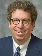
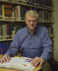
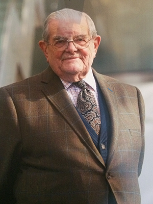
By Avindra Nath, MD, and James Sejvar, MD

headshot_JJS-3

James Sejvar, MD

headshot_Avi

Avindra Nath, MD

In recent years, there has been an emergence of several major viral infections with devastating neurological consequences, including West Nile virus, dengue, chikungunya, enterovirus D68, Ebola and now Zika virus. Increased global travel and climate change, leading to changing patterns of vector distribution and behavior are among the major reasons for the emergence of these infections. Zika virus is the most recent epidemic that is having devastating effects on human populations in affected regions, and is rapidly spreading across the South American continent.

Epidemiology

Zika virus was first identified from a primate in 1947 in the Zika forest of Uganda.1 The first human cases occurred in Africa and then in Southeast Asia in the 1960s.2,3 During the intervening years, Zika virus was associated with isolated cases or small outbreaks mainly in Africa. In 2007, there was an outbreak in Yap, the Federated States of Micronesia, where nearly three-quarters of the population was infected.4, 5 This represented the largest outbreak of Zika virus infection to that point. In 2013, there was an epidemic in French Polynesia, which was associated with a reported increase in cases of the autoimmune peripheral nerve disorder Guillain-Barre syndrome, although a causal association between Zika virus and Guillain-Barre syndrome was never established.

In December 2014, Zika virus was first detected in Brazil. Although it is unknown how it was introduced into Brazil, some hypothesize that a traveler attending the 2014 football/soccer World Cup introduced the virus. The outbreak in Brazil was fast moving and large. Tens of thousands of people became ill, and likely millions of people were infected. Similar to French Polynesia, shortly after the beginning of the Zika virus outbreak, clinicians began reporting larger-than-expected numbers of Guillain-Barre syndrome. Many of these people had reported a febrile rash illness compatible with Zika in the days or weeks before their weakness onset. In addition, clinicians in Brazil noted a 20-fold increase in microcephaly in 2015, compared to previous years, with microcephalic babies born approximately eight to nine months after the first recognition of Zika virus. Some of the infants’ mothers reported a rash illness compatible with Zika virus infection while pregnant, leading to the suspicion that the microcephaly was somehow associated with Zika virus infection.

Nearly 90 percent of the cases of microcephaly occurred in the northeastern region of the country,6, 7 areas experiencing some of the heaviest burdens of Zika virus infection as well. French Polynesian health authorities reported an unusual increase in central nervous system malformations in babies born during a Zika virus outbreak on the islands from 2014 to 2015.6 The infection has now spread across most of South America and Mexico. To date, few cases have been reported in the United States among travelers returning from Zika virus-affected regions.8, 9

Virology and Pathophysiology

Zika virus is a positive-sense, single-stranded RNA virus (genome 10.7 K nucleotides) belonging to the flaviviridae family, which includes dengue, yellow fever, Japanese encephalitis, St. Louis encephalitis and West Nile virus. It has the ability to cross the placenta and cause developmental brain abnormalities in children, suggesting that the virus likely infects neural stem cells. The severity of brain malformations may be related to the stage of fetal development at the time of infection. Microcephaly would be the most common manifestation, but if infection were to occur in earlier stages of fetal development, anencephaly or lissencephaly may occur.

The pathophysiology of ascending paralysis and myelitis in adults is unknown. However, mice injected with the virus can develop paralysis, suggesting direct invasion by the virus, although an immune-mediated, post-viral syndrome is also possible. It remains unknown if once infected and recovered if an individual develops long-term immunity or not, and if recurrent infections or relapses can occur. Questions regarding long-term viral persistence in tissue reservoirs also remain unanswered.

Transmission

The virus is transmitted by the Aedes species of mosquitoes 10, in particular Aedes aegypti, the vector involved with transmission of dengue, a closely related flavivirus. Additionally, experimental evidence suggests the virus can be transmitted by Asian tiger mosquitoes (Aedes albopictus) 11, 12, which can survive in cold temperatures. Most arboviruses have an intermediary host or “reservoir.” For West Nile virus, birds, particularly corvids, serve as these reservoirs. For Venezuelan, Western and Eastern equine encephalitis viruses, horses serve this role, and for Japanese encephalitis virus, it is primarily pigs. However, the transmission of Zika virus generally occurs directly between humans and mosquitos. There is some evidence that human-to-human transmission may occur through sexual intercourse, and the virus has also been detected in saliva, so the potential for oral transmission also exists. The virus has been isolated from the amniotic fluid of pregnant women and blood and tissues of newborns, suggesting materno-fetal transmission.13 So far, an intermediary host has not been identified.

Clinical Manifestations

Zika_Photo1

Female Aedes aegypti mosquito

The majority of Zika virus infections ­­­— 80 percent — are clinically asymptomatic.4 Among persons who develop symptoms, Zika virus infection is generally considered to be mild, causing fever, rash and body aches. Some may develop conjunctivitis. Symptoms usually last one week.

The full spectrum of neurological complications from this viral infection remains unknown. The epidemiological association between microcephaly and the infection seems strong. In Brazil, annual reported rates of microcephaly would generally be somewhere around 150 cases per year. Reportedly, between October 2015 and January 2016, more than 3,500 babies were born with the condition. CT brain scans show evidence of widespread calcification. Other malformations, such as anencephaly and lissencephaly, might also occur. It remains uncertain if other organs may be involved in addition to the brain. However, the differential diagnosis of microcephaly is broad. Hence, when presented with a patient with microcephaly, it remains important to consider other common causes, such as genetic, craniostenosis, and infections, such as toxoplasmosis, rubella, varicella zoster virus and cytomegalovirus. Intrauterine cerebral anoxia, exposure to drugs, alcohol and other toxins, malnutrition and metabolic disorders such as phenylketonuria can also cause microcephaly. Patients with microcephaly often have developmental delay, difficulty with gait and balance, mental retardation, seizures and hyperactivity.

Guillain-Barre syndrome appears to be a recurring possible complication of Zika virus infection. Following the introduction of Zika virus into French Polynesia, clinicians began reporting larger-than-expected numbers of Guillain-Barre syndrome cases on the island.14 Following the introduction of Zika virus to Brazil in December 2014, again, reports surfaced of large numbers of Guillain-Barre syndrome cases. In Brazil, few cases of Guillain-Barre syndrome had laboratory confirmation of Zika virus, but currently the primary method of diagnostic testing is through the detection of viral RNA through polymerase chain reaction. In Guillain-Barre syndrome, by the time the clinical features of limb weakness develop, it is unlikely that there would still be circulating virus, and, as such, detection of viral RNA would not be expected. Less commonly, some patients have been thought to have a myelitis or polio-like manifestations. Currently, it is unclear if these are all related or if indeed both spinal cord and peripheral nerves can be involved. Thus, in Brazil, epidemiologic evidence and the close temporo-spatial clustering of both Guillain-Barre syndrome and Zika virus cases provides intriguing circumstantial evidence for an association.

In other cases in which the virus was newly introduced, reported increases of Guillain-Barre syndrome cases have invariably appeared, including in Colombia, Venezuela and, more recently, El Salvador, which reported 46 Guillain-Barre syndrome cases in a five-week period from December 2015 to early January 2016. That is nearly three times more than the country would normally see in that timeframe. Laboratory substantiation of an association between Zika virus and Guillain-Barre syndrome has proved challenging, however. As noted, by the time of onset of weakness, the virus would be expected to be cleared from the body, and molecular techniques to identify the virus or viral RNA would not be expected to be positive. Detection of Zika virus-specific antibodies would provide evidence of current or prior infection. However, that method also has its challenges. Dengue virus is a closely related flavivirus to Zika, and invariably co-circulates in all areas currently associated with Zika virus. However, dengue virus infection has also rarely been associated with Guillain-Barre syndrome, and laboratory testing by serology is challenging due to the substantial cross-reactivity of antibodies between Zika virus and dengue virus.

Since these viruses are carried by the same mosquito vector and co-circulate at the same times of the year, it can be challenging to differentiate between infection with the two viruses.15 Development of a robust serologic assay that can reliably differentiate Zika virus from dengue and other closely related flaviviruses will be crucial in order to provide laboratory evidence of Zika-associated Guillain-Barre syndrome, as well as other late complications of Zika virus. Currently, the nature of the neuropathy is not known, as results of electrodiagnostics to determine the clinical sub-type of Guillain-Barre syndrome possibly associated with Zika virus has been rarely reported. It would be important to know if it is axonal or demyelinating and if it is immune mediated. This could affect treatment and prognosis. Recovery from demyelinating neuropathies is generally better than those due to axonal injury. Isolated reports suggest that the neuropathy may be demyelinating and may respond to treatment with intravenous immunoglobulin.14

Laboratory Diagnosis

Viremia occurs only during the first few days of the illness, but if blood samples are obtained during that time, virus can be detected by polymerase chain reaction.16 Following this phase, IgM antibodies can be demonstrated by ELISA or Western blot analysis. Previous epidemics have noted that there is cross reactivity between antibodies to Zika and other arboviruses such as dengue.5 The Centers for Disease Control and Prevention (CDC) has issued guidelines for the testing of infants born with possible Zika virus infection.17

Treatment and Prevention

Currently, there is no effective treatment or vaccine against the virus. Hence, prevention is key with control of mosquito populations and prevention of mosquito bites. Travel advisories have been issued for pregnant women not to travel to areas experiencing Zika virus outbreaks. For individuals who suffer from the neurological consequences of the infection, long-term supportive and symptomatic treatment is key. The socio-economic impact of the infection, particularly if the association between Zika virus and microcephaly holds true, will likely be huge and felt for decades. While the large number of cases of microcephaly is tragic, whatever the eventual cause turns out to be, it will result in large numbers of children with developmental disorders and begs for the need to train personnel in a wide variety of health disciplines, including neurology, rehabilitation, specialized nursing, social services, etc., to care for and treat this population. Ongoing surveillance for Zika virus in the Americas and elsewhere, to monitor its continued spread, as well as documentation of infection among travelers returning from affected areas will be critical. Development of more robust serologic assays that can differentiate Zika virus from other closely related flaviviruses will be an important tool to substantiate an association between Zika virus and devastating neurologic conditions, such as Guillain-Barre syndrome and microcephaly. Ultimately, the long-term epidemiologic pattern of Zika virus will be important to monitor.

References:

  1. Weinbren, M.P. and M.C. Williams, Zika virus: further isolations in the Zika area, and some studies on the strains isolated. Trans R Soc Trop Med Hyg, 1958. 52(3): p. 263-8.
  2. Simpson, D.I., Zika Virus Infection in Man. Trans R Soc Trop Med Hyg, 1964. 58: p. 335-8.
  3. Olson, J.G., et al., Zika virus, a cause of fever in Central Java, Indonesia. Trans R Soc Trop Med Hyg, 1981. 75(3): p. 389-93.
  4. Duffy, M.R., et al., Zika virus outbreak on Yap Island, Federated States of Micronesia. N Engl J Med, 2009. 360(24): p. 2536-43.
  5. Lanciotti, R.S., et al., Genetic and serologic properties of Zika virus associated with an epidemic, Yap State, Micronesia, 2007. Emerg Infect Dis, 2008. 14(8): p. 1232-9.
  6. Control, E.C.f.D.P.a., Rapid risk assessment: Zika virus epidemic in the Americas: potential association with microcephaly and Guillian Barre syndrome. 10 December 2015.
  7. Bogoch, II, et al., Anticipating the international spread of Zika virus from Brazil. Lancet, 2016.
  8. Control, C.f.D., CDC telebriefing: Zika virus travel alert. 15 January 2016.
  9. Hennessey, M., M. Fischer, and J.E. Staples, Zika Virus Spreads to New Areas – Region of the Americas, May 2015-January 2016. MMWR Morb Mortal Wkly Rep, 2016. 65(3): p. 55-8.
  10. Li, M.I., et al., Oral susceptibility of Singapore Aedes (Stegomyia) aegypti (Linnaeus) to Zika virus. PLoS Negl Trop Dis, 2012. 6(8): p. e1792.
  11. Grard, G., et al., Zika virus in Gabon (Central Africa)–2007: a new threat from Aedes albopictus? PLoS Negl Trop Dis, 2014. 8(2): p. e2681.
  12. Wong, P.S., et al., Aedes (Stegomyia) albopictus (Skuse): a potential vector of Zika virus in Singapore. PLoS Negl Trop Dis, 2013. 7(8): p. e2348.
  13. Pan American Health Organization and World Health Organization. Epidemiological Alert: Neurological syndrome, congenital malformations, and Zika virus infection. Implications for public health in the Americas. http://www.paho.org, 1 December, 2015.
  14. Oehler, E., et al., Zika virus infection complicated by Guillain-Barre syndrome–case report, French Polynesia, December 2013. Euro Surveill, 2014. 19(9).
  15. Carod-Artal, F.J., et al., Neurological complications of dengue virus infection. Lancet Neurol, 2013. 12(9): p. 906-19.
  16. Faye, O., et al., One-step RT-PCR for detection of Zika virus. J Clin Virol, 2008. 43(1): p. 96-101.
  17. Staples, J.E., et al., Interim Guidelines for the Evaluation and Testing of Infants with Possible Congenital Zika Virus Infection – United States, 2016. MMWR Morb Mortal Wkly Rep, 2016. 65(3): p. 63-7.
Avindra Nath, MD, is intramural clinical director and a senior investigator with the Section of Infections of the Nervous System, National Institute of Neurological Diseases and Stroke, National Institutes of Health, Bethesda, Maryland. James Sejvar, MD is a neuroepidemiologist with the division of High-Consequence Pathogens and Pathology, National Center for Emerging and Zoonotic Infectious Diseases, Centers for Disease Control and Prevention, Atlanta, Georgia. The authors have no financial relationships relevant to this article to disclose. The findings and conclusions in this article are those of the authors and do not necessarily represent the official position of the Centers for Disease Control and Prevention or National Institutes of Health.


Teaching Courses Expand Education at WCN

By Wolfgang Grisold

Table 1: Teaching Courses The free early morning teaching courses have remained the most visited teaching courses, despite the early hour. Teaching courses on education, advocacy and career received little attendance, yet these topics are inherent topics of the WFN. Also, palliative care received little attention as teaching courses. This shows that the topic needs promotion within the scientific sessions. All teaching courses will be available on the WFN website after the Congress.

Table 1: Teaching Courses The free early morning teaching courses have remained the most visited teaching courses, despite the early hour. Teaching courses on education, advocacy and career received little attendance, yet these topics are inherent topics of the WFN. Also, palliative care received little attention as teaching courses. This shows that the topic needs promotion within the scientific sessions. All teaching courses will be available on the WFN website after the Congress.

The WCN took place Oct. 31-Nov 5, 2015, at the CasaPiedra Conference Center in Santiago, Chile, with 3,500 delegates from 112 countries participating and creating a joint sense of neurology worldwide. The lectures from the World Health Organization (WHO) and the presentation of the new WHO Atlas of Neurology showed the disparity of neurological workforces around the world and documented the importance of the WFN activities in the work with global organizations. One of the goals and aims of the WFN is to spread neurological development and neurological science and update participants on current issues, but also to highlight new neurological fields of development.

Scientific sessions and topics are an excellent venue and bring participants close to the leading persons in the field. This concept was successfully used in plenary lectures and scientific main topic sessions.

The WFN also has long kept the tradition of offering teaching courses at its congress. This concept has been broadened since Marrakesh, as the WFN teaching courses now are being offered on all days of the congress. The teaching courses were co-chaired by Sergio Castillio (local committee, Chile) and Wolfgang Grisold (teaching course committee, WFN). The topics presented include many general and common topics, such as stroke, dementia, epilepsy and others.

Also, several courses with hands-on opportunities were offered to include and engage participants in practical issues, such as EMG, ultrasound and botulinum toxin treatment. Experience at the WCN in Marrakesh and Vienna has shown that these types of highly interactive teaching courses, despite often technical issues and difficulties, are well appreciated by attendees.

In Chile, 36 teaching courses were held. The list (Table 1) shows the topics presented, which helped to disseminate educational news and expert knowledge. Teaching courses offer attendees the advantage of being in a smaller group and interacting with renowned experts. This facilitates a climate of interaction by allowing questions and discussion of opinions.

Dr. Freedman lectures on education.

Dr. Freedman lectures on education.

One special component of the WCN teaching courses are the early morning free teaching courses, a one-hour teaching session presented by a single expert in the field. This expert has the privilege to create his or her talk in the form of a lecture, deviating from the usually shorter teaching course lectures, and allowing the speaker to emphasize and shape the message that he or she considers important. Despite the early time of these teaching courses, the number of participants was between 100 and 200 per course (and 339 for the topic of epilepsy). The topics included MS (95), chronic inflammatory demyelinating polyneuropathy (125) and the top 10 advances in neurology (95).

Cynthia L. Harden presents a morning teaching course on epilepsy.

Cynthia L. Harden presents a morning teaching course on epilepsy.

Knowledge, skills and tutoring from experienced lecturers cover many aspects of neurology. The issue of palliative care is a topic with an increasing scientific basis1. Still palliative care is often confounded with end of life care, or worse, a situation of helplessness. The WFN has established a Research Group on this topic, and the Teaching Course on palliative care was an important step in establishing this important topic into our future curricula.

Dr. Nitrini speaks on education in Latin America.

Dr. Nitrini speaks on education in Latin America.

All neurologists, working in any part of the world, also are aware of the importance of training, lecturing and teaching, and also that the diversity of careers from hospital to consultant level and from academic career to nonacademic career is wide. For this reason, a teaching course on education in neurology covered the topics on how to establish a curriculum, as well as how to integrate neurologic knowledge and procedures into the national situation, politics and legislation. It is important to define new learning strategies and teaching methods, to eventually support or replace the traditional apprenticeship model of neurologic education. Also the classic concept of knowledge, skills and competence will need to include aspects of professionalism and attitude. Knowledge is acquired in an incremental, almost pyramidal way. Yet, individuals and institutions also will need to adapt to new developments, and this procedure of systemic approach to replace old content or procedures is termed unlearning

Several speakers and participants gather for a photo opp.

Several speakers and participants gather for a photo opp.

One lecture by Dr. Freedman focused on the merging and growing possibilities of e-learning. He presented his own experience with video conferences worldwide and gave important clues to enlarge the scope of this project.

The local flavor of Latin American neurology was presented by Prof. Nitrini of Brazil, who outlined educational activities in Latin America. He discussed the rise of impact factors among publications from Latin America in the past years.

Within the topic of applied teaching and education, translation was discussed by Prof. Camfield of Canada. This term describes the often difficult transfer of young patients from pediatric neurology into adult neurology. Translation also has been well described in other fields of pediatrics and demonstrates a clash of concepts between different concepts of patient care and involvement of careers.

Prof. Raad Shakir outlines the role of the WFN in the Palatucci course.

Prof. Raad Shakir outlines the role of the WFN in the Palatucci course.

Patient advocacy is an emerging concept, which empowers physicians to not only be involved in the provision of the best medical care, but also to advocate for patients in the micro- and macro-environment. This concept in neurology is carried forward by the American Academy of Neurology (AAN) Palatucci courses in the United States, and the AAN and WFN implemented a joint AAN-WFN one-day course in Chile. Topics on advocacy, press work and presentations were given to a small, but soon to be powerful group. Also WFN President Raad Shakir presented his concept of worldwide engagement of the WFN in health matters.

A good turnout attends a day programming.

A good turnout attends a day of programming.

Last but not least, some of the most important targets of education are patients and caregivers. Despite the importance of these groups, patient awareness days are, surprisingly, often not included in congresses. Since WCN 2015, the WFN implemented a patient day into it congresses. This year’s topics included stroke, epilepsy, MS and dementia, and WFN Past-President Vladimir Hachinski served as convenor and engaged in the discussion.

Footnote

  1. Oliver DJ, Borasio GD, Caraceni A, de Visser M, Grisold W, Lorenzl S, Veronese S, Voltz R. “A consensus review on the development of palliative care for patients with chronic and progressive neurological disease.” Eur J Neurol. (Oct 1, 2015): doi: 10.1111/ene.12889).


European ‘Wanderjahr’: Postgraduate Training in Nervous Diseases for Americans in the 1880s

By Peter J. Koehler

Thomas Neville Bonner (1923–2003)

Thomas Neville Bonner (1923–2003)

“Medical students constitute the only class of students who in any considerable number follow the good old German custom of supplementing their regular course of study (Lehrjahre) by a season of travel (Wanderjahre) for the purpose of seeing how people in other places perform that work which is to occupy the remainder of their lives.” — Henry Hun, 1883

Since 2010, we have published a number of short papers on international relationships and exchange in the neurological community. It is of interest to note that the leading centers of medical education changed over the past centuries and thereby advanced international exchange. Italian (Padua) and French (Paris, Montpellier) universities were popular in the 16th and 17th centuries. Leiden (the Netherlands with Boerhaave and Albinus) became popular in the early 18th century, and later, during that century, the center moved to Edinburgh (with Whytt, Cullen and the Monros). Then, in the beginning of the 19th century, it was clearly Paris that, following the revolution, became a center of integrated medicine and surgery, attracting foreign students.

Henry Hun's Guide to American Medical Students in Europe

Henry Hun’s Guide to American Medical Students in Europe

Of particular interest in the second half of the 19th century is the great number of foreign (American, Russian, Japanese, Scandinavian, etc.) students and physicians, who visited German-speaking countries between 1870 and 1914. The American medical historian Thomas Neville Bonner (1923–2003) stated that in this period, over 10,000 American physicians studied in Vienna, where some of the courses were given in English. He called it “the German magnet” and noted that “at least a third and perhaps a half of the best known men (and women) in American medicine of this era received some part of their training in a German (or Austrian) university: (Bonner, p.23).

The well-known American internist William Henry Welch, one of the founding professors of Johns Hopkins Medical School, opined that it was “conventional Mecca of American practitioners” and advised to “stick to Germany, where I find all the opportunities for learning pathology which I could desire.” It is of no surprise that American medical education was influenced by returning physicians. Johns Hopkins University Medical School was organized according to the German model. In this period of increasing specialization in medicine, it was important for physicians to improve their knowledge in Europe.

Henry Hun

Henry Hun

As improvement of medical practice was considered more important than research, smaller numbers of (mostly younger) Americans visited other German universities, including Leipzig (Carl Ludwig), Heidelberg, Breslau and Strasburg, with the purpose to do scientific work. Although a minority, the latter persons were important for the origin of medical research in the U.S. The majority went for postgraduate training, in particular for improving skills in a clinical specialty. Some of the courses were organized to train the practical use of the ophthalmoscope, microscope, laryngoscope and stethoscope. In April 2011, I discussed the European peregrination of Bernard Sachs between 1878 and 1884.

Features of Specialization in Medicine/Neurology

  • Education/Medical Curriculum
  • Neurological Practice/Special Hospitals
  • Instruments
  • University Chairs
  • Societies
  • Journals/Monographs
  • Success of Specialization Determined by:
  • Economic Reward
  • Social Prestige
  • Ideology of Progress
  • Influence of Public, etc.

A Guide for Medical Students in Europe

Theodore Meynert

Theodore Meynert

In this issue, I wish to discuss a particular book that was published to help American students and physicians finding their way in Europe, notably Henry Hun’s Guide to American Medical Students in Europe (1883). Henry Hun (1854–1924) was a lecturer on diseases of the nervous system at the Albany Medical College. His father, Thomas Hun, one of the founders of this college, stayed in Europe for his postgraduate studies for six years (1830s), and although professor of the Institutes of Medicine, he gave lectures on the nervous system (in a period before specialization in neurology started). Henry’s older brother Edward (1842–1890) became chair of diseases of the nervous system in Albany and was among the original members of the American Neurological Association, Which was founded in 1875. Following graduation from Harvard Medical School, Henry Hun followed postgraduate courses in several European cities for over two years. He became professor of diseases of the nervous system at Albany in 1884. In the preface to his Guide, he noted that “Every year, a large number of Americans go to Europe to complete their medical studies. Unfortunately the great majority of these students have very little definite information about the different universities or about the way in which medicine is taught abroad, and on this account they lose much valuable time in getting to work.” The book has 151 pages, and, although we know he himself visited Vienna, Heidelberg, Berlin, Paris and London, most of the book is on German (speaking) universities and hospitals. As he did not visit all places described in the book himself, he received information from colleagues, acknowledging them in the preface: “For a large part of the information in the book, I am indebted to the kindness of many friends.” He admitted that some American centers provide good opportunities too.

Some students go to New York or the other large cities of America, but by far the greater number go to Europe, and especially to Germany. The reason for this is not difficult to understand. In our large cities, and especially in New York, there are certain clinics and opportunities of study that are probably unsurpassed in the world. But there is, undoubtedly, no place where a student can attend so many excellent clinics with so little loss of time, or where he can so well train his eyes and hands in methods of diagnosis and treatment, as in Vienna; while, if he is less anxious for clinical study, and wishes to train himself in laboratory work and methods, he can nowhere accomplish this so well as in Germany (Hun, p.1-2).

Expenses, language and cultural activities

The Guide offered all kind of information, including the expenses to be expected. “By exercising the very strictest economy, a medical student could study in Germany for a year (one semester being spent in Vienna, the other in a smaller town) for between $700 and $800.” As for Berlin: ‘The cost of living in the best pensions varies from 120 to 200 marks ($30 to $50) per month” (Hun, p.35). And of course, the language needed consideration. “Even though the student has a fair knowledge of the German language, and can read it without much difficulty, he will find it greatly to his advantage to live in a German family for a couple of months, and to work at the language before he commences to attend lectures.” However, he noted “the majority of the instructors can speak English more or less perfectly.” He even paid attention to the cultural activities, such as art and music. “If Munich or Dresden be selected as the cities in which to learn German, the spare hours of the day may be devoted to the picture galleries,” and “the Germans are a very music-loving people, and if the student is interested in music he can combine the study of that branch with the study of German.”

Neuroanatomy and Nervous Diseases

Koehler_05

Wilhelm Erb

Most important of all, were his advises about specialization and for the aim of this essay, neuroanatomy and nervous diseases will be discussed. With respect to Vienna, he advised to go to the well-known neuroanatomist and psychiatrist Theodore Meynert.

Prof. Meynert lectures every day, except Saturday, from 12 to 1. On three days, he gives systematic lectures on the functions of the brain and their disorders. These lectures are very interesting, but very difficult to understand. Twice a week he exhibits patients. On Saturday, from 10 to 12, he demonstrates the anatomy of the brain. He also allows students to work in his laboratory on the finer anatomy of the nervous system. His work and lectures are very interesting, but he is very irregular in his attendance (p.19).

“But the study of nervous diseases is not satisfactory in Vienna,” although Moriz Benedikt gave good courses on electro-diagnosis and therapeutics. Hun considered Berlin and Heidelberg (Friedreich, Erb) the most important universities for nervous diseases. In Berlin the use of electricity in diagnosis and treatment was given by Martin Bernhardt (of the Bernhardt-Roth syndrome, a.k.a. meralgia paresthetica) and Ernst Julius Remak (son of Robert Remak). Hun expressed mixed feelings about the course of nervous disease by Carl Wernicke. They were “very good, but rather abstruse lectures on the general principles of nervous diseases, without much regard to any special diseases. He does not show many patients.” He also mentioned the lectures given by Carl Westphal, demonstrating that psychiatric and nervous diseases at most German universities were still taught as one discipline.

On the first two days, he shows insane patients. Pn the last day, he shows patients with nervous disease. He devotes the first part of each exercise to a systematic lecture, and, in the last part, he exhibits two or three patients. Prof. Westphal devotes himself rather to showing the disease in its clinical aspect than to discussing the nature of the process taking place in the brain, and in this respect the course is in decided contrast to that of Prof. Meynert in Vienna (Hun, p.44).

As for physiology, he, of course, referred to the famous physiologist Emile du Bois-Reymond, who “gives experimental lectures on physiology, and he allows students to work in his physiological laboratory. He has a very handsome and well-arranged lecture-room and laboratory.” Hun also referred to Hermann Munk.

In Heidelberg Nikolaus Friedreich (of the well-known hereditary ataxia) and Wilhelm Erb were Hun’s favorites. “Prof. Friedreich used to hold an excellent medical clinic. He visited the wards daily with the students. He assigned cases to the students and criticized their examinations. He also gave systematic lectures on the theory and practice of medicine. His successor, Prof. Erb, will probably conduct the clinic in the same way, and will doubtless devote much time to the discussion of nervous diseases’.

French and English centers only form a small part of Hun’s book. Various Paris hospitals are described, but for nervous diseases the Salpàªtriè
re was mentioned noting that “Prof. Charcot was last year appointed professor of nervous diseases, and this year he will hold a clinic of diseases of the nervous system at the Salpàªtriè
re on Thursday and Sunday.” In London, of course the National Hospital (for the paralyzed and epileptic) was mentioned including “Physicians, Dr. Ramskill, Radcliffe, Hughlings-Jackson and Buzzard; physicians for outpatients, Drs. H. Charlton Bastian, AY. R. Gowers, and D. Ferrier Assistant Physicians, Drs. J. A. Ormerod and P. Horrocks.”

Well-known Scientists who Visited European countries, German speaking in particular

  • Moses Allen Starr: Erb, Meynert, Nothnagel (Charcot)
  • James Jackson Putnam: von Rokitansky, Meynert (H. Jackson)
  • William Gibson Spiller: Obersteiner, Oppenheim, Edinger (Dejerine, Gowers)
  • Switzerland
  • Monakow -> Gudden

Russia

  • Kozhevnikov -> Germany
  • Korsakow -> Meynert
  • Sechenov -> MD in Vienna, Helmholtz, Du Bois-Reymond, Ludwig
  • Bekhterev -> Flechsig (Leipzig), Du Bois-Reymond, Meynert, Westphal, Charcot, Wundt
  • Pavlov -> Ludwig (Leipzig), Heidenhain (Breslau)

Sweden

  • Henschen -> Ludwig and Cohnheim
  • England
  • Sherrington -> Goltz
  • Head -> Ewald Hering (Prague)

Italy

  • Luciani -> Leipzig

Holland

  • Ariëns Kappers -> Edinger

It is clear from the above that American neurology in the late 19th century was influenced by European, in particular German, knowledge in a high degree. Around the turn of the century, interest in American medical knowledge led to the start of migration in opposite direction. Several high-ranking German visitors, among whom neuroscientists, visited the U.S., including Ehrlich, Forel, Freud, Helmholtz,

Hirschberg, Robert Koch, Carl Ludwig, Waldeyer, Sauerbruch and many others would follow.

Further reading:

Bonner TN. American Doctors in German Universities. Lincoln: Nebraska University Press, 1963.


CALL FOR SUBMISSIONS
Special Issue on ‘Neurological Diseases in South America’

This is a call for submissions of papers to a forthcoming special issue of eNeurologicalSci (eNS) on “Neurological Diseases in South America,” which will be published in July 2016. The guest editors of this issue welcome submissions of original manuscripts and reviews that deal with basic, clinical and epidemiological studies addressing research on neurological disorders in South America.

In particular, we invite research focusing on regional specific features related to epidemiology, diagnosis, clinical manifestations, and treatment strategies and outcomes. All submissions will be peer reviewed and selected for publication based on scientific merit, novelty, timeliness and topical balance.

Before submission, authors should carefully read over the journal’s Author Guidelines.

Prospective authors should submit an electronic copy of their complete manuscript through the journal Manuscript Tracking System.

For more information, visit the website.

Editor-in-Chief

Bruce Ovbiagele, MD, MSc, FRCP (London), FAAN, Pihl Professor and Chairman of Neurology, Medical University of South Carolina

Special Issue Guest Editors

Paulo Caramelli, Universidade Federal de Minas Gerais, Belo Horizonte, MG, Brazil

Adriana Conforto, Hospital das Clinicas/Sao Paulo University and Hospital Israelita Albert Einstein, Sao Paulo, Brazil

Renato J. Verdugo, Faculty of Medicine, Clinica Alemana-Universidad del Desarrollo, Santiago, Chile

Ricardo Allegri, Neurological Research Institute Raú
l Carrea, Buenos Aires, Argentina


Editor’s Update and Selected Articles from the Journal of the Neurological Sciences

By John D. England, MD, Editor-in-Chief

John D. England

John D. England

Following the advice of the Editorial Board members for the Journal of the Neurological Sciences (JNS), we have reorganized the contents index for each issue. This has been done to enhance perusal of the contents by readers of the journal.

The contents are now organized into orderly section headings of Editorials, Reviews, Original Articles, Short Communications, Letters to the Editor and Book Reviews. We still welcome unique and informative case reports, but most of these are now published as Letters to the Editor.

Additionally, we have reserved space for a President’s Column, which is written by the president of the World Federation of Neurology. The inaugural President’s Column, entitled “Neurodegenerative Noncommunicable Diseases (Neurology NCDs). Where are we now?” by President Raad Shakir, was published in the Sept. 15, 2015, issue of JNS. Of course, all of these articles are accessible with the online version of JNS.

In our ongoing attempt to enhance accessibility of JNS articles to members of the World Federation of Neurology, we have selected two more free-access articles, which are profiled in this issue of World Neurology.

  1. Anna Rostedt Punga, et al. from the department of neuroscience in Uppsala, Sweden, summarize data from 71 patients with autoimmune myasthenia gravis patients, which suggest that the immuno-microRNAs miR-150-5p and miR-21-5p are a biological marker for the disease. They compared sera from 71 patients with myasthenia gravis, 23 patients with other autoimmune disorders and 55 healthy controls. The levels of miR-150-5p and miR-21-5p were significantly elevated in the sera from patients with myasthenia gravis compared to both healthy controls and patients with other autoimmune diseases. Additionally, both of these microRNAs were significantly reduced in the patients with myasthenia gravis on immunosuppressive medications compared to the patients with myasthenia gravis who were not on immunosuppressive medications. If corroborated by additional studies, this report suggests that circulating miR-150-5p and miR-21-5p may be a disease-specific biological marker for autoimmune myasthenia gravis. A.R. Punga, M. Andersson, M. Alimohammadi, T. Punga, “Disease Specific Signature of circulating miR-150-5p and miR-21-5p in Myasthenia Gravis Patients,” J.Neurol.Sci. 356 (2015) 90-96.
  2. In an accompanying editorial, Fredrik Piehl and Maja Jagodic explain what microRNAs do and comment on their potential as novel biological markers and drug targets for inflammatory neurological diseases. This short paper provides an excellent primer on microRNAs. As the authors summarize, miRNAs are important regulators of biological processes and are the most abundant class of gene regulatory molecules. Understanding how miRNA expression is altered in various diseases is an important and evolving area of research. Their study should result in important new insights into disease mechanisms and perhaps lead to new avenues of treatment. F. Piehl, M. Jagodic, “MicroRNAs as Promising Novel Biomarkers and Potential Drug Targets for Inflammatory Neurological Diseases,” J. Neurol. Sci. 356 (2015) 3-4.
John D. England, MD, is editor-in-chief of the Journal of the Neurological Sciences.


Fellowship Awardee from India Presents Research

Sudip Paul's poster investigation looks at “Wavelet-Based Analysis as a Tool to Evaluate the Degree of Neuronal Insult in Animal Model of Ischemic Stroke.”

Sudip Paul’s poster investigation looks at “Wavelet-Based Analysis as a Tool to Evaluate the Degree of Neuronal Insult in Animal Model of Ischemic Stroke.”

Sudip Paul, assistant professor, department of biomedical engineering, North-Eastern Hill University (NEHU), Shillong, India, was given the opportunity to present his research as a poster presentation. He presented “Wavelet-Based Analysis as a Tool to Evaluate the Degree of Neuronal Insult in Animal Model of Ischemic Stroke” at the 45th Annual Meeting of Society for Neuroscience Conference, held Oct. 17–21, 2015. He was awarded the Junior Traveling Fellowship 2015 by the World Federation of Neurology (WFN) with an amount of GBP 1000 toward his travel to the United States.

Paul works in the field of electrophysiology and is concerned with brain signal analysis in the living system.

Sudip Paul, WFN Junior Traveling Fellowship awardee, travels to the 45th Annual Meeting of the Society of Neuroscience in Chicago to present his research.

Sudip Paul, WFN Junior Traveling Fellowship awardee, travels to the 45th Annual Meeting of the Society of Neuroscience in Chicago to present his research.

“I was so much benefited in the form of exposure to my research field, which provided me with an opportunity to learn about different aspects of brain signal acquisition and its interpretation to derive a fruitful result from the experimentation. This also provided me with a platform to be exposed to the cutting-edge advancement in the field of neuroscience,” he said.

Paul, who interacted with stalwart scientists working in this area, added that he was thankful to the NEHU administration and departmental faculty members for their support and especially to Dr. Tapas Kumar Sinha and the constant research activity provided by him.


Global Neurology: Lessons Learned From Cambodia

By Soma Sahai-Srivastava, MD

Soma Sahai-Srivastava

Soma Sahai-Srivastava

Cambodia is a Southeast Asian country of 15 million people with a per capita annual income of $1,080. For 100 years until 1953, it was a French colony, followed by formation of a kingdom. In 1970, civil war led to the rise of Pol Pot and his Khmer Rouge communist agenda, a classless society with no economy. Under Pol Pot (1975–1979), the educated class was targeted, and 2 million to 3 million Cambodians were executed. Only a handful of doctors remained, as many died or left the country.

In my many trips to Cambodia in the last decade as a solo volunteer, I realized there are no short-term volunteer opportunities for neurology clinical care, unlike other specialties, such as emergency medicine, pediatrics and surgery. No neurology outpatient clinics or inpatient specialized programs existed until recently. There were only three neurologists in the country reported in 2012 (Loo, 2012). Currently there is no adult or pediatric neurology residency program. Pediatric patients with epilepsy, autism or developmental disorders are treated in a tertiary care mental health clinic by psychiatrists. The first internal medicine residency program launched in 2011.

Residents at the University of Health Sciences undergo a two-week hands-on session covering internal medicine, psychiatry and neurosurgery.

Residents at the University of Health Sciences undergo a two-week hands-on session covering internal medicine, psychiatry and neurosurgery.

I realized that our first priority was clinical education and curriculum development rather than patient care, since neuroscience education at all levels —undergraduate, medical school and postgraduate — is lacking in modern Cambodia. There are only four pathologists in the country; none are neuropathologists. Anatomy teaching does not include 3-D models or cadaver or animal dissection. With the help of an educational two-year grant award from the WFN, a neurology outreach program for Cambodian health care professionals was convened July 25–Aug. 10, 2015, in Cambodia.

Our first goal was the development of appropriate undergraduate and residency neurology curriculum in collaboration with the only government academic medical center, the University of Health Sciences (UHS), which sets the curriculum for undergraduate and postgraduate education in Cambodia for all academic institutions. We met with educational leaders and reviewed the current curriculum, especially in context of a standard Western neurology curriculum. Challenges to establi

At the University of Puthisastra, medical students take part in three days of neurology education.

At the University of Puthisastra, medical students take part in three days of neurology education.

shing a standard neuroscience curriculum were identified and included lack of cadaver training and neuroanatomists in Cambodia. We decided to establish of two neurology teaching modules for their Train-the-Trainers program— a basic neurosciences module and a clinical neurology module.

Our second goal was to provide workshops on basic clinical neurology skills to health care professionals in all the major cities in Cambodia. We conducted a two-week hands-on session for 50 residents at UHS in Phnom Penh, covering internal medicine, psychiatry and neurosurgery. Tools used for these seminars included PowerPoint presentations, a brain model for basic neuroanatomy, a neurological examination video, neurology tool kits for neurological examination, and pre-test and post-test outcome evaluations. We produced a video on neurological examination and used brain models as a teaching tool. Fifty neurology tool kits were distributed to the residents, the contents of which included a Snellen eye chart, penlight, dermatome chart, tongue blade, reflex hammer, tuning fork and pins. We also taught at the University of Puthisastra, where 150 medical students received three days of neurology education.

Teaching rounds were held for a neurology inpatient team comprising internal medicine residents and faculty from several hospitals, including Calmette Hospital, Kossamak Hospital and Khmer Soviet Friendship Hospital in Phnom Penh. In addition, we visited the Children’s and Adolescent Mental Health Referral Center at Chey Chumneas Hospital in the Takhamu-Kandal Province, where we held a half- day seminar and hands-on teaching session on neurological examination along with distribution of neurology tool kits. In the beautiful colonial city of Battambang, we conducted teaching rounds in the outpatient clinic over two consecutive days for children up to 18 years with neurological disorders, including epilepsy and developmental disorders. This program was held at the Outpatient Free Clinic of the Battambang Catholic Church. We also trained physiotherapists in the department of physiotherapy on basic neurological skills.

We then traveled to Siem Reap, where we met with the Deputy Director of the Apsara Authority, the government agency that supervises all national monuments and will be setting up future workshops at the 400-square-kilometer Angkor Heritage Complex (one of the seven ancient wonders of the world) in Siem Reap for health care professionals.

Some of the lessons learned during our trip:

  1. Barriers to global neurology in developing countries include lack of an organized clinical setup for patient care.
  2. It is important to understand the current state of neuroscience curriculum to determine the needs.
  3. Often one needs to return to the drawing board with basic neuroscience.
  4. Initiate clinical education of existing medical docs (our focus: psychiatry, internal medicine and neurosurgery).
  5. Developing many levels of peer-to-peer relationships improves networking and learning (e.g., resident to resident, faculty to faculty).

We will return to Cambodia in June 2016 for two weeks with the following objectives:

  1. To provide an additional year of continuity to our neurology basic training program and provide neurology toolkits for Cambodian health care professionals.
  2. To train Cambodian neurology trainers in two neurology modules: basic and clinical neurosciences.
  3. To establish and deliver the first neurology skill lab for international program students at UHS.
  4. To establish and deliver a revised curriculum and course syllabus design for a neurology module undergraduate medical students.
  5. To develop curriculum for neurology residency training, with the goal of assisting in readiness in establishing the first residency program in the fall of 2016.
  6. To develop culturally, geographically and medically relevant assessment exercises that include formative assessment (i.e., pre-test, post-test) and summative exams with rating scores that are consistent with learning objectives.
Soma Sahai-Srivastava, MD, is associate professor of neurology, medical director of the neurology clinics, and director of the Headache Center at the University of Southern California, Los Angeles.


BOOK REVIEW: Palliative Care in Amyotrophic Lateral Sclerosis: From Diagnosis to Bereavement, 3rd Edition

Ed. David Oliver, Gian Domenico, Wendy Johnston
New York: Oxford University Press
326 Pages, with index

By Colin Quinn, MD

BookReview“Nothing we can do.” This is a commonly used phrase when discussing the management of amyotrophic lateral sclerosis with recently diagnosed patients. It is often uttered at a time when patients are at their most vulnerable. They have just discovered they have a relentlessly progressive neurodegenerative disease and simultaneously they are left with the impression that they are on their own because there is “nothing we can do.”

In more than 20 concise and thoughtful chapters, Palliative Care in Amyotrophic Lateral Sclerosis provides an excellent guide for clinicians and patients regarding the particular needs of patients with ALS and their families, from diagnosis to death.

For many patients, the journey with ALS begins at diagnosis, and this is the focus of the opening chapter. As there is not yet a distinct biomarker for ALS, exclusion of other conditions is a critical part of the diagnostic approach, and is well described here. Up-to-date information regarding new genes associated with ALS and new understandings of the pathology behind ALS are briefly discussed.

The chapters which immediately follow include an honest and data-driven discussion covering both the purpose and challenges in delivering palliative care to ALS patients and an approach to breaking the news of an ALS diagnosis.

The subsequent chapters are devoted to a wide range of specific issues regarding the needs of patients, from frontotemporal dementia to frozen shoulder. All chapters provide clinically relevant information (“90% [of patients with ALS] are able to die peacefully”) and detailed descriptions of the variety of approaches to deal with the problems facing this patient population. The tone is nonjudgemental and inclusive of a variety of points of view, particularly regarding physician-assisted suicide and complementary and alternative medicines.

The chapters are written by authors with a depth and breadth of experiences on particular topics. This is demonstrated by a clear understanding of the variety of challenges facing patients and the pros and pitfalls of interventions. One minor criticism of this multiple-author approach is that there is often overlap between chapters and, at times, this means that the information in one portion of the book maybe somewhat inconsistent or incomplete when compared to another section. For example. in Chapter 7 (Control of Symptoms: Dysphagia), scopolamine is the first medication listed as a potential intervention for sialorrhea, however in Chapter 9 (Pain, Psychological distress and Other Symptoms), scopolamine is not mentioned at all.

This one minor critique aside, this should be required reading for anyone involved in the management of patients with this ALS. While there may be no cure for this disease, there is a great deal we can do.

Oliver Sacks: A Bright Star in the Neurological Sky

By Orrin Devinsky, MD

Oliver Sacks (left) and Orrin Devinsky.

Oliver Sacks (left) and Orrin Devinsky.

Oliver Sacks is likely the world’s best-known neurologist. From the early roots of a pure subspecialty in the early 19th century to now, it would be hard to identify another neurologist who has touched as many lives. He did it all outside of the mainstream of traditional academic or private practice neurology. He forged his own path.

Born in 1933 in London, his early life was an intellectual cauldron of family and friends. His mother was a surgeon and anatomist. His father was a general practitioner who taught Sacks that every patient was a special story. His aunts, uncles and cousins included inventors, chemists, United Nations diplomats, Nobel laureates, celebrated cartoonists, directors, writers, etc. His two best childhood friends were Jonathan Miller and Eric Korn. Miller trained as a neurologist, but went on to become an acclaimed opera director, actor, author, television presenter, humorist and documentarian. Korn was a polymath who loved science, theater, poetry and literary criticism. He was an antique bookseller who helped recreate Darwin’s library at Down House.

This idyllic childhood was destroyed by the blitz of London in World War II. Sacks was sent to a boarding school in the country, where a cruel headmaster reigned. He went on to college at Oxford, where he first encountered his literary mentor, W.H. Auden. After medical school at Oxford and an internship at Middlesex Hospital in London, Sacks came to America with his motorcycle. Shortly after arriving, he sent his parents a one-word telegram, “Staying.” In 1965, after completing an internship at Mount Zion in San Francisco and a residency in neurology and neuropathology at the University of California-Los Angeles, Sacks moved to New York, which would be his home until his death 50 years later.

He attempted a career in neuropathology, but clumsy hands held him back, and he moved to his passion, clinical medicine and writing. His first book, Migraine, was published in 1970. He interwove the neurological history of migraine with his case studies and his own migrainous experiences. While working at a chronic hospital in the late 1960s, Sacks cared for patients with postencephalitic parkinsonism, who had been left for custodial care. He convinced the hospital’s administration to give them the chance to be treated with L-dopa. He prevailed, and the complex stories of triumph and defeat remain among the most fascinating and beautifully written case studies in medicine. Awakenings propelled Sacks into a literary arena no neurologist had ever reached before. Twenty years later, Penny Marshall would direct the movie in which Robin Williams played Sacks, and Robert De Niro played one of the postencephalitic patients.

His career continued to rise and through his many books — from The Man Who Mistook His Wife for a Hat to Musicophilia, from An Anthropologist from Mars to Hallucinations, he created a remarkable neuroliterary legacy. In addition, he published more than 50 papers and letters in the mainstream academic literature. His detailed case studies — from achromatopsia to temporal lobe epilepsy — gave new insights to neurologic disease and brain mechanisms. His last year of life was remarkably productive with the release of his memoir, On The Move, and several extended essays in the New Yorker and the New York Review of Books. In his final months of life, he wrote his most provocative and compelling pieces — three essays in the New York Times that revealed his terminal diagnosis and how he planned to live what time remained to him. These essays resonated with readers around the world.

On a personal side, Oliver Sacks loved his patients, family and friends. He understood how to love in a simple and primal way — to see all of nature as alive and interesting and intersecting. Jewish in heritage, his religion was science. His passions were endless — from eating fresh herring and smoked salmon — to venerating colors, such as indigo and orange, and life’s creations, from lemurs and ferns to cycads and sea urchins. In being himself, following his heart, exposing his feelings and thoughts, he made this world richer and more special.

The legacy of a man is impossible for his friends and contemporaries to measure. Yet for Sacks, it is a safe bet that his writings will instruct future generations on how to practice medicine, how to write honestly and boldly, how to see diversity and differences as wondrous, how to live and how to die. Ben Jonson gambled well and said that Shakespeare was “not of an age, but for all time.” Sacks was certainly not of our age — he loved fountain pens and paper, read books and wrote letters, disliked talking on the phone, and felt that the Internet, texting and emailing, for all they had added to our world, also subtracted from the richness of experience and education. Sacks was certainly not of our world. He was more at home in water than on land and preferred reading a dictionary to watching television, and watching a mineral of tantalum and tungsten to reading a novel.

Sacks’ social network of friends, colleagues and correspondents had no clear limits, including from animal people like Jane Goodall and Katy Payne to Nobel laureates in chemistry, physics, and economics. Neuroscience and neurology were his home, and he was close to Stan Prusiner, Eric Kandel, Gerald Edelman, Francis Crick, D. Carleton Gajdusek and many others. With some, like Jun Wada, whom he had not seen in more than 50 years, he continued to correspond. He loved Wada’s letters with their pressed maple and ginkgo leaves. He was friendly with fern enthusiasts and lichen lovers, and with actors like Robin Williams, Dustin Hoffman, and Robert De Niro. He loved the herring mavens of Norway and Iceland and Houston Street’s Russ and Daughters.

Neurology owes a debt to Oliver Sacks. He helped rudder the ship, which was heading on a course of abstraction, of pinpointing the exact location of a problem in the nervous system, oft-times at the expense of caring for patients.

As a medical student in the 1980s, I was attracted to neurology as a way to study the brain and behavior. Yet neurologists were the butt of endless jokes. For stroke, MS, ALS, brain tumors and neuropathy, we could tell you where the problem was and maybe even what caused it, but we couldn’t help the patient. At best, it was said, we could only refer them to physical or speech therapy.

Sacks never accepted this paradigm. Many patients ask doctors what they would do if they were in the patient’s situation. Oliver always observed from the patient’s perspective. In doing so, he gave neurology two priceless gifts. First, he made caring for the whole patient an essential element of practice and showed that it was essential not only for reasons of human dignity but for its therapeutic effect as well. Whatever special insights his education and training brought, he respected the patient and family’s views as equally priceless in understanding illness and people and charting a course of caring.

While I was in medical school researching Tourette syndrome, a pediatric neurologist told me I must read Awakenings — that it was the most informative and insightful account of tics in the neurological literature. I was blown away. My doubts about what neurology could be dissolved. And my understanding of what a physician could be transformed.

One of the greatest blessings in my life has been my friendship with Oliver and what he has taught me. His medical lessons were many: to listen, to hear, to find the story and to see the life; to see yourself as a partner, an explorer and a healer; to doubt accepted answers whenever your mind will let you; to remember the curiosity and dreams that lead you to become a doctor; and to hold that gift for as long and tightly as you can. His life lessons were more profound. His life was infused with the joy and creativity and the simplicity and honesty of a child, and layered on to that was an intensity and depth of knowledge across a dizzying array of disciplines, and an intellectual passion that could charm and amaze in the same moment. Most of us speak and think in single notes; Oliver thought and wrote in chords.

Oliver loved Darwin, and their lives had many parallels. Darwin honed his craft, his theory and his reputation on an eight-year project in which he precisely dissected, classified and redefined our understanding of barnacles while suffering a severe illness with GI symptoms and headaches. Oliver suffered migraine headaches and wrote his first book precisely describing, analyzing and organizing migraines. A few years later Darwin published his major work, The Origin, and Oliver had his, Awakenings. Darwin was a London boy whose first major trip was to sail around the South America on a boat; Oliver left London and flew across North America on a motorcycle. Darwin loved the comfort of his family and world at Down House; Oliver had his in NYC’s west village. Darwin loved the Royal Botanical Gardens at Kew; Oliver, the New York Botanical Garden in the Bronx. Darwin’s mind was unparalleled in churning countless facts into general theories; Oliver’s mind churned science and experience into gorgeous tapestries of humanity.

We must celebrate this man whose playful heart, sweet smile, poetic pen and magical mind will remain a bright star in the neurological sky.

Oliver connected to our world at so many levels, and connected to many other worlds that escape our notice. He heard chords while we barely heard notes, and he let us listen through his ears. He was deeply loved and will be missed terribly.

Orrin Devinsky, MD, is a professor of neurology, neurosurgery and psychiatry at the New York University School of Medicine and director of the New York University Comprehensive Epilepsy Center.


NIH-Led Effort Details Global Brain Disorders Research Agenda in November 19, 2015, Nature Supplement

Note: This National Institutes of Health press release highlights the publication of a supplement on Brain Disorders Across the Lifespan — Research to Achieve Nervous System Health Worldwide.

Chart shows comparison of disability associated life years (DALYs) between high-income and low- and middle-income countries. The data were derived from the World Health Organization and the Global Burden of Disease 2010 Study.

Chart shows comparison of disability associated life years (DALYs) between high-income and low- and middle-income countries. The data were derived from the World Health Organization and the Global Burden of Disease 2010 Study.

Infants are starved of oxygen during difficult births. Children’s cognitive function is permanently damaged due to malnutrition or exposure to infections or toxins. Adults suffer from crippling depression or dementia. The breadth and complexity of these and other brain and nervous system disorders make them some of the most difficult conditions to diagnose and treat, especially in the developing world, where there are few resources. A National Institutes of Health (NIH)-led collaboration has studied these complex issues that occur across the lifespan and today published a supplement to the journal Nature that lays out a research strategy to address them.

“We may be at a tipping point for research related to global brain disorders,” according to an introductory article authored by co-editors Dr. Donald Silberberg, of the University of Pennsylvania, Philadelphia, and Dr. Rajesh Kalaria, of Newcastle University in the United Kingdom. “Over the past few decades, exciting basic science discoveries have been made, effective interventions have been developed and advances in technology have set the stage for a research agenda that can lead to unprecedented progress in this field.”

More than 40 scientists collaborated to produce nine review articles that detail research priorities for different aspects of brain disorders in low- and middle-income countries (LMICs). The most strategic opportunities involve cross-disciplinary studies of the relationship among environmental, developmental and genetic factors on brain disorders, the co-authors note. Advances in genomics provide new clues for mental disorders research, including predispositions for substance abuse and addiction, which could be harnessed to improve diagnosis and identify tailored treatments. The miniaturization of diagnostic technologies and other mobile health advances could improve surveillance, assessment and treatment of mental and nervous system disorders in LMICs, where cell phones are widely used.

Cover of supplement to journal Nature, titled Brain Disorders Across the Lifespan: Research to Achieve Nervous System Health Worldwide.

Cover of supplement to journal Nature, titled Brain Disorders Across the Lifespan: Research to Achieve Nervous System Health Worldwide.

To address infection-related nervous system morbidity, scientists should produce accurate estimates of disease burden, develop point-of-care assays for infection diagnosis, improve assessment tools for cognitive and mental health impairment and study ways to improve infection prevention and treatment. In addition, the authors note that because LMIC populations suffer exposures to toxins due to poorly regulated mining or other industries, there are opportunities to advance scientific understanding of brain responses to environmental challenges.

The authors also advocate for longitudinal studies that would be conducted across the lifespan in LMICs, to study the unique circumstances and risk factors in childhood, adolescence, adulthood and old age. Regional variations in the challenges posed by brain disorders mean that research priorities need to be addressed country-by-country, and by regions within countries. To explore these many research gaps, local scientific capacity must be developed, as these questions are best addressed by indigenous scientists who can seek context-sensitive solutions.

Although they cause nearly one-third of the global burden of disease, brain and nervous system disorders have been largely absent from the global health agenda, according to authors. As the population ages, these disorders will make up a growing proportion of illness and disability. This rise will be steeper in LMICs, where early life trauma, infectious disease and malnutrition contribute to the development of these disorders, the co-authors of the study predict. Although developing countries bear a disproportionately large share of these problems, they have minimal resources to cope with the challenges.

“This burden significantly affects the ability of children and adolescents to thrive and live out their true potential, and the ability of young adults to be productive economically and support their families, as well as the opportunity for older adults to age in safe and nurturing settings,” the co-authors observe.

The tide is changing, the supplement’s authors acknowledge, with mental health, substance abuse and chemical exposures among the priorities included in the new Sustainable Development Goals, announced by the United Nations last September.

The project, led by the Center for Global Health Studies at the NIH’s Fogarty International Center, grew from a meeting of grantees and other scientific experts, convened in February 2014.

While advances in brain imaging, nanoscience and genetics hold much promise for research discoveries, more resources are needed, according to Fogarty Director Dr. Roger I. Glass, who contributed a foreword to the publication. “We hope this supplement inspires other scientists and funding partners to join us in addressing the full spectrum of research, training, implementation and policy questions needed to alleviate global suffering from mental and neurological disorders that occur across the lifespan.”

The journal supplement is open-access and available at www.nature.com/brain-disorders.

The Fogarty International Center addresses global health challenges through innovative and collaborative research and training programs and supports and advances the NIH mission through international partnerships. For more information, visit www.fic.nih.gov.

About the National Institutes of Health (NIH): NIH, the nation’s medical research agency, includes 27 Institutes and Centers and is a component of the U.S. Department of Health and Human Services. NIH is the primary federal agency conducting and supporting basic, clinical, and translational medical research, and is investigating the causes, treatments, and cures for both common and rare diseases. For more information about NIH and its programs, visit www.nih.gov.