From Animal Spirits to Brain-Computer Interface

Figure 1. Human physiology according to Galen (129-c.199).

A look back at the relationship between electricity and the brain on the 100th anniversary of the first human EEG recording.

by Peter J. Koehler

This year marks the centennial of the first registration of a human electroencephalogram (EEG). Why is it that just a few hundred years ago physicians were still thinking in terms of animal spirits (spiritus animalis) flowing through cerebral ventricles and hollow nerves, when today we can create brain-computer interfaces to help disabled persons control their wheelchairs?1

Figure 2. Ventricular or cell doctrine from the 1508 edition of Gregor Reisch’s Margarita philosophica.

“Neurophysiology” Before the 18th Century

The doctrine of pneuma (spiritus in Latin, a very fine and volatile material principle of life) states that natural spirits (spiritus naturalis) arise in the liver, according to the physiology of Galen (129-c.199). These are converted into vital spirits (spiritus vitalis) after passing through tiny pores between the right and left parts of the heart. After passing through the rete mirabele — a vascular plexus of blood vessels at the base of the brain surrounding the pituitary gland that is present in mammals, but not in humans — they become animal spirits (spiritus animalis),2 flowing through the brain cavities and nerves. (See Figure 1.) Greco-Roman medicine, with the even older humoral pathophysiology in addition to this pneuma doctrine, was influential for many centuries.

Figure 3. Cornelis Stalpart van der Wiel contested the localization of the soul in the pineal gland.

In the Middle Ages, the material part of the soul was still localized in the brain cavities — a system also known as ventricular or cell doctrine. It was assumed that in the first cell — our first and second ventricles — the information from the senses comes together
(Figure 2; sensorium commune). This is also where fantasy and imagination reside. In the second cell (today’s third ventricle), thinking and judgment were localized, and in the third cell memories were stored. The spiritus animalis could reach the muscles through nerves, which were assumed to be hollow.

In Traité de l’Homme, written in the 1630s but published posthumously in 1664, René Descartes (1596-1650) described his reasons for localizing the material part of the soul in the pineal gland (glandula pinealis) rather than in the ventricles. In the following 150 years, a debate arose as to whether this gland was the seat of the soul, particularly because of the discovery of stones (calcifications) often found in this gland at autopsy. Some denied it for this reason, such as the Dutch physician Cornelis Stalpart van der Wiel (1620-1702, Figure 3).3

Figure 4a. Leyden jar (left) at the Museum Boerhaave, Leiden, and Figure 4b. Electric eel from Albertus Seba’s Thesaurus: Naaukeurige beschryving van het schatryke kabinet der voornaamste seldzaamheden der natuur [Accurate description of the treasury of the principal rarities of nature] (Locupletissimi rerum naturalium thesauri accurata descriptio (4 Vols. 1734-65)).

Others reasoned instead that people with such stones exhibited behavioral disorders. For example, a case involved a young woman who was convicted of infanticide of her newborn in the 17th century. She was decapitated and at autopsy a pineal gland stone was found that would have explained her behavior.

In the 18th century, electric fish, specifically the electric eel, found in Dutch colonies in the West Indies — present-day Surinam and British Guiana — played a role in the realization that electricity is connected to nerve function. These fish can build up a potential of 700 to 800 volts, much more than the electric ray (torpedo), which had already been described in Ancient Greece.

Dutch colonists, who had felt the effects of a Leyden jar with Pieter van Musschenbroek (1692-1761) from 1745 onward, recognized this feeling when they encountered the electric eel in the West Indies. Experiments were conducted, and correspondence took place with the Hollandsche Maatschappij der Wetenschappen in Haarlem (Netherlands), which had been founded in 1752. The final proof of the electrical properties occurred when sparks were drawn at demonstrations before the Royal Society in London in the 1770s. Thus arose the concept of animate electricity from Luigi Galvani (1737-1798) which, incidentally, was opposed by Alessandro Volta (1745-1827).4,5

Figure 5a. (left) Title page of Dubois Reymond’s book (second volume), on which we see a drawing of a horse falling paralyzed into a stream by the discharge of an electric eel. Figure 5b. Apparently the drawing was made by him, as we read (to the left below): “EdBRdel” (E dBR delineavit).

Neurophysiology in the 19th Century

Finally, it was Emile Dubois Reymond (1818-1896) who was able to demonstrate the action potential (he called it “negative variation”) in a peripheral nerve with a sensitive galvanometer (1848). With this, he provided irrefutable proof that nerves were actually “electric,” which greatly influenced thinking about the etiology and treatment of nervous diseases. Shortly thereafter, the inventor of the ophthalmoscope (1851), Hermann von Helmholtz (1821-1894), was able to determine the velocity of the signal that propagates along the sciatic nerve of a frog.

Meanwhile, various physicians had begun to apply electrotherapy. In fact, this had been possible since the aforementioned invention of the Leyden jar, which allowed electricity to be brought outside the walls of the laboratory. With electromagnetic equipment and increased interest in the electrical properties of the nervous system, the use of electrotherapy gained interest.

This interest in electricity gradually shifted to the central nervous system, especially when the physician-electrotherapist Eduard Hitzig (1838-1907) got the idea of stimulating the brain of laboratory animals after a chance observation.6,7 With his sensitive stimulation method, he and Gustav T. Fritsch (1838-1927) were the first to be able to elicit muscle contractions in the limbs of a dog (1870; Figure 6). Due to the Franco-German war, however, their publication was hardly noticed. Three years later, it could be confirmed by David Ferrier (1843-1928), who did more detailed neurophysiological research in several species of animals.

Figure 7. Berger did not publish his first human registration. This is the protocol of his registration on June 28, 1924, with a drawing showing the area of the skull defect and position of the electrodes.

Electrodiagnostics

Shortly thereafter, Richard Caton (1842-1926) in Liverpool was able to observe “feeble currents of varying direction” with electrodes on the cortex of a rabbit using a sensitive coil galvanometer combined with optical magnification.9 Several other researchers engaged in similar studies, including Adolf Beck (1863-1942) in Kraków. In 1890, Beck observed in laboratory animals that spontaneous fluctuations ceased after sensory stimulation. He also observed desynchronization of cortical activity upon sensory stimulation.

In 1901, Leiden physiologist Willem Einthoven (1860-1927) invented the string galvanometer, which shortly thereafter enabled him to record the ECG. He received the Nobel Prize for this in 1924. Hans Berger (1873-1941), who was interested in psycho-physical correlation through a telepathy (he did not use that term) incident, used this instrument — and later the Edelmann string galvanometer — for the registration of brain waves in humans. This year marks the centennial of his first registration from the human cortex in someone with a skull defect. (See Figure 7.)

It was not until five years later, in 1929, that he began to publish papers about it.10 In fact, recognition came only after confirmation by Edgar D. Adrian (1889-1977) and Bryan Matthews (1906-1986), who presented their work to the British Physiological Society in May 1934. In an article in Brain in the same year, they proposed the eponym Berger rhythm in reference to the alpha rhythm, which they believed could be found occipitally. Berger’s role during the Nazi period — some sources originally claimed he was forced by the Nazis to retire and commit suicide because he disagreed with the regime — turned out to be less courageous than previously suggested.11

Figure 8. Hans Berger and the EEG of his 16-year-old daughter Ilse at rest (top), during a math problem (middle) and after she had solved it (bottom). The recordings were published in Berger’s 12th report of 1937.

The discovery of EEG had a huge impact, both on science and the general public. The latter has been fascinatingly described by medical historian Cornelius Borck in his book Hirnströme. Eine Kulturgeschichte der Elektroenzephalographie.13 His book was later translated into English.14

Brain Organ

Some years ago, I received a box with historical material from the son of Dutch clinical neurophysiologist Willem Storm van Leeuwen (1912-2005). The box contained recordings from the 1950s carrying the name “Brain organ.” After digitization, I was able to hear the sonified EEG signal, a registration made by Jan Willem Storm van Leeuwen in the 1950s. The sound changed tone with the opening and closing of the eyes. He published a paper on it in 1958.15

Figure 9. Albert Einstein’s EEG being registered and compared with other scientists at Massachusetts General Hospital in 1950.

As Storm Van Leeuwen had trained in Bristol with William Grey Walter (1910-1977) and in Cambridge with Adrian in the late 1940s, it is likely that he got the idea from the latter, who had written that by listening to the nerve discharge during an experiment, he had been able to save a lot of time and photographic material. Moreover, Adrian sometimes found it an advantage to be able to both hear and see the rhythm as he took the EEG from himself.16 Listening provided a useful complement to visual analysis, as several EEG phenomena could be more easily identified with hearing.

Cybernetic theory-inspired physicist Edmond Dewan (b. 1931) — a friend of the pioneer in that field, the mathematician and philosopher Norbert Wiener (1894-1964) — designed a “brainwave control system” in the 1960s that allowed him to turn off a bedside lamp without using motor skills.

Figure 10. Alvin Lucier during a performance of Music for Solo Performer, which can be found on YouTube (Alvin Lucier – “Music For Solo Performer” (1965) – YouTube).

As an amateur organist, Dewan befriended experimental composer Alvin Lucier (1931-2021), who was inspired to create the first composition based on brainwaves. Music for Solo Performer was first performed in 1965. Sitting on a chair with his eyes closed, Lucier’s EEG was transmitted to numerous speakers scattered around the auditorium. Because the amplified alpha rhythm was below human audible range, the speakers were placed directly opposite various percussion instruments, which were then activated by vibration. As Lucier tried to refrain from mental activity, percussion sounds gradually emerged in the room (Figure 10). These then suddenly stopped again when he opened his eyes, engaged in mental exercises, or when his attention was drawn to sounds from the audience.

Brain-computer music interfaces became very popular. In 1995, when I attended a meeting of the European Club for the History of Neurology in Oslo, Norway, the opening ceremony took place in the Town Hall. The Norwegian composer Arne Nordheim (1931-2010), a pioneer of electronic music in Norway, produced music from signals routed to a computer from the EEG recording of a mother and her epileptic child. Today, such an experiment would probably raise eyebrows and ethical questions, but at the time, it was reported in Norwegian newspapers.

The sonification of brain waves took a new turn toward the late 1960s, when cybernetic theories and scientific breakthroughs gave rise to the field of biofeedback, in which biological processes were measured and fed back to the same individual to gain control over those processes. The sonification of EEG provided an interaction between neurophysiology and experimental music. Although initially applied by neurophysiologists as an adjunct visual EEG analysis, experimental composers turned to brainwave sonification to explore the sonic boundaries between mind and machine.17

Brain-Computer Interfaces

During the past decades, the EEG signal became important as a diagnostic tool for communication with patients suffering from disorders of consciousness.18 In the field of rehabilitation of paralyzed patients, intracortical brain-computer interfaces are being used to restore movement and communication by decoding movement signals from the brain. Brain-computer interfaces enabled functional restoration of movement and communication, including robotic arm control, reanimation of paralyzed limbs through electrical stimulation, cursor control, translating attempted handwriting movements into text, and decoding speech.19

EEG became a promising tool for bridging the gap between mind and machine, enabling the integration of mental and computer processes into one comprehensive system. Berger probably would have been thrilled.

Acknowledgement

I am grateful to Ragnier Stien for making available the program of the performance in Oslo. •


Literature

Vansteensel MJ, Pels EGM, Bleichner MG, Branco MP, Denison T, Freudenburg ZV, Gosselaar P, Leinders S, Ottens TH, Van Den Boom MA, Van Rijen PC, Aarnoutse EJ, Ramsey NF. Fully Implanted Brain-Computer Interface in a Locked-In Patient with ALS. N Engl J Med. 2016 Nov 24;375(21):2060-2066.

McHenry LC. Garrison’s History of Neurology. Springfield, Illinois, Thomas, 1969, p. 13.

Koehler PJ. Brain stones. World Neurology 2017 (January): 6-7.

Koehler PJ, Piccolini M, Finger S. The “Eels” of South America: Mid-18th-Century Dutch Contributions to the Theory of Animal Electricity. Journal of the History of Biology 2009;42(4):715-763.

Koehler PJ. Medical observations by European physicians in the colonies. Philippe Fermin’s observations in 18th century Surinam. World Neurology July 2016.

Hagner M. The electrical excitability of the brain: toward the emergence of an experiment. J Hist Neurosci. 2012 Jul;21(3):237-49.

Koehler PJ. Eduard Hitzig’s experiences in the Franco-Prussian War (1870-1871): the case of Joseph Masseau. J Hist Neurosci. 2012 Jul;21(3):250-62.

Fritsch G, Hitzig E. Über die elektrische Erregbarkeit des Grosshirns. Archiv für Anatomie, Physiologie und Wissenschaftliche Medicin 1870;300-32.

Caton R. The electric currents of the brain. British Medical Journal 1875;2:278.

Berger H. Über das Elektroenzephalogramm des Menschen. Arch. Psychiat Nervenkrankh 1929;87:527-70.

Zeidman LA, Stone J, Kondziella D. New revelations about Hans Berger, father of the electroencephalogram (EEG), and his ties to the Third Reich. J Child Neurol. 2014 Jul;29(7):1002-10.

Berger H. Über das Elektroenzephalogramm des Menschen XII. Arch. Psychiat Nervenkrankh 1937;106:165-87.

Borck C. Hirnströme: Eine Kulturgeschichte der Elektroenzephalographie. Göttingen, Wallstein, 2005.

Borck C. Brainwaves: A Cultural History of Electroencephalography. London/New York, Routledge, 2018.

Storm van Leeuwen W, Bekkering DH. Some results obtained with the EEG-spectrograph. EEG Clin. Neurophysiol 1958; 10:563–70.

Adrian ED, Matthews BHC. The Berger rhythm: potential changes from the occipital lobes in man. Brain 1934; 57: 355–85.

Lutters B, Koehler PJ. Brainwaves in concert: the 20th century sonification of the electroencephalogram. Brain. 2016 Oct;139(Pt 10):2809-2814.

Galiotta V, Quattrociocchi I, D’Ippolito M, Schettini F, Aricò P, Sdoia S, Formisano R, Cincotti F, Mattia D, Riccio A. EEG-based Brain-Computer Interfaces for people with Disorders of Consciousness: Features and applications. A systematic review. Front Hum Neurosci. 2022 Dec 5;16:1040816.

Deo DR, Willett FR, Avansino DT, Hochberg LR, Henderson JM, Shenoy KV. Brain control of bimanual movement enabled by recurrent neural networks. Sci Rep. 2024 Jan 18;14(1):1598.

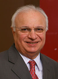
Prof. Vladimir Hachinski Awarded the 2024 Ryman Prize

On the left, Christopher Luxon, Prime Minister of New Zealand, presenting the Ryman Prize for the “world’s best development, advance, or achievement that enhances quality of life for older people” to Prof. Vladimir Hachinski. Congratulations to Prof. Hachinski.

The First International Congress of the Nepalese Academy of Neurology

More than 250 participants and speakers bring updates on Parkinson’s disease, migraines, and more.

Marianne de Visser

Rajeev Ojha

Rajeev Ojha and Marianne de Visser

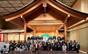
I was fortunate to be invited to the First International Congress of Nepalese Academy of Neurology May 18-19 in Kathmandu, Nepal. Sessions on major neurological disorders, such as stroke, movement disorders, demyelinating disorders, epilepsy, headache, and cognitive neurology filled two conference days. There were also sessions on neuromuscular disorders and general neurology. Plus, there were posters and oral communications presented by early-career neurologists and residents. Dr. Rajeev Ojha, co-author of this report, was instrumental as one of the organizers of the congress, and he invited me to participate in the congress.

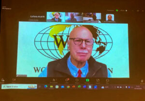
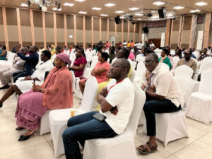
When I was on stage for my talk, I conveyed a message from WFN President, Prof. Wolfgang Grisold, in which he congratulated the president of the Nepalese Academy of Neurology on this unique event. It was a splendidly organized congress in a beautiful hotel in the center of Kathmandu, thanks to the assistance of the upcoming neurological generation and volunteers from the medical school. There were more than 250 participants and many speakers from abroad, including India, Malaysia, the Netherlands, Spain, and the United States.

Prof. Marianne de Visser giving a lecture during the neuromuscular session of congress on the second day.

During breaks between the sessions, there was plenty of time for networking. We were able to discuss how training in neurology and neurological care was organized in Nepal. The standard of care in Nepal is impressive considering that according to the World Bank it is a low- and middle-income country. MRI and endovascular interventions are readily available. However, the cost of thrombolysis and mechanical thrombectomy is high, and the insurance service is not available to most of the general public.

Prof. Shen-Yang Lim, co-lead of monogenic portal development and underrepresented populations for the East Asia branch of the Global Parkinson´s Genetics Program (GP2), congratulated Nepal on being connected to the GP2, which is a collaborative research program to understand the genetic architecture of Parkinson´s Disease. Dr. Avinash Chandra from Nepal highlighted how oral abortive treatments for migraine, such as rizatriptan and sumatriptan, have been available for a few years and recently zolmitriptan has been made available in nasal spray.  On the other hand, local speaker Dr. Niraj Gautam spoke about the unavailability of monoclonal antibodies used in the treatment of multiple sclerosis in Nepal. Prof. Lekha Pandit, from India, and Prof. Xavier Montalban, from Spain, presented an excellent diagnosis update and a discussion about the management of multiple sclerosis, respectively.

Right Honorable Vice President of Nepal, Mr. Ram Sahaya Prasad Yadav, congratulating first Nepalese neurologist Prof. Dinesh Bikram Shah during the inauguration ceremony of First Nepalese International Congress 2024.

The organizing committee included a neuromuscular session in the program. I selected myositis as a topic. Another speaker in that session was Prof. Nalini Atchayaram, from India, who gave a presentation on Hirayama disease, a disorder that should not be missed because it is treatable by surgery and may mimic ALS. She cited studies composed of hundreds of patients with this disease who, if diagnosed early, usually had excellent outcomes.

The other speakers gave excellent presentations on the diagnostic approach of muscular dystrophy (Dr. Saraswati Nashi, also from India) and on ocular myasthenia gravis (Prof. Rabindra Shrestha, from Nepal). Part of the congress was dedicated to an amazing cultural event with dancing and singing by local dancers in traditional costumes from different regions across Nepal.

All in all, this was an extremely inspiring and memorable congress that also put early career neurologists in the spotlight. During the breaks, many neurologists were willing to share the history of neurology becoming a separate discipline in Nepal and how they managed to establish three training centers across Nepal. There is an urgent need for training more neurologists (currently four residents per annum), since Nepal currently has 35 neurologists for a population of 31 million inhabitants.

I am indebted to the organizers of the congress, in particular to NAN President Prof. Kumar Oli and Secretary General Prof. Rabindra Shrestha. To my esteemed colleague and co-author, Rajeev Ojha, and to everyone who made my first visit to Nepal an incredible experience, I would like to express my sincere gratitude. •


Marianne de Visser, MD, PhD, FEAN, is a neurologist at Amsterdam University Medical Center in the Netherlands, and emeritus professor of neuromuscular diseases. Rajeev Ojha, MD, DM, FEBN, is a lecturer in the department of neurology at Tribhuvan University Institute of Medicine in Kathmandu, Nepal.

Infantile Spasms: Collaborating for Awareness, Management, and Education

Partnerships could lead to new treatments and improved patient care.

Christina Briscoe Abath, MD, EdM, and Keryma Acevedo, MD

Keryma Acevedo, MD

Christina Briscoe Abath, MD, EdM

Although it has gone by many other names, infantile epileptic spasms syndrome (IESS) has been described since 1841. It is the most common infantile-onset epilepsy syndrome, and first-line treatments are over 40 years old. Steroids (prednisolone or ACTH) have been used since the 1950s, and vigabatrin has been used since the 1980s. The newest effective treatment for some children, epilepsy surgery, has been prescribed for epileptic spasms since 1990.1 There has been consensus about the initial treatment for over a decade, as well as recognition that the lead time to standard treatment affects long-term developmental outcomes.2,3 Treatment initiation of first-line therapy is considered a priority for the clinical team.

Despite this, most children in the world continue to have long delays in diagnosis. Although present in most settings, these delays are not equally distributed. In a large series from Bangladesh, the median lead time to a standard therapy was 5 months.4 Even in Boston (U.S.), the average lead time to standard therapy was 29 days.5 In the U.S., BIPOC (Black, Indigenous, and People of Color) infants, and those whose caregivers speak another language, are more likely to have delays to neurology referral.6,7

Just as disturbing, not all children who are correctly diagnosed will receive appropriate treatment despite consensus about standard therapies. At quaternary care centers in the U.S. with pediatric epileptologists, Black/Non-Hispanic children and those with public insurance are less likely to receive a standard treatment course.8

In many countries, standard therapies are not always readily available. In Chile, for example — despite access to pediatric neurologists and similar delays to diagnosis as in Boston — challenges with access to standard treatments in the public system mean that between 2015-2022, only 67% of children received a standard treatment course.5 Moreover, anti-seizure medications (ASMs) for IESS are not covered by the National Epilepsy Plan, resulting in an extremely heavy economical overload for families.9,10

On top of these issues, ACTH and vigabatrin have not been consistently available, and prednisolone has not been incorporated in the country’s list of approved drugs. This means that prednisolone is not available for purchase in Chile, resulting in families needing to go to Perú or other countries to buy the medication if indicated by their clinical team.11 Efforts are underway to address these challenges with the Ministry of Health.

Hypsarrhythmia, initially described by Gibbs and Gibbs in 1952 (spelled as hypsarhythmia), is the classical interictal background pattern found in children with infantile epileptic spasms syndrome (IESS). It is high voltage, chaotic, and has embedded multifocal epileptiform abnormalities. Hypsarrhythmia may be state dependent, and only present in sleep. While no longer required for IESS diagnosis, when present it can suggest a diagnosis when accompanied by history concerning for epileptic spasms in children of the appropriate age (1-24 months).

Partnering Together for Solutions

New therapies and research to understand mechanisms of disease for IESS are certainly needed, since only around half of children will respond to the first standard therapy and only around 70% will respond to early sequential or dual therapy.12,13 For this, significant increases in research for pediatric epilepsy are needed, given significant disparities in funding.14 The U.S. National Institutes of Health (NIH) spent approximately $68 per person with epilepsy in 2023 ($232 million for 3.4 million individuals in the U.S.).15,16 In contrast, ALS receives $9,512 per person, Alzheimer’s $583 per person, and Parkinson’s disease $270 per person.16 Inadequate funding for drug research and development may be one reason why there have been no new first-line medications approved for IESS since the 1980s.

However, the percentage of children who do respond means that most children will still benefit from what we currently know and can apply appropriately.13 Given the high morbidity and mortality of IESS — with a 5-10% chance of response to a non-standard therapy — standard therapies are most likely cost-effective for public systems. Research should be done to support this supposition. We do know that epilepsy surgery is cost-effective for epilepsy, though cost-effectiveness specifically for epileptic spasm surgery has not been specifically assessed to our knowledge.17,18

Intervention is needed at every level of health systems to effectively address these gaps in diagnosis and therapy. In May 2022, the Intersectoral Global Action Plan on Epilepsy and Other Neurological Disorders (IGAP) was unanimously approved at the 75th World Health Assembly, providing an invaluable tool to advocate for treatment and access around the world.19,20

Joint efforts are needed to improve the standard of care for IESS, including early diagnosis, training general practitioners and pediatricians about adequate referral for suspicious clinical events, education of parents of children at high risk for IESS (Down Syndrome, Tuberous Sclerosis, Hypoxic Ischemic Encephalopathy), and access to standard treatments.

Families of children who have been affected by IESS are uniquely motivated to address these challenges. Partnering with these families to advocate for awareness and access to therapies is also needed. At a micro level, social media campaigns can promote epileptic spasm awareness. At the middle level, medical and other professional schools should incorporate pediatric epilepsy education (more common than other conditions that receive more attention). At a macro level, health systems should prioritize access to cost-effective epilepsy medication and therapies (including epilepsy surgery).

Much remains to be done around the world. In the U.S., one of the authors (CB) has been fortunate to start to work with the Infantile Spasms Action Network, parents of children with IESS, and other neurologists who are passionate about closing the equity gaps. We have created a module for primary care providers to recognize epileptic spasms and are working on a series of grassroots initiatives (a children’s book and a stuffed bear with EEG electrodes) to promote pediatric epilepsy awareness in Spanish, French, and English.

In Chile, the Chilean League Against Epilepsy (LICHE) is a non-profit organization that has several pharmacies distributed around the country that not only provide social benefits and discounts in ASMs, but also act as an intermediary to import medications to collaborate in the management of epilepsies, partnering with patients and their neurologists. As a PAHO/WHO Collaborating Centre, LICHE is committed with advocacy and education for patients, their families, caregivers and the community.

We believe collaboration, funding, and advocacy will be essential to making progress for children with IESS around the world. Please reach out to us with ideas, comments, and questions at:  christina.briscoeabath@childrens.harvard.edu and kacevedo@uc.cl. •


Christina Briscoe Abath, MD, EdM, is incoming instructor of neurology, Boston Children’s Hospital. Keryma Acevedo, MD, is associate professor, Section of Neurology, Division of Pediatrics, Pontificia Universidad Católica de Chile, and president of the Chilean League Against Epilepsy

References

1. Chugani HT, Shewmon DA, Shields WD, et al. Surgery for Intractable Infantile Spasms: Neuroimaging Perspectives. Epilepsia. 1993;34(4):764-771. doi:10.1111/j.1528-1157.1993.tb00459.

2. O’Callaghan FJK, Lux AL, Darke K, et al. The effect of lead time to treatment and of age of onset on developmental outcome at 4 years in infantile spasms: Evidence from the United Kingdom Infantile Spasms Study. Epilepsia. 2011;52(7):1359-1364. doi:10.1111/j.1528-1167.2011.03127.

3. Pellock JM, Hrachovy R, Shinnar S, et al. Infantile spasms: A U.S. consensus report. Epilepsia. 2010;51(10):2175-2189. doi:10.1111/j.1528-1167.2010.02657.

4. Abath CB, Chandra Saha N, Hoque SA, et al. Clinical characteristics of children with infantile epileptic spasms syndrome from a tertiary-care hospital in Dhaka, Bangladesh. Heliyon. 2023;9(3):e14323. doi:10.1016/j.heliyon.2023.e14323.

5. Briscoe Abath C, Vega S, Castro Villablanca F, Moya J, Garrido C, Hadjinicolaou A, Marin J, Munoz C, Soto C, Quintanilla C, Singh A, Margarit C, Andrade L, Leal L, Santana Almansa A, Gupta N, Acevedo K, Harini C. Proyecto Recaida: multi-center collaboration for infantile epileptic spasms syndrome relapse prediction and prognosis. American Epilepsy Society Conference 2023. Published December 2023. Accessed January 15, 2024. https://aesnet.org/abstractslisting/proyecto-recaida-multi-center-collaboration-for-infantile-epileptic-spasms-syndrome-relapse-prediction-and-prognosis.

6. Hussain SA, Lay J, Cheng E, Weng J, Sankar R, Baca CB. Recognition of Infantile Spasms Is Often Delayed: The ASSIST Study. The Journal of Pediatrics. 2017;190:215-221.e1. doi:10.1016/j.jpeds.2017.08.009.

7. Briscoe Abath C, Gupta N, Hadjinicolaou A, et al. Delays to Care in Infantile Epileptic Spasms Syndrome: racial and ethnic inequities. Epilepsia. n/a(n/a). doi:10.1111/epi.17827.

8. Baumer FM, Mytinger JR, Neville K, et al. Inequities in Therapy for Infantile Spasms: A Call to Action. Annals of Neurology. 2022;92(1):32-44. doi:10.1002/ana.26363.

9. Jr JE. Seizures and Epilepsy. OUP USA; 2013.

10. Engel J. What can we do for people with drug-resistant epilepsy?: The 2016 Wartenberg Lecture. Neurology. 2016;87(23):2483-2489. doi:10.1212/WNL.0000000000003407.

11. Ramani PK, Briscoe Abath C, Donatelli S, et al. Initial combination versus early sequential standard therapies for Infantile Epileptic Spasms Syndrome-Feedback from stakeholders. Epilepsia Open. 2024;9(2):819-822. doi:10.1002/epi4.12895.

12. Knupp KG, Leister E, Coryell J, et al. Response to second treatment after initial failed treatment in a multicenter prospective infantile spasms cohort. Epilepsia. 2016;57(11):1834-1842. doi:10.1111/epi.13557.

13. Mytinger JR, Parker W, Rust SW, et al. Prioritizing Hormone Therapy Over Vigabatrin as the First Treatment for Infantile Spasms. Neurology. 2022;99(19):e2171-e2180. doi:10.1212/WNL.0000000000201113.

14. Meador KJ, French J, Loring DW, Pennell PB. Disparities in NIH funding for epilepsy research. Neurology. 2011;77(13):1305-1307. doi:10.1212/WNL.0b013e318230a18f.

15. Epilepsy Data and Statistics | CDC. Published March 30, 2023. Accessed January 15, 2024. https://www.cdc.gov/epilepsy/data-research/facts-stats/?CDC_AAref_Val=https://www.cdc.gov/epilepsy/data/index.html.

16. RePORT. Accessed January 15, 2024. https://report.nih.gov/funding/categorical-spending#/.

17. Widjaja E, Li B, Schinkel CD, et al. Cost-effectiveness of pediatric epilepsy surgery compared to medical treatment in children with intractable epilepsy. Epilepsy Research. 2011;94(1):61-68. doi:10.1016/j.eplepsyres.2011.01.005.

18. Ngan Kee N, Foster E, Marquina C, et al. Systematic Review of Cost-Effectiveness Analysis for Surgical and Neurostimulation Treatments for Drug-Resistant Epilepsy in Adults. Neurology. 2023;100(18):e1866-e1877. doi:10.1212/WNL.0000000000207137.

19. Grisold W, Freedman M, Gouider R, et al. The Intersectoral Global Action Plan (IGAP): A unique opportunity for neurology across the globe. Journal of the Neurological Sciences. 2023;449. doi:10.1016/j.jns.2023.120645.

20. Wilmshurst JM. ICNA and the Global Regional Initiative Program: Aligning with IGAP towards providing child neurology services where they are most needed. Developmental Medicine & Child Neurology. n/a(n/a). doi:10.1111/dmcn.15836.

Neurology Update in Kazakhstan

Neurologists from around the world converge in Almaty.

By Aida Kondybayeva

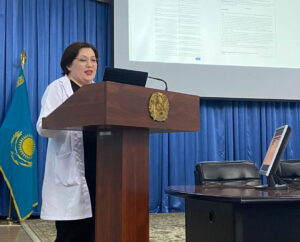
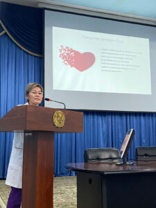
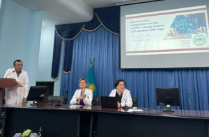
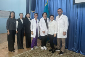
The VI International Educational Forum: Neurology Update in Kazakhstan, took place May 17-18, 2024, at the DoubleTree by Hilton Hotel in Almaty, Kazakhstan. The forum has become an annual tradition for neurologists not only from various regions of Kazakhstan but also from Kyrgyzstan, Azerbaijan, and Uzbekistan, gathering more than 700 doctors online and in person. The forum was supported by the Asfendiyarov Kazakh National Medical University and the Central City Clinical Hospital of Almaty.

The forum featured world-renowned neurologists, including:

  • Paul Boon, MD, PhD, FEAN, president of the European Academy of Neurology, Belgium;
  • Andrei Alexandrov, MD, professor of neurology at the University of Arizona, United States;
  • Thanh Nguyen, MD, FRCPc, FSVIN, FAHA, president of the Society of Vascular and Interventional Neurology, United States;
  • Dieter Ritmacher, PhD, vice dean for research and postgraduate education, professor of neurosciences, at the Nazarbayev University School of Medicine in Astana, Kazakhstan;
  • Valery Feigin, MD, PhD, professor, National Institute for Stroke and Applied Neurosciences, School of Clinical Sciences at the Auckland University of Technology in New Zealand;
  • Celia Oreja-Guevara, MD, vice chair and senior neurologist of the department of neurology at the University Hospital San Carlos, associate professor of neurology at the Universidad Complutense, and head of the Multiple Sclerosis Center at the University Hospital San Carlos in Madrid, Spain;
  • Ugur Uygunoglu, MD, professor, department of neurology at Istanbul University, Cerrahpasa School of Medicine;
  • Natalia Khachanova, department of neurology and clinical neurophysiology at the Pirogov National Medical and Surgical Center, and neurologist of the MS department of City Clinical Hospital No. 24.

The forum also featured a number of Kazakh speakers, including:

  • Prof. Zhannat Idrisova, Asfendiyarov Kazakh National Medical University;
  • Prof. Gulnar Kabdrakhmanova, Marat Ospanov West Kazakhstan Medical University;
  • Ruslan Belyaev, Karaganda Medical University;
  • Aida Kondybayeva, MD, PhD, Asfendiyarov Kazakh National Medical University;
  • Karlygash Kuzhibayeva, head of the Center for Multiple Sclerosis, Autoimmune, and Orphan Diseases of the Nervous System, in Almaty;
  • Tatyana Kaymak, neurologist at “SanClinic” in Semey; Adil Bisembaev, Private Clinic Almaty.

Next year, the International Educational Forum: Neurology Update in Kazakhstan 2025, will be held on
April 25-26.

We look forward to seeing you in Almaty in 2025! •


Aida Kondybayeva, MD, PhD, FEAN, is head of the Scientific and Educational Center for Neurology and Applied Neuroscience at Asfendiyarov Kazakh National Medical University, and chair of the Educational Committee at Kazakhstan National Association of Neurologists “Neuroscience” and Institutional Delegate at the European Academy of Neurology from Kazakhstan.

Meet the WFN Trustee Candidates

Get to know the candidates for WFN trustee in their own words.

Six candidates have been recommended by WFN’s Nomination Committee for the upcoming election of a trustee at the virtual Council of Delegates (COD) meeting in September and have presented their statements. They are in alphabetical order:

Fernando Cendes

Valery L. Feigin

Miguel Osorno Guerra

Brian Sweeney

Barbara Tettenborn 

Tissa Wijeratne

According to the WFN guidelines, further nominations can be submitted by five member societies 30 days prior to the election date. The deadline for additional nominations will be Friday, July 26, 2024.

Electronic voting will occur over three weeks starting Aug. 26, 2024.

Click the names above to read each candidate’s statements.

Candidate Statement for WFN Trustee: Fernando Cendes

Fernando Cendes

It is with great enthusiasm that I present my candidacy for the position of elected trustee at the WFN 2024 elections. I have the commitment, expertise, and background necessary. I am deeply committed to supporting the WFN’s mission and am eager to contribute my skills and experience to this important work.

I am a former Fellow and completed my PhD at the Montreal Neurological Institute and Hospital, McGill University, Canada (1991-1997), where I have an appointment as a part-time adjunct professor.

I have been a full-time neurology professor since 1997 and am currently the head of the Neurology Department at the University of Campinas, São Paulo state, Brazil. Our university hospital is a referral center for about four million people with complex neurological diseases. In addition to patient care, I teach and train undergrad and graduate students and neurology residents.

I am the principal investigator of The Brazilian Research Institute for Neuroscience and Neurotechnology (BRAINN), which is one of the Research, Innovation, and Dissemination Centers (RIDC) sponsored by FAPESP (São Paulo Research Foundation) with a 12-year operational grant. This center investigates the basic mechanisms leading to epilepsy and stroke, combining genetics, neurobiology, pharmacology, neuroimaging, computer sciences, robotics, physics, and engineering.

My service as an educator in neurology goes beyond my institution in the form of numerous lectures, teaching seminars, invited conferences worldwide, and participation in various international commissions in the International League Against Epilepsy and other societies. I also served as a member of the program subcommittee of the Global Alliance for Chronic Diseases and a delegate to the WFN for the Brazilian Academy of Neurology.

I am the newly appointed editor-in-chief of Epilepsia, and I serve on several editorial boards. My research is focused on epilepsy, neuroimaging, and clinical neuroscience, with more than 500 papers published. •

Candidate Statement for WFN Trustee: Valery L. Feigin

Valery L. Feigin

I am a professor of neurology and epidemiology and director of the National Institute for Stroke and Applied Neurosciences at Auckland University of Technology in New Zealand. I am also a Fellow of the Royal Society of New Zealand; an affiliate professor of the Department of Global Health at the University of Washington (U.S.); visiting professor of Capital Medical University (China); a member of the WHO Technical Advisory Group on NCD-Related Research and Innovation; founder and ex-officio co-chair of the GBD Stroke and Neurology Groups; editor-in-chief of Neuroepidemiology (IF 5.7) and co-chair of the Global Policy Committee of the World Stroke Organization (WSO) where I also served as a member of the Board of Directors, co-chair of the Research Committee, Guidelines Committee, Member-at-Large, and Executive Committee.

I have been working with the WFN for more than a decade as an active neurology advocate (including the Soriano Award Lecture and other public lectures and interviews), and served as a member of the Neuroepidemiology Speciality Group. Together with seven other WHO Stroke Advisory Group members for ICD-11, we justified and argued successfully for the reclassification of stroke as a neurological disease, which was endorsed for global use by the U.N. in 2020.

My 500-plus journal publications (h-index 136) have been cited more than 330,000 times, and used for evidence-based guidelines, health-care planning, priority setting, and resource allocation across the globe.

My motivation to become a trustee of the WFN is to enhance the role of the WFN in the global awareness of neurological disorders and implementation of evidence-based prevention and management strategies, workforce development, and organization of neurological services to reduce the burden of neurological disorders, with strong emphasis on low- and middle-income countries. Given my position on the Global Burden of Disease Study, I would also like to ensure that more WFN members are involved as co-authors/lead authors in the GBD Study collaboration. •

Candidate Statement for WFN Trustee: Miguel Osorno Guerra

Miguel Osorno Guerra

I am honored to be nominated as trustee of the WFN. We are in a time where innovation, collaboration, and organization must meet the diverse needs of different countries.

My name is Miguel Osorno Guerra. My professional experience in both public and private institutions in Mexico has given me insight into the significant deficiencies and challenges in neuroscience education, particularly in economically disadvantaged areas.

As secretary and later president of the Mexican Academy of Neurology (MAN), I contributed to redesigning the Journal of Neuroscience and consolidating the textbook “Elementary Neurology” for medical schools, now in its third edition. Our guiding principle was “what every doctor should know about neurology.” My organizational skills in academic events and congresses, both nationally and internationally, have significantly contributed to the dissemination of neurological knowledge.

I am also a postgraduate professor, working closely with young neurologists, encouraging them to develop research projects. This mentoring is crucial in fostering a new generation of skilled and innovative neurologists.

Our collaboration with the WFN included promoting the certification of three Mexican hospitals and participating in the WFN’s educational programs, enabling young neurologists from Latin America to enhance their skills and positively impact their communities.

My commitment to neurology education aims to improve the quality of neurological care and develop comprehensive programs for addressing neurological diseases. Through my work with the WFN, I have learned two key lessons:

Individual and isolated efforts have limited impact.

Several regions, including Latin America, need better integration with the WFN and should play a larger role in the future.

If elected, I pledge to work diligently with the WFN to achieve our shared goal of improving neurological patient care worldwide. •

Candidate Statement for WFN Trustee: Brian Sweeney

Brian Sweeney

Thank you for considering my application to be a trustee of the WFN. I have had a lifelong commitment to neurology as a clinician and educator. Having trained in medicine and neurology in Ireland and the U.K., I returned home to Ireland in 1996 as one of three neurologists in my province.

I have been a teacher all of my working life, and an administrator as the national specialty director of neurology for the Royal College of Physicians Ireland, a member of the Specialist Certificate Examination Committee of the Association of British Neurologists (ABN) and Royal College of Physicians U.K. As a member of the European Union of Medical Specialists, I have had the privilege of training and working with neurologists from all over the world – Africa, Asia, Australia/New Zealand, Europe, and North and South America.

I have been the senior neurologist in my region of Ireland, national lead for neurology for the Health Service Executive, Ireland, and national specialty director for neurology training and dean of the Irish Institute of Clinical Neuroscience.

My experience of working in and trying to develop an understaffed and underfunded neurology service in Ireland from the 1990s through the 2020s gives me insight into the challenges and advances facing neurology worldwide in the 21st century. These include amazing diagnostic and therapeutic developments in areas like stroke, neuroinflammation, neuroradiology and genetics on one hand, with major challenges providing the staffing and other resources to allow people with neurological illness to access these innovations on the other.

My vision is for neurology to keep its clinical soul while embracing new technologies such as AI to better provide care to our patients and their families. •