Oxford University Press, 2015
By Murray Grossman, MD
 Neuroimaging is an important adjunct to clinical neurology. We cannot easily examine the brain directly. The neurologic exam is designed to allow us to make reasonable inferences about abnormalities of neurologic functioning, and neuroimaging helps us visualize the brain. This significantly enhances our ability as clinicians to diagnose the cause of a neurological abnormality and monitor response to an intervention. This is particularly true in neurodegenerative diseases, where the neurologic exam has been immensely assisted by neuroimaging. Indeed, with advances in neuroscientific knowledge, novel imaging techniques have been developed to provide additional insights into neurological functioning in health and disease.
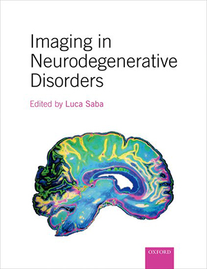
Prof. Luca Saba has edited a comprehensive textbook Imaging in Neurodegenerative Disorders (Oxford University Press, 2015, 562 pages including an index). As the title indicates, this volume focuses on neurodegenerative diseases. While neurodegeneration has been an elusive domain of neurology, recent advances in imaging have contributed importantly to advancing clinical and scientific knowledge in this area.
The book consists of 10 sections. The introduction focuses on specific background knowledge areas, such as epidemiology, health economic considerations and molecular biology. A lengthy chapter is devoted to symptoms associated with neurodegenerative diseases, and readers might have found it easier to have portions of this chapter included in sections devoted to imaging of patients with the corresponding conditions.
Imaging techniques are presented in section two, which chapters devoted to computed tomography, magnetic resonance imaging, nuclear medicine and positron emission tomography. Although recent advances in techniques related to positron emission tomography are discussed, it is unfortunate that there is not an equivalent chapter on advances related to magnetic resonance imaging.
The heart of the text is in the subsequent seven sections. Each is devoted to a specific domain within neurodegeneration. Two large sections are devoted to disorders of cognition and movement disorders. The Cognition Section includes chapters on Alzheimer’s disease and an authoritative chapter on frontotemporal dementia by Jennifer Whitwell.
The Movement Section includes chapters considering Parkinson’s and Huntington’s diseases. This area of neurology is undergoing an important revolution. As treatments emerge for the underlying histopathologic abnormalities, classification based on phenotype gradually is giving way to classification based on targets of treatment, namely, pathology.
Phenotype-based classification has resulted in the inclusion of the chapters considering dementia with Lewy bodies and corticobasal syndrome in the Cognition Section, while conditions caused by similar histopathologic abnormalities, such as progressive supranuclear palsy (a tauopathy) and multisystem atrophy (a synucleinopathy), are placed in the Movement Section.
The section on strength contains a single authoritative chapter by Martin Turner, who considers imaging in amyotrophic lateral sclerosis. A considerable portion of this chapter is devoted to extramotor brain involvement. This appropriately acknowledges that up to half of patients with amyotrophic lateral sclerosis have cognitive deficits, and suggests that the title of the section might be adjusted.
Additional sections include chapters discussing coordination, demyelinating disorders, trauma and the peripheral nervous system. The final section is devoted to neuroimaging after therapy. This is a crucial consideration as therapies emerge for neurodegenerative diseases. It might have been useful here to consider quantitative measurement in neuroimaging since many of these techniques are being used as endpoints in treatment trials.
Imaging in Neurodegenerative Disorders is a comprehensive text that has an appropriately broad scope. Chapters are devoted to all the key areas in neurodegeneration, and the chapters are comprehensive. The book is generally well illustrated, although occasional images seem to have lower resolution than is needed to illustrate a finding optimally. I recommend this text to students of neurodegeneration who are interested in a comprehensive introduction to neuroimaging.
