IAPRD and PSN Join Hands: First Movement Disorder Course and Botox Workshop

By Abdul Malik, MBBS, DCN, MD

PSNConference

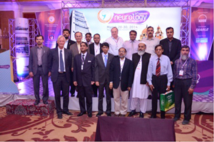
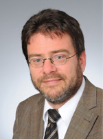
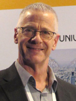
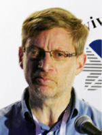
PSN conference organizers and faculty with Erik Wolters, Daniel Truong and R. Pfeiffer.

The specialty of neurology shows remarkable growth in last decade in Pakistan. The 21st meeting of the Pakistan Society of Neurology (PSN) was organized in collaboration with the International Association of Parkinson’s and Related Disorders (IAPRD) March 28-30 in Karachi, Pakistan.

There were eight scientific sessions and a half-day Botox Hands-On Workshop in this conference. The speakers from Netherlands, the United States, Saudi Arabia and Pakistan shared their experiences pertaining to neurology. The core of the discussion was the advances in movement disorders and the newer therapies now emerging. The guest faculty from the IAPRD had given a detailed overview on the topic.

An ample demonstration on patients was given with the title of “Botulinum Toxin Hands-On Use in Dystonia” by Prof. Daniel Truong from The Parkinson and Movement Disorder Institute (U.S.). He also delivered a lecture on Clinical Approach and Management of Dystonias. Prof. Ronald Pfeiffer, vice chair of the Department of Neurology at the University of Tennessee Health Science Center (U.S.) gave us an updated overview on Autonomic Dysfunction in Parkinson’s Disease and Drug-Induced Movement Disorders. The keynote speaker was Prof. Erik Wolters, IAPRD president, who is working in the Universities of Maastricht and Zurich. He delivered his lectures on the topics of Behavioral Dysfunction in Parkinson’s Disease and Parkinson’s Disease — Revisited.

More than 200 exceedingly participative audiences from all parts of the country attended all of the scientific sessions. In the inaugural session of the three-day conference, the Sardar Alam, outgoing Pakistan Society of Neurology (PSN) president, welcomed the delegates and presented the report of the last two years of the society’s works. Prof. M. Wasay, chairman of the organizing committee, stated the statistics of neurology care and neurologist in Pakistan. He said that for every one million people only one neurologist is available.

In the scientific sessions, Asif Moin, from Saudi Arabia, delivered the talk on utilization of SPECT and PET in epilepsy; Qasim Bashir discussed initial experience in establishing an interventional neurology/neuroendovascular surgery program in Lahore: procedural types and outcomes; Qurat Khan from AKU deliberated on introduction to behavioral neurology; Bushra Afroze talked on biotinidase deficiency — clinical presentation, diagnosis and treatment while neurodegeneration in children: diagnostic issues in developing countries was presented by Tipu Sultan from Children Hospital Lahore. A unique and thought-provoking session of this was in the Neurology Training and Advocacy session. This session highlighted the glimpses on post-graduate neurology in Pakistan by Sarwar Siddiqi, psychiatric care and interface with neurology by Prof. Iqbal Afridi, neurosurgery care and interface with neurology by Prof. Junaid Ashraf and advocacy for neurological care in Pakistan Prof. Rasheed Jooma.

There were 15 original oral presentations and 20 poster presentations from all major institutes of the country. This year, PSN officially had given best oral presentation and poster presentation awards. There was also an inaugural dinner in which the primary guest was Prof. Asghar Butt, vice president of CPSP. Prof. Masood Hameed Khan, vice chair of DUHS was honored during the social portion of the evening.

The conference was a great success in terms of participation, and the quality of training and education was superb.

The World of Neurology Comes Together at the AAN Annual Meeting

AAN_0733
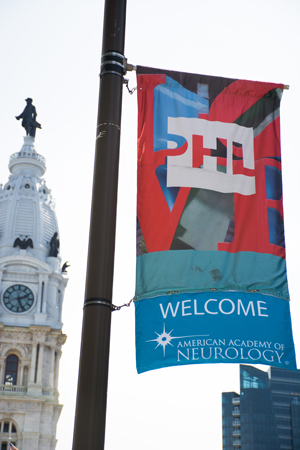
The 2014 Annual Meeting of the American Academy of Neurology (AAN) attracted more than 13,000 neurology professionals to Philadelphia, a city rich in its own neurology-related history. More than 2,500 presentations of cutting-edge research were available to attendees from across the world. The number of non-U.S. attendees was nearly 4,000.

Science and continuing medical education are the main draws to the Annual Meeting, and this year was no exception. One of the highlights was the Presidential Plenary Session, the AAN’s premier lecture awards for clinically relevant research. James L. Bernat, MD, FAAN, gave the Presidential Lecture on “Challenges to Ethics and Professionalism Facing the Contemporary Neurologist.” The George C. Cotzias Lecture featured Stefan M. Pulst, MD, FAAN, speaking on “Degenerative Ataxias: From Genes to Therapies.” The Sidney Carter Award in Child Neurology went to Darryl C. De Vivo, MD, FAAN, who expounded on “Rare Diseases and Neurological Phenotypes.” Finally, David M. Holtzman, MD, FAAN, shared “Alzheimer’s Disease in 2014: Mapping a Road Forward,” as the Robert Wartenberg lecturer.

The Hot Topics Plenary Session presented recent research findings and clinical implications regarding identification of a unique molecular and functional microglia signature in health and disease; emerging concepts in chronic traumatic encephalopathy; functional connectivity and functional imaging in movement disorders; and the global epidemic in stroke.

Abstracts related to new therapeutic developments, clinical applications of basic and translational research, and innovative technical developments were shared and discussed at the Contemporary Clinical Issues Plenary Session. This year’s topics were acute ischemic stroke, Friedreich’s ataxia, insomnia, epilepsy, functional (psychogenic) disorders and the Parkinson’s disease Progression Marker Initiative.

At the Frontiers in Translational Neuroscience Plenary Session, attendees heard about the clinical aspects of tissue environments for brain repair; opportunities and challenges of robot-assisted and facilitated neurorecovery; network-based neurodegeneration; using fixed circuits to generate flexible behaviors; advances in the Human Connectome Project; and the nightlife of astrocytes.

The Controversies in Neuroscience Plenary Session featured pairs of experts debating three of the most current and controversial issues in neurology: “Does preventing relapses protect against progressive MS?,” “Is intervention for asymptomatic AVM useful?” and “Should neurologists prescribe marijuana for neurological disorders?”

The Clinical Trials Plenary Session addressed important topics that affect patient care identified from submitted abstracts that had recently been presented at other society meetings, including stroke, MS, migraine, intracranial hypertension and glioblastoma. The week concluded with the Neurology Year in Review Plenary Session, which examined advances in MS, headache/pain, neuromuscular diseases, movement disorders, neurocritical care and Alzheimer’s disease.

More science was available at numerous platform sessions, seven poster sessions and a new, fast-paced series of Poster Blitz presentations.

Another new feature at this year’s meeting was an International Lounge, which provided an opportunity for foreign guests to mingle, network with leading neurologists from the U.S. and other countries, and relax before the next session.

A significant portion of the meeting was devoted to helping U.S. neurologists understand the many changes initiated by Congress and federal regulatory agencies as part of health care reform. Neurologists, like other physicians, are under tremendous pressure to reduce treatment costs without compromising the quality of care.

The AAN works hard to guide members through these issues using a variety of resources and tools, while advocating on their behalf with members of Congress and administration officials in Washington, DC.  U.S. Senator Bob Casey (D-PA) spoke with neurologists concerned about cuts to federal health programs for the elderly and the economically disadvantaged.

Another Pennsylvania legislator, Congressman Chaka Fattah of the U.S. House of Representatives, spoke at the Brain Health Fair about his initiative to support brain research. This fair is a free public event presented by the American Brain Foundation. The fair typically draws 1,000 to 2,000 people from the region who are living with brain disease or caring for a family member or friend. They hear about new research and treatments from neurologists, health organizations and the medical industry.

Media from across the globe gathered to provide powerful coverage of neurology’s largest meeting. Reporters and photographers shared the latest news in brain research with their readers and listeners back home. The AAN pressroom was lively with representatives from major national and international news outlets and neurology trade publications.

AAN President Timothy A. Pedley, MD, FAAN, remarked on the strong media interest in the meeting.

“The frequently tragic and debilitating nature of so many brain diseases provides a focus for our attention when we come together at the Annual Meeting to share our experiences and chart our progress,” said Pedley. “The scale of media coverage and public interest reminds us of the tremendous responsibility we have to work even harder to discover more effective treatments and, eventually, cures or preventive strategies for the neurologic diseases that can be so devastating to our patients.”

Plans are already under way for the 2015 Annual Meeting, to be held April 18-25 in Washington, DC. Until then, the AAN will continue to connect neurologists with the best in science and education through several gatherings, including The Sports Concussion Conference in July; the AAN Fall Conference in October; and Breakthroughs in Neurology 2014: Translating Today’s Discoveries into Tomorrow’s Clinic, to be held in December.

As always, the world is invited.

Experiments Into Readiness for Action: 50th Anniversary of the Bereitschaftspotential

By Lüder Deecke

Figure 1: Original experimental setup (A) and first results (B) in Freiburg, Germany, at the University Hospital of Neurology with Clinical Neurophysiology, Hansastr. 9a, Freiburg, known popularly as

Figure 1: Original experimental setup (A) and first results (B) in Freiburg, Germany, at the University Hospital of Neurology with Clinical Neurophysiology, Hansastr. 9a, Freiburg, known popularly as “Neurophys.”


A: The centerpiece of the laboratory — the Mnemotron CAT Computer (Computer of Average Transients) 400 B and Mosely Autograf (XY-Plotter). Prof Richard Jung, head of the hospital, had been offered a post at the Max Planck Institute in Munich. For rejecting this, his home University of Freiburg gave him the CAT Averager as a present for staying. The two inserted photos show Hans Helmut Kornhuber on the left (age 36 in 1964) and Lüder Deecke on the right (age 26 in 1964).


B: Above: Changes in brain potentials with voluntary movements of the left hand based on unipolar recordings of the precentral region versus the nose (Subject G.F., average of 512 movements). Onset of movement at the arrow (0 sec) – left of the arrow brain activity prior to movement onset — right of the arrow brain activity after the onset of movement. Note the negative potential at readiness and the positive potential after the action. The graph shows higher amplitudes over the contralateral (right) hemisphere. Negative polarity is up.

Below: The bipolar serial recording with sagittal electrodes with voluntary movements of the right hand (average from 400 movements. Subject B.C.). The graph shows phase reversal around the precentral electrode: The premotor negativity and the postmotor positivity are strongest over the central region. In the fronto-precentral recording negativity of the precentral electrode is down, in the pre-centro-occipital recording negativity of the precentral electrode is up.


A special session inaugurated and chaired by Mark Hallett, Bethesda, Maryland, at the International Congress of Clinical Neurophysiology (ICCN2014) in March 2014 in Berlin celebrated the 50th anniversary of the Bereitschaftspotential. The session included lectures by Lüder Deecke, Vienna: “Experiments Into Readiness for Action — Bereitschaftspotential;” Hiroshi Shibasaki, Akio Ikeda, Kyoto, Japan: “Generator Mechanisms of BP and Its Clinical Application;” Gert Pfurtscheller, Graz, Austria: “Movement-Related Desynchronization and Resting State Sensorimotor Networks;” and Ross Cunnington, Brisbane, Australia: “Concurrent fMRI-EEG and the Bereitschafts-BOLD-Effect.” The session was well accepted.

In this paper, I would like to give an outline of the history of the Bereitschaftspotential and a selection of the main research results of our experiments into readiness for action.

The History of the  Bereitschaftspotential

In 1964, my mentor Hans Helmut Kornhuber (1928-2009) and I discovered the readiness potential (Kornhuber and Deecke, 1964). We submitted the full paper in the same year. It was published in the first 1965 issue of “Pflügers Archiv” (Kornhuber and Deecke, 1965).

We described a novel method, reverse averaging, for recording brain electrical  activity prior to voluntary movement in humans by noninvasive means and presented the first fundamental results obtained with this method. We found that a negative electrical cortical potential consistently preceded human voluntary movement and named it the Bereitschaftspotential (BP) or readiness potential. (See Figure 1B.)

The BP is the electrophysiological sign of planning, preparation and initiation of volitional acts. How did the idea come up to record brain potentials preceding human voluntary movements in the EEG? It began on a Saturday in May 1964 when Kornhuber invited his doctoral student L.D. for lunch into the ‘Gasthof zum Schwanen’ at the foot of the Schlossberg hill in Freiburg, Germany (near the Black Forest).

We sat in the beautiful garden and discussed our frustration with the fact that the brain was investigated — at that time — only as a responsive apparatus, i.e. as a mere reacting system. Neurophysiologists were engaged worldwide only in what was called “the responsive brain” (later culminating in a book with this title by McCallum & Knott, 1976). We felt that it would be far more exciting to investigate what is going on in our brain before we make a voluntary movement. No sooner said than done.

We went back to the lab and started immediately planning the experiment. However, we soon ran into an important problem: Brain potentials in the EEG are the result of averaging. For us to get the results we needed, the averaging process must be triggered by the movement or action itself. But how can you trigger on an event that comes as unpredictably and spontaneously as a human voluntary movement?

It was Hans Kornhuber, who found the solution: We would store the EEG on magnetic tape along with the electromyogram (EMG) of the movements and then play the tape backward in the time-reversed direction from the present back to the past, i.e. using reverse averaging with the start of the movement as the trigger. At that time, magnetic tape recorders only had a high-speed rewind, and programmable computers were not yet available, so we were literally removing the tape reels from the recorder, turning them around, and placing them back on the recorder. By these means, we found a brain potential, which was electrically negative and started already 1½ to 1¼ seconds prior to the movement or action. Negativity in the brain means activity.

This method of recording of the readiness potential — Bereitschaftspotential by reverse averaging was the basis of my doctoral thesis, which I completed in 1965 at the University of Freiburg with Korn-huber as my doctoral supervisor. The experiments that led to the discovery of the Bereitschaftspotential were inspired by a positive concept of will, i.e. individual humans have, indeed, their own will and own decision-making power, so that we are capable of designing our lives largely by ourselves and use goal-oriented action to create our own future.

Kornhuber lectured on human freedom in a seminar for students of all faculties. He conducted a survey among his listeners as to who is freer: humans or chimpanzees, chimpanzees or rhesus monkeys, rhesus monkeys or cats, cats or salamanders, salamanders or spiders, and so on until down to the earthworm. The seminar participants answered the question of freedom unambiguously, namely according to the position of the animal in the evolution.

Obviously, we interpret evolution as a process making organisms freer and freer; one also can say making them more and more autonomous. Similarly, we consider an adept, e.g. Shaolin monk, who works hard on his own personality, to become more and more free. Kornhuber was of the opinion and demonstrated that one can make freedom a topic of scientific investigation. Not to deal with freedom only philosophically but also to explore it using scientific means (Kornhuber, 1978; 1984; 1987; 1988; 1992; 1993).

Immanuel Kant, the great German philosopher of the enlightenment (who coincidentally was born in the same town, Königsberg, as Kornhuber) was the first to distinguish between two fundamental aspects of freedom, namely freedom from and freedom to. Freedom to is the much more important aspect here, and this distinction was taken up and further developed by Kornhuber. The scientific breeding ground for the experiments toward the Bereitschaftspotential was thus already prepared well in advance. Rather than being a serendipitous discovery, the Bereitschaftspotential was therefore the result of a new branch of research planned by Kornhuber and myself.

The Laboratory: Original  Experimental Setup  in Freiburg/Germany

Figure 2: Chairma n and speakers in the session

Figure 2: Chairman and speakers in the session “Bereitschaftspotential – 50 Years After Its Discovery” at the International Congress of Clinical Neurophysiology 2014 (ICCN2014) on March 20, 2014, in Berlin. From left to right: Gert Pfurtscheller, Graz, Austria; Ross Cunnington, Brisbane, Australia; Lüder Deecke, Vienna; Hiroshi Shibasaki, Kyoto, Japan; and Mark Hallett, Bethesda, Maryland (Chairman).


During the experiments, the subject sat in a Faraday cage for electrical shielding. The EEG was recorded using a Schwarzer-EEG Type E 502 with tube amplifiers and the EMG by a Tönnies-EMG and Stimulation Unit. Data were stored using a Telefunken four-channel magnetic tape recorder M 24-4, frequency-modulated. The tape reels were turned around for reversed averaging. The center-piece of the experimental setup is shown in Figure 1A: Mnemotron CAT Computer 400 B with Mosely Autograf. (See Figure 1: How  Kornhuber and I came to the opportunity to use this then-ultra-modern device for our experiments.)

Figure 1B gives one of our first results, a slowly increasing ramp-up of negativity was recorded, which was stronger over the contralateral hemisphere. Negativity culminated at movement onset (and after the action returning to positivity), which we called the Bereitschaftspotential. In the lower set of graphs in Figure 1B, this negativity was demonstrated by a bipolar recording to show phase reversal around the precentral electrode. We called the pre-movement negativity the “Bereitschaftspotential” and in our English summary of Kornhuber and Deecke (1965) offered the English translation “readiness potential.” Somehow the tongue-twister Bereitschaftspotential was preferred and is now a German word in the English language.

We instructed our subjects to make their movements:

  • at irregular intervals
  • out of free will
  • of their own accord

The movements we analyzed were therefore entirely self-initiated without external simuli. We dislike the term “self-paced,” suggesting regular pace, while it is so important to make the movements at irregular intervals, which make them more volitional, and regular pace makes them more automatic. At that time (1964), this was a remarkable instruction for subjects, to which I think we owe our success. And, thus, a brain potential evolved completely different from W. Grey Walter’s (1924-1971) expectance wave or CNV (both having been first published in 1964). By the way, Walter came from Bristol in 1964 to make summer vacation in the Black Forest, which was fashionable for British at the time. He visited Richard Jung, and Kornhuber and I showed Walter our first results. It was Walter who in a later publication coined the term “opisthochronic averaging” for our methodology. Our first movements under study were simple movements (rapid flexions of the forefinger).

Another methodological prerequisite is to investigate monophasic movements, i.e. that the flexed finger remains in the flexed position until the end of the analysis epoch. Using wrist extension and flexion in one flick of the hand is not good, since this employs two movements instead of one.

By comparing active movements with analogous passive ones, we aimed to show that the BP occurs prior to active movements only. To initiate passive movements, the experimenter pulled a string that was fixed to the subject’s finger and ran over a pulley, so that pulling would cause the subject’s finger to flex. Indeed, we did not detect any BP prior to such passive movements, but recorded evoked potentials after movement onset elicited by the passive movement. Post-movement onset potentials also occurred in the active state. We referred to these as “reafferent potentials” because the term “evoked potentials” should, by definition, be reserved for potentials that are elicited by external stimuli.

Citation Classics

Our first full paper (Kornhuber and Deecke, 1965) became a Citation Classic on Jan. 22, 1990. Eugene Garfield of the journal database Current Contents (CC) “awarded” this label to papers that were frequently cited. For a paper written in German, this does not occur too often. Garfield gave a translation of the German title: “Changes in Brain Potentials With Willful and Passive Movements in Humans: the Readiness Potential and Reafferent Potentials.” As part of receiving Citation Classic status, we were asked to write up how we arrived at our discovery (Citation Classic Commentary), and we gave our commentary the title: “Readiness for Movement – The Bereitschaftspotential Story” (Kornhuber HH & Deecke L (1990).

The Citation Classic Commentary was published both in CC Life Sciences and in CC Clinical Medicine. Three more of our papers became Citation Classics: No. 2 was Deecke, Scheid, Kornhuber (1969). Here we continued our investigation into the cerebral activity preceding willful movement, and also compared finger movements with arm movements. The 1963 Nobel Laureate for medicine, Sir John Eccles was interested in our work. In his 1977 book, “The Self and Its Brain,” which he co-auhored with Karl R. Popper, he wrote about our research: “There is a delightful parallel between these impressively simple experiments and the experiments of Galileo Galilei who investigated the laws of motion of the universe with metal balls on an inclined plane.”

In his previous book,  “The Understanding of the Brain” (Eccles JC 1973), he wrote (page 108): “In an initial investigation by Grey Walter, the subject was trained to perform a movement after a double stimulus sequence: a conditioning, then a later indicative stimulus. An expectancy wave was observed as a negativity over the cerebral cortex before the indicative stimulus. Essentially, this wave is produced by the conditioned expectancy of the indicative stimulus and not by a voluntary movement. The problem is to have a movement executed by the subject entirely on his own volition, and yet to have accurate timing in order to average the very small potentials recorded from the surface of the skull. This has been solved by Kornhuber and his associate who used the onset of the movement to trigger a reverse computation of the potentials up to 2 seconds before the onset of the movement. The subject initiates these movements “at will” at irregular intervals of many seconds. In this way, it was possible to average 250 records of the potentials evoked at various sites over the surface of the skull, as shown by the numbers in Figure 4-3 and the corresponding traces. (Figure 4-3 is taken from Deecke, Scheid. Kornhuber [1969] and shows the comparison between finger and arm movements.) These experiments at least provide a partial answer to the question: What is happening in my brain at the time I am deciding on some motor act?”

Citation Classic No. 3, Deecke, Grözinger, Kornhuber (1976), resulted from my habilitation thesis, a requirement to become a professor at a German university. This paper in Biological Cybernetics, “Voluntary Finger Movement in Man: Cerebral Potentials and Theory,” comprises a lot of experiments performed in Ulm with many figures and a comprehensive analysis of the BP and its components. Three different brain potentials preceding voluntary rapid finger flexion were distinguished:

  1. Early negative activity of the Bereitschaftspotential — widespread
  2. Pre-motion positivity, PMP – widespread
  3. Motor potential (MP) – unilateral, restricted to the contralateral motor cortex.

Two years later, Citation Classic No. 4 (Deecke & Kornhuber, 1978) was published. This paper in Brain Research, “An electrical sign of participation of the mesial ‘supplementary’ motor cortex in human voluntary movements” first suggests that the early component of the BP, BP1 or BPearly is generated by the SMA and — as we found later — also by the cingulate motor area (CMA). Both being clinicians, Kornhuber and I also investigated patients and were early on able to link Parkinson’s disease (PD) with the Bereitschaftspotential. Working with patients means investigating lesion experiments made by nature, and if carefully studied, their pathological state can tell us a lot about the normal function.

Kornhuber had worked on the basal ganglia and cerebellum and found that the basal ganglia are an important component of the cortico-basal ganglia-thalamo-cortical loop. This loop was known by neurosurgeons long ago and created the prerequisites for the thalamotomies in classical stereotaxic surgery. The cortico-basal ganglia-thalamocortical loop was later confirmed and called motor loop by Alexander et al (1986), and by Mahlon DeLong (1990). Nowadays, we have to “think in loops,” and the motor loop is the key to the understanding of Parkinson’s disease. In our Citation Classic No. 4 (Deecke & Kornhuber 1978), we found differences in the BP between Parkinson’s disease patients and normals. One of the best examples of how the BP in Parkinsonian patients looks like came from an elderly woman among our subjects — she was a duchess — suffering from Parkinson´s disease who was trying hard in our BP experiment to meet our performance criteria. The main feature was that we found a high amplitude BP over the vertex but not very much of a BP in the contralateral and ipsilateral precentral leads due to her PD. Experiments were carried out in patients with bilateral Parkinsonism selected for pronounced akinesia but minimal tremor, and in age-matched healthy control subjects. The vertex maximum of the Bereitschaftspotential had previously been explained by volume conduction. It was argued that a vertex electrode collects activity from both motor cortices. In bilateral Parkinsonism, however, we see a bilateral reduction of the Bereitschaftspotential in the precentral area, whereas over other cortical areas (in particular vertex and mid-parietal), it was not significantly reduced. This publication therefore established the SMA participation in the generation of the BP, and our findings were confirmed by CD Marsden (1938-1998) (Marsden et al 1996). Barrett, Shibasaki & Neshige (1986) reported that the BP is normal in PD. But Dick et al (1989) found that the BP is abnormal in PD. Our group repeated the experiments and confirmed the findings of Dick et al 1989. Harasko, van der Meer et al 1996 cited in Lang and Deecke 1998 ibidem Figure 5 on page 235. Results: In the PD patients (N=8), the BP starts later and is initially lower in amplitude as compared to the normal controls (N=8). After the onset of movement, the amplitudes are equal and even sometimes larger in the PD patients as compared to the controls. Thus, Parkinsonian patients start later with their BP, but then keep up with the normal controls.

In May 1979, Kornhuber and I organized the MOSS V congress (one of the series of the EPIC congresses) in ReisensburgCastle, near Ulm, Germany. Afterward, we edited a book of the conference proceedings: Progress in Brain Research Vol 54 (Kornhuber & Deecke [Eds] 1980). The next conference on the Bereitschaftspotential was an international symposium in April 1988 in Vienna. which I organized in honor of Kornhuber’s 60th birthday. The proceedings also were published as a book (Deecke, Eccles, Mountcastle [Eds] 1990).

The Dispute With Libet

In October 1988, Sir John Eccles invited scientists working on the motor system to contribute to a study week in the Vatican, titled “The Principles of Design and Operation of the Brain” and a resulting book (Eccles & Creutzfeldt [Eds] 1990). I met Benjamin Libet at earlier conferences, and he also attended the Vatican Study Week. We liked each other and became friends, although not being of the same opinion regarding the freedom of human will. He worked with Wundt’s event clock and asked his subjects to remember the position of a red dot on the clock at the instant when the “conscious urge to move” occurred to him or her. Libet made a big claim in his Vatican lecture (Libet 1990) by saying that freedom is firmly linked with consciousness, the state of full awareness. He brings it to the point in his own words: “The unconscious initiation of a freely voluntary act.” Libet is correct in this statement, but his interpretation is not correct. He takes it for granted that in our unconscious or preconscious inner world there is no freedom, and thus concluded that we have free will in the control of the movement but not in its initiation. This is because the  “W” in his paradigm (conscious wish) comes later than the start of the readiness potential.

Kornhuber and I,  joined by the philosopher Daniel C. Dennett, are of the opinion that there are conscious and unconscious agendas in the brain, and both are important. The conscious and unconscious agendas of the brain were the subject of a workshop at the European Neurological Society (ENS) Congress 2010 in Berlin, where Dennett and Adrian Owen were the speakers. I also expressed our view in a paper in Brain Sciences (Deecke L, 2012). Libet’s paradigm is a mixture of cerebral and subjective events, and the problem lies in the subjective event (W).

It is hard to trace back split seconds, and 200 msec is very, very short. I tried the Libet paradigm myself with students, and we were not able to accurately perform in his paradigm with Wundt’s event clock position that has to be recalled retrospectively. Wundt’s event clock has been designed for sensory psychophysical experiments.

In a voluntary movement paradigm (BP-paradigm), it is like a “foreign body,” because it is an external stimulus and thus disturbs the “volitionality,” so to say, the willfullness of the movement or ac-tion. In conclusion, Kornhuber and I feel that the importance of consciousness has been underestimated by the behaviorists. It is not an epiphenomenon. If after brain injury, consciousness is regained, the lesion can (partially) be compensated, however never without consciousness. Conscious awareness is a shining light of freedom, although it should not be overestimated either.

For most of the vital functions, consciousness is not necessary, and it is not the only sign of freedom. The experiment of Libet et al (1983), which showed that the BP is not, right from the beginning, accompanied by a consciousness about the intention of movement is taken at present as the main argument for advocating a total determinism, a complete unfreedom of humans. This position is not tenable. What would be essential to study is the original planning and decision. This, however, has been completed already before the beginning of the experiment, when subjects gave their informed consent to the experimenter’s instructions. Repetitions of stereotyped simple movements are not suitable for such an investigation.

Thoughts of planning and motivation are as we all know performed in the light of consciousness. The conscious awareness to want to make a movement does occur, in investigations of the BP, about 200 ms prior to the muscle contraction — Libet’s W. This is roughly the same time span needed for a motor reaction upon an expected auditory stimulus. Although the decision to act already has been made earlier, consciousness is switched on in order to be able to make changes to the movement if necessary — changes that can go as far as not executing the action at all (Libet’s veto), and to be able to learn from the success of the movement. In both cases, the following brain areas are activated: the SMA, the pre-SMA, the anterior portion of the cingulate cortex (CMA) and a part of the motor cortex (Cunnington et al 2000), for which the basal ganglia do the groundwork (Kornhuber and Deecke 2012). However, with the self-initiated movement — but not with externally triggered movements — additionally the basal ganglia are activated before the movement (Cunnington et al, 2002). This preparatory process for the spontaneous movement, through which the readiness for movement in the SMA builds up, remains unconscious for the first 400 ms, as Libet has demonstrated.

It is not unusual for something to happen unconsciously in the brain as well as in sensory systems. In the motor system, processes that are initially conscious can become unconscious through automatiza-tion (also cf. Wu et al 2004). The switching on of consciousness shortly before the movement is a great expenditure for the brain and shows that even such “unimportant” repeated movements needed for the averaging procedure are controlled, if they are voluntary.

Consciousness is known to be restricted and its time is valuable, only important events get access to it. As measurements of the “channel capacities” of the senses in psychophysical experiments have shown, there is a selection/filtering of the important matters between the information flow in our senses and that in our consciousness. This sophisticated selection also is unconsciously organized and represents an enormous compression of information of at least 104; information flow through the receptors and afferent nerves is at least 105 bit/sec (by order of magnitude), whereas only 10 bit/sec show up in consciousness. The will, however, always takes part, albeit sometimes merely to the degree that it delegates as much as possible to unconscious routines and expert systems of the brain. The unconscious processes therefore do not lessen freedom; on the contrary, they form its primary basis.

Magnetoencephalography (MEG)

In 1981, Hal Weinberg invited me as a distinguished visiting professor to the Simon Fraser University in Greater Vancouver, Canada, and we were the first to record the MEG equivalent of the BP, the “Bereitschaftsfeld, BF” (Deecke, Weinberg Brickett 1982). We investigated the Bereitschaftsfeld accompanying foot movements as did Riita Hari and her group (Hari et al 1983). We also investi-gated slow magnetic fields of the brain preceding speech in Vancouver (Weinberg et al 1983).

Using our own MEG in Vienna, we later investigated the problem of why the SMA (and CMA) activity was not so visible in the MEG (the onset time of the BF was not earlier than 600 msec). The answer is that the two SMAs on the mesial surface of the brain are opposing each other, and if they are both active — which is the case most of the time — their activities cancel each other. We were using two strategies to overcome the problem of cancellation. The first, published by Lang et al (1991), involved investigating a patient with a stroke in one of his SMAs (the right in this case). The patient had only one SMA left, and we were able to record early starting magnetic fields of his left SMA — he performed right-sided movements. The paper’s abstract summarizes this: “Previous studies by magnetoencephalography (MEG) failed to consistently localize the activity of the supplementary motor area (SMA) prior to voluntary movements in healthy human subjects. Based on the assumption that the SMA of either hemisphere is active prior to voluntary movements, the negative findings of previous studies could be explained by the hypothesis that magnetic fields of current dipole sources in the two SMAs may cancel each other. The present MEG study was performed in a patient with a complete vascular lesion of the right SMA. In this case, it was possible to consistently localize a current dipole source in the intact left SMA starting about 1,200 msec prior to the initiation of voluntary movements of the right thumb.”

Our second strategy was to use a multichannel whole scalp MEG instrument on healthy subjects. In Vienna, we had a CTF system with 143 channels and used a sophisticated analysis (two-dipole and three-dipole model in the same subject) to study the Bereitschaftsfield (BF) prior to tapping movements (Erdler et al 2000). The paper’s abstract states: “Despite the fact that the knowledge about the structure and the function of the supplementary motor area (SMA) is steadily increasing, the role of the SMA in the human brain, e.g., the contribution of the SMA to the Bereitschaftspotential, still remains unclear and controversial. The goal of this study was to contribute further to this discussion by taking advantage of the increased spatial information of a whole-scalp MEG system enabling us to record the magnetic equivalent of the Bereitschaftspotential 1, the Bereitschaftsfeld 1 (BF 1) or readiness field 1. Five subjects performed a complex, and one subject a simple, finger-tapping task. It was possible to record the BF 1 for all subjects. The first appearance of the BF 1 was in the range of -1.9 to -1.7 s prior to movement onset, except for the subject performing the simple task (-1 s). Analysis of the development of the magnetic field distribution and the channel waveforms showed the beginning of the Bereitschaftsfeld 2 (BF 2) or readiness field 2 at about -0.5 s prior to movement onset. In the time range of BF 1, dipole source analysis localized the source in the SMA only, whereas dipole source analysis containing also the time range of BF 2 resulted in dipole models, including dipoles in the primary motor area. In summary, with a whole-head MEG system, it was possible for the first time to detect SMA activity in healthy subjects with MEG.”

fMRI – functional Magnetic  Resonance Imaging

In 2003, Marjan Jahanshahi and our chairman of the session, Mark Hallett, edited a book titled, “The Bereitschaftspotential — Movement-Related Cortical Potentials” (Jahanshahi M & Hallett M 2003). The closing chapter is written by Deecke and Kornhuber (Deecke & Kornhuber, 2003). The book and chapter for the first time compile all of the evidence that the BP or better “BP-like movement-related activity” can be recorded from the basal ganglia. This cannot be done by EEG or MEG, but requires the fMRI. And not just the routine fMRI but an fMRI improved in such a way that the temporal resolution is high enough to justify the term “event-related fMRI” and in our special case “movement-related fMRI.” This was achieved by Cunnington et al (1999) who showed that it was feasible to record a BP equivalent in the haemodynamic response of the fMRI. Using single-event fMRI in combination with fuzzy clustering analysis, it is possible to analyze the “Bereitschafts BOLD effect” in the form of the haemodynamic response time course. This haemodynamic response resembles the BP or BF but is delayed in time (Cunnington et al 2002).

The movement-related fMRI experiments of Cunnington et al (2002) had great localizatory power and were particularly valuable for mapping the mesial surfaces of the hemispheres, where SMA and CMA are located. In this paper, self-initiated movements were compared with externally triggered movements. In both conditions, the following structures were active: pre-SMA, SMA proper and CMA (cingulate motor area, i.e. part of the anterior cingulate gyrus. The border between pre-SMA and SMA proper is the VAC line (vertical anterior commissure line). Although both pre-SMA and SMA proper are active in self-initiated movements and also in externally triggered ones, there are slight differences: The activity with self-initiated movements is slightly more anterior. It is true that the pre-SMA has somewhat greater involvement in self-initiated movements, but it is clear that it is not the pre-SMA alone that is active (Shibasaki & Hallett 2006) but that both pre-SMA and SMA proper are always activated in voluntary movement.

Using the fMRI, it also was possible to show that the SMA/CMA is active, when a movement is not executed but only imagined. Kasess et al (2008) compared movements that are actually executed with those that are only imagined. They obtained the interesting result that SMA/CMA were active in both imagination and execution, however the motor cortex, M1, was active in executed movements only. In this context, we may report on earlier work using DC-EEG (Uhl et al 1990) and Single Photon Emission Computed Tomography (SPECT) (Goldenberg et al (1989) in mental imagery. We were able to show that the frontal cortex (of which the SMA/CMA form a part) is necessary to bring about the mental imagery (Lang et al 1988; Uhl et al 1990).

Mental imagery is the term in the psychological literature meaning our ability to see something in our mind’s eye. In Uhl et al 1990, we investigated the slow cerebral potentials (DC potentials) accompanying mental imagery. The interesting result was that — with the willful attempt to see something in your mind’s eye (mental imagery) — the frontal cortex was activated first and only thereafter the posterior (sensory) areas of the brain were activated. The strong initial DC negativity of the frontal cortex demonstrated that these frontal areas fulfill an important role in mental imagery: They show that mental imagery is an act of volition, and these frontal areas are needed for the act of bringing about the imagery. In the SPECT study (using the same subjects), analogous results were obtained. These experiments have shown that our “motivational brain” is not only involved when it “exerts itself” in the form of movement or action, but also when it comes to “endogenous acts” (pure mental acts) such as mental imagery, learning with mental rehearsal and thinking. We all know from introspection that the generation of an image in our “mind´s eye” may need considerable effort, and when we increase the effort we achieve a sharper image.

Another important finding of the fMRI studies was that movement-related activity also was recorded from the basal ganglia (Cunnington et al 2002). This finding is important and makes perfect sense in view of the cortico-basal ganglia-thalamocortical loop (cf. Kornhuber, 1974a; Alexander et al (1986); DeLong, 1990). The activity traveling through this loop comes from the SMA/CMA and goes to the M1. On its way, it not only informs the basal ganglia that a movement is about to be initiated but also draws upon the expertise of the basal ganglia as large stores of (over)learned movements and skills. The basal ganglia do the groundwork for the motor cortex M1. In this context, the “chunking hypothesis” of the Hallett group is attractive (Gerloff et al, 1997). These findings are exactly in line with our SMA hypothesis, where we envisage the SMA as a job distributor and supervisor. The SMA organizes sequential tasks in such a way that it breaks down the sequences into handy pieces and reserves the appropriate time slots for their launch. This is what we understand by spatial and temporal coordination. The interesting finding of Cunnington et al (2002, Figure 21) was, however, that activity in the lentiform nucleus (at the junction between the putamen and the external pallidum) was found for self-initiated movements only. For externally triggered movements, there was no evidence of increased activation within the basal ganglia.

A last word about the CMA, the cingulate motor area: Kornhuber and I have reported on this area since the 1990s, but the researcher who worked most intensively on its function is Jun Tanji (Tanji 1994). The cingulate motor areas, located in the banks of the cingulate sulcus, constitute a portion of the cingulate cortex of primates. The rostral cingulate motor area (CMAr) is crucial for reward-based planning of motor selection, whereas the caudal (CMAc) is not (Shima et al 1991). Cunnington recently expanded on this, and investigated concurrent fMRI-EEG and the Bereitschafts-BOLD-effect (cf. his abstract at the end).

To conclude, let me shortly report on the visual hand-tracking experiments of brothers Wilfried and Michael Lang (Lang et al 1983; Deecke et al 1984). In these tracking experiments, designed by them, the temporal course of the moving stimulus was known to the subject while the direction of the moving stimulus (which changes suddenly at a certain time) was unpredictable: The SMA showed antici-patory behavior in that the BP declined ½ sec before the expected change, whereas the directed attention potential (which had its maximum over the parietal area) continued to remain high until 200 msec after the direction change of the stimulus, when the sensory processing was completed. Thus, the frontal lobe, after deciding what to do, delegated further action to posterior cortical areas, which are competent to use visual stimuli and to decide in detail how to perform the tracking task.

From these and similar experiments and from previous results on lesions (Kleist, 1934; Shallice, 1991), Kornhuber developed a theory on the components of volition and their functional local-ization in the frontal lobes (Kornhuber, 1984; Lang et al, 1983, 84; Deecke et al, 1985). One of the stages of volition is planning, and for planning, a working memory is required. The brain is a coop-erative system, but one with strategic organization. There is little doubt that in humans the prefrontal-orbital cortex controls the highest level of planning and decision-making. The frontal lobe is the most humane part of humans. It is due to our frontal lobe that we have the personality we have, equipped with what we call reasoned free will.

This is all laid out in a book that was recently published in English in the U.S., which may be recommended as further reading (Kornhuber & Deecke 2012).

I am ending with the photo taken at the session with Mark Hallett’s camera, and with the abstracts of the other speakers. They are published in Clin. Neurophysiol. and can be found at www.ICCN2014.

S4 Generator Mechanism of BP and Its Clinical Application

Hiroshi Shibasaki, Akio Ikeda (Kyoto/JP)

Since discovery of the slow negative electroencephalographic (EEG) activity preceding self-initiated movement by Kornhuber and Deecke in 1964, various source localization techniques in normal subjects and epicortical recording in epilepsy patients have disclosed the generator mechanisms of each identifiable component of the movement-related cortical potentials (MRCPs). Regarding simple movements, the initial slow segment of BP (early BP) begins about 2 sec before the movement onset in the pre-supplementary motor area (pre-SMA) with no movement site-specificity and in the SMA proper with some somatotopic organization, and shortly thereafter in the lateral premotor cortex bilaterally with relatively clear somatotopy. About 400 ms before the movement onset, the steeper negative slope (late BP) occurs in the contralateral primary motor cortex (M1) and lateral premotor cortex with precise somatotopy. Both early and late BPs are influenced by complexity of the movements while late BP is influenced by discreteness of finger movements. Volitional motor inhibition or muscle relaxation is preceded by BP, which is quite similar to that preceding voluntary muscle contraction. Regarding movements used for daily living such as grasping and reaching, BP starts from the parietal cortex, more predominantly of the dominant hemisphere. BP has been applied for investigating pathophysiology of various movement disorders.

Early BP is smaller in patients with Parkinson disease, probably reflecting the deficient thalamic input to SMA. BP is smaller or even absent in patients with lesions in the dentato-thalamic pathway. Because BP does not occur before involuntary movements, BP is used for detecting the participation of the “voluntary motor system” in the generation of apparently involuntary movements in patients with psychogenic movement disorders.

S5 Movement-Related Desynchronization and Resting State Sensorimotor Networks

Gert Pfurtscheller (Graz/AT)

Preparation for a voluntary movement is not only accompanied by the “Bereitschaftspotential” (BP) and the pre-movement desynchronization (ERD) of central alpha and beta band rhythms but also by a concomitant heart rate (HR) deceleration. The intimate connection between brain and heart was enunciated by Claude Bernard more than 150 years ago (Darwin 1999, pp. 71-72, originally published 1872) and is based on central commands pro-jecting to cardiovascular neurons in the brain stem and modulating the HR.

One interesting question is, why do BP, ERD and HR changes start already some seconds prior to movement onset? It has been documented that the resting state sensorimotor network can oscillate at  ~ 0.1 Hz observed in EEG, NIRS-HbO2/Hb and fMRI-BOLD signals (Vanhatalo et al PNAS 2004, Sasai et al Neuroimage 2011). This suggests that the ongoing brain activity can display slow/ultraslow excitability fluctuations in the range of ~10 sec, and voluntary movements are most likely initiated if the excitability in resting state sensorimotor networks reaches a specific threshold. Remarkable is that a close coupling can exist between cerebral and cardiovascular ~0.1 Hz oscillations.

S97 Concurrent fMRI-EEG  and the Bereitschafts-BOLD-Effect

*Ross Cunnington

1University of Queensland, School of Psychology & Queensland Brain Institute, Brisbane, Australia

The cortical correlates of voluntary actions precede movement by up to 1-2 sec, as evident in the Bereitschaftspotential. This sustained activity prior to movement is strongly influenced by attention and by arousal levels, showing less activity when actions are relatively unattended and when arousal level is low. While fMRI has revealed key regions that contribute to pre-movement activity, the relationship between activity in these regions and the Bereitschaftspotential is not well understood. By using concurrent EEG and fMRI measurement and single-trial correlation analysis, we find that the cingulate motor area in the mid cingu-late cortex plays a key role in driving sustained activity in the higher motor areas prior to voluntary action. Specifically, we find that trials in which early Bereitschaftspotential activity is large are associated with greater activity in the mid cingulate cortex, and a greater influence of the mid cingulate cortex on sustained activity in the supplementary motor area. This key role of the cingulate motor area in driving sustained activity of the supplementary motor prior to movement can explain how factors such as attention and arousal level also have such a strong influence on early neural activity of the Bereitschaftspotential.

Thanks go to Dr. Volker Deecke, senior lecturer, Centre for Wildlife Conservation, University of Cumbria, UK, for his comments and help with the English.

References:

Alexander GE, DeLong MR, Strick PL (1986) Parallel organization of functionally aggregated circuits linking basal ganglia and cortex. Ann Rev Neurosci 9: 357-381.

Barrett G, Shibasaki H, Neshige R (1986) Cortical potential shifts preceding voluntary movement are normal in Parkinsonism. Electroenceph Clin Neurophysiol 63:340-348

Cunnington R, Windischberger C, Deecke L, Moser E (1999) The use of single event fMRI and fuzzy clustering analysis to examine haemodynamic response time courses in supplementary motor and primary motor cortical areas. Biomed Technik 44 (Suppl 2): 116-119

Cunnington R, Windischberger C, Deecke L, Moser E (2002) The preparation and execution of self-initiated and externally triggered movement: A study of event-related fMRI. NeuroImage 15: 373-385

Cunnington R, Windischberger C, Deecke L, Moser E (2003) The preparation and readiness for voluntary movement: a high-field event-related fMRI study of the Bereitschafts-BOLD response. NeuroImage 20: 404-412

Deecke, L., Scheid, P., Kornhuber, HH 1969. “Distribution of readiness potential, pre-motion positivity and motion potential of the human cerebral cortex preceding voluntary finger movements.” Exp. Brain Res. 7, 158-168, criteria met for Citation Classic.

Deecke, L., Grözinger, B., Kornhuber, HH 1976. “Voluntary finger movement in man: Cerebral potentials and theory.” Biol Cybern 23, 99-119, criteria met for Citation Classic.

Deecke, L., Kornhuber, HH 1978. “An electrical sign of participation of the mesial “supplementary” motor cortex in human voluntary finger movement.” Brain Res. 159, 473-476, criteria met for Citation Classic.

Deecke, L; Weinberg, H.; Brickett, P. (1982). Magnetic fields of the human brain accompanying voluntary movement. Bereitschaftsmagnetfeld. Exp. Brain Res. 48: 144–148.

Deecke L, Boschert J, Weinberg H, Brickett P (1983) Magnetic fields of the human brain (Bereitschaftsmagnetfeld) preceding voluntary foot and toe movements. Exp Brain Res 52: 81-86

Deecke L, Eccles JC, Mountcastle (Eds) (1990) From Neuron to Action. An Appraisal of Fundamental and Clinical Research. Springer Publisher. BerlinHeidelbergNew York. xii 677 pp ISBN 3-540-52072-4

Deecke L, Lang W (1990) Movement-related potentials and complex actions: Coordinating role of the supplementary motor area. In: Eccles JC, Creutzfeldt O (Eds): The principles of design and operation of the brain. Pontificiae Academiae Scientiarum Scripta Varia 78, pp 303-336, RomeVatican (1990)

Deecke L, Heise B, Kornhuber HH, Lang M, Lang W (1984) Brain potentials associated with voluntary manual tracking: Bereitschaftspotential, conditioned pre-motion positivity, directed attention potential and relaxation potential. Anticipatory activity of the limbic and frontal cortex In: Karrer R, Cohen J, Tueting P, eds., Ann NY Acad Sci Vol 425: 450-464

Deecke L, Kornhuber HH, Lang W, Lang M, Schreiber H (1985) Timing functions of the frontal cortex in sequential motor- and learning tasks. Hum Neurobiol 4: 143-154.

Deecke L, Kornhuber HH (2003) Human freedom, reasoned will and the brain: The Bereitschaftspotential story. In: M Jahanshahi, M Hallett (Eds) The Bereitschaftspotential, movement-related cortical potentials. Kluwer Academic / Plenum Publishers New York, pp 283-320 ISBN 0-306-47407-7

Deecke L (2012) There are conscious and unconscious agendas in the brain and both are important our will can be conscious as well as unconscious. Brain Sci 2, 405-420

DeLong MR (1990) Primate models of movement disorders of basal ganglia origin. Trends Neurosci 13: 281-285

Dick JPR, Rothwell JC, Day BL, Cantello R, Buruma O, Gioux M, Benecke R, Berardelli A, Thompson PD, Marsden CD (1989) The Bereitschaftspotential is abnormal in Parkinson‘s disease. Brain 112:233-244

Eccles, J. C. (1973) The understanding of the brain. New York. McGraw-Hill. xv 238 pp

Eccles JC, Creutzfeldt O (Eds) (1990) The principles of design and operation of the brain. Pontificiae Academiae Scientiarum Scripta Varia 78. Rome. Vatican

Erdler M, Beisteiner R, Mayer D, Kaindl T, Edward V, Windischberger C, Lindinger G, Deecke L (2000) Supplementary motor area activation preceding voluntary movement is detectable with a whole scalp magnetoencephalography system. NeuroImage 11: 697-707

Gerloff C, Corwell B, Chen R, Hallett M, Cohen LG (1997) Stimulation over the human supplementary motor area interferes with the organization of future elements in complex motor sequences. Brain 120: 1587-1602

Goldenberg G, Podreka I, Uhl F, Steiner M, Willmes K, Deecke L (1989) Cerebral correlates of imagining colors, faces and a map – I. SPECT of regional cerebral blood flow. Neuropsychologia 27:1315-1328

Hari R, Antervo A, Katila T, Poutanen T, Seppänen M, Tuomisto T and Varpula T (1983) Cerebral magnetic fields associated with voluntary limb movements in man. Nuovo Cimento 1983, 2D: 484–494.

Jahanshahi M Hallett M (Eds) The Bereitschaftspotential, movement-related cortical potentials. Kluwer Academic / Plenum Publishers New York, viii 334 pp (2003) ISBN 0-306-47407-7

Kant, I. 1785. “Grundlegung zur Metaphysik der Sitten.” Riga: Hartknoch.

Kant, I. 1795. “Zumewigen Frieden.” Königsberg: Nicolovius.

Kasess CH. Windischberger C, Cunnington R, Lanzenberger R, Pezawas L, Moser E (2008) The suppressive influence of SMA on M1 in motor imagery revealed by fMRI and dynamic causal modeling. NeuroImage 40(2) 828-837

Kleist K (1934) Gehirnpathologie. Barth, Leipzig Kornhuber HH, Deecke L (1964) Hirnpotentialänderungen beim Menschen vor und nach Willkürbewegungen, dargestellt mit Magnetbandspeicherung und Rückwärtsanalyse. Pflügers Arch.281: 52.

Kornhuber HH, Deecke L (1965) Hirnpotentialänderungen bei Willkürbewegungen und passiven Bewegungen des Menschen: Bereitschaftspotential und reafferente Potentiale. Pflügers Arch 284: 1-17 ‘Citation Classic’

Kornhuber, HH 1974. “Cerebral cortex, cerebellum and basal gangalia: an introduction to their motor functions.” In: Schmitt, F.U., Worden, F.G. eds.: The Neurosciences. Third Study Program.CambridgeMass.: MIT Press. 267-280.

Kornhuber HH: (1974) The vestibular and the general motor system. In: HH Kornhuber (Ed.) Handbook of Sensory Physiology VI/2 Vestibular System Part 2 Springer Berlin pp 581-620

Kornhuber HH, Deecke L (Eds): Motivation, motor and sensory processes of the brain: Electrical potentials, behavior and clinical use. Amsterdam, Elsevier, Prog Brain Res Vol 54, 811 pp (1980)

Kornhuber HH, Deecke L, Lang W, Lang M (1989) Will, volitional action, attention and cerebral potentials in man: Bereitschaftspotential, performance-related potentials, directed attention potential, EEG spectrum changes. pp 107-168 in HershbergerWA, ed., Volitional action. Conation and control. Elsevier Science/North Holland.

Kornhuber HH, Deecke L (1990) Readiness for movement — The Bereitschaftspotential story. Citation Classic Commentary. Current Contents Life Sciences 33 (4): 14.

Kornhuber HH, Deecke L (1990) Readiness for movement – The Bereitschaftspotential story. Citation Classic Commentary. Current Contents Clinical Medicine 18 (4): 14

Kornhuber, HH (1978) “Motorische Systeme und Sensomotorische Integration.” In: Stamm, R.A., Zeier, H. eds.: Die Psychologie des 20. Jahrhunderts. Bd. VI. Zürich: Kindler. 750-762.

Kornhuber, HH 1984 “Attention, readiness for action, and the stages of voluntary decision.” Exp. Brain Res. Suppl. 9, 420-429.

Kornhuber, HH 1987. “Handlungsentschluß, Aufmerksamkeit und Lernmotivation im Spiegel menschlicher Hirnpotentiale, mit Bemerkungen zu Wille und Freiheit.” In: Heckhausen, H., Gollwitzer, P.M., Weinert, F.E. eds.: Jenseits des Rubikon: Der Wille in den Humanwissenschaften. Berlin, Heidelberg: Springer. 376-401.

Kornhuber, HH 1988. “The human brain: from dream and cognition to fantasy, will, conscience and freedom.” In: Markowitsch, H.J. ed.: Information processing by the brain. Toronto, Bern, Stuttgart: Hans Huber Publ. 241-258.

Kornhuber, HH 1992. “Gehirn, Wille, Freiheit.” Rev. Metaphys. et Morale. 2. 203-223.

Kornhuber, HH 1993. “Prefrontal cortex and homosapiens: on creativity and reasoned will.” Neu-rol. Psychiat. Brain Res 2, 1-6.

Kornhuber HH, Deecke L (2012) The will and its brain — an appraisal of reasoned free will. University Press of America, Lanham, MD, USA, xvi 116 pp ISBN978-0-7618-5862-1

Lang W, Lang M, Kornhuber A, Deecke L, Kornhuber HH (1983) Human cerebral potentials and visuo-motor learning. Pflügers Arch Eur J Physiol 399: 342-344.

Lang W, Lang M, Heise B, Deecke L, Kornhuber HH (1984) Brain potentials related to voluntary hand tracking, motivation and attention. Hum Neurobiol 3: 235 – 240.

Lang W, Lang M, Podreka I, Steiner M, Uhl F, Suess E, Müller C, Deecke L (1988) DC-potential shifts and regional cerebral blood flow reveal frontal cortex involvement in human visuomotor learning. Exp Brain Res 71: 353-364

Lang W, Cheyne D, Kristeva R, Beisteiner R, Lindinger G, Deecke L (1991) Three-dimensional localization of SMA activity preceding voluntary movement. A study of electric and magnetic fields in a patient with infarction of the right supplementary motor area. Exp Brain Res 87: 688-695

Lang W, Deecke L (1998) Psychophysiologie der Motorik. Chapter 5 in: F Rösler (Ed) Volume 5 Ergebnisse und Anwendungen der Psychophysiologie. Enzyklopädie der Psychologie, Serie I Biologische Psychologie. Hogrefe Göttingen, pp 225-283

Libet B, Gleason CA, Wright EW, Pearl DK (1983) Time of conscious intention to act in relation to onset of cerebral activity (readiness-potential). Brain 106: 623-642

Libet B (1990) Cerebral processes that distinguish conscious experience from unconscious mental functions. In: Eccles JC, Creutzfeldt O (Eds): The principles of design and operation of the brain. Pontificiae Academiae Scientiarum Scripta Varia 78, pp 186-202, RomeVatican

Marsden CD, Deecke L, Freund H-J, Hallett M, Passingham RE, Shibasaki H, Tanji J, Wiesendanger M (1996) The functions of the supplementary motor area: Summary of a workshop. Advances in Neurology, Vol. 70: Supplementary Sensorimotor Area, HO Lüders (Ed) pp 477-487

McCallum, WC, Knott JR (1976) The responsive brain. John Wright & Sons Ltd. Bristol 272 pp.

Popper KR, Eccles JC (1977) The self and its brain. An argument for interactionism. Springer International. Berlin, Heidelberg, LondonNew York 597 pp.

Shallice, T., Burgess, P. 1991. “Higher-order cognitive impairments and frontal lobe lesions in man.” In: Levin, H.S., Eisenberg, H.M., Benton, A.L. eds.: Frontal lobe function and dysfunction. New York, Oxford: OxfordUniversity Press, 125-138.

Shibasaki H, Hallett M (2006) What is the Bereitschaftspotential? Clin Neurophysiol 117:2341-2356

Shima K, Aya K, Mushiake H, Tanji J (1991) Two movement-related foci in the primate cingulate cortex observed in signal-triggered and self-paced forelimb movements. J Neurophysiol 65:188-202

Tanji J (1994) The supplementary motor area in the cerebral cortex. Neurosci Res 19: 251-268

Uhl F, Goldenberg G, Lang W, Lindinger G, Steiner M, Deecke L (1990) Cerebral correlates of imagining colours, faces and a map – II. Negative cortical DC-potentials. Neuropsychologia 28:81-93

Walter WG, Cooper R, Aldridge VJ, McCallum WC, Winter AL (1964) Contingent negative vari-ation: An electric sign of sensori-motor association and expectancy in the human brain. Nature 203: 380-384.

Weinberg H, Brickett P, Deecke L, Boschert J (1983) Slow magnetic fields of the brain preceding movements and speech. Proceedings of the IV Int Workshop on Biomagnetism, Rome Sept, 1982 Il Nuovo Cimento 2: 495-504

Wu T, Kansaku K, Hallett M (2004) How self-initiated memorized movements become automatic: a functional MRI study. J Neurophysiol 91:1690-1698.

Kornhuber HH & Deecke L, The Will and its Brain, University Press of America publisher, 2012.


Electroencephalogram Examples and Guides

By Elaine Wyllie, MD

EEG-Book-COVER-A
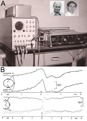
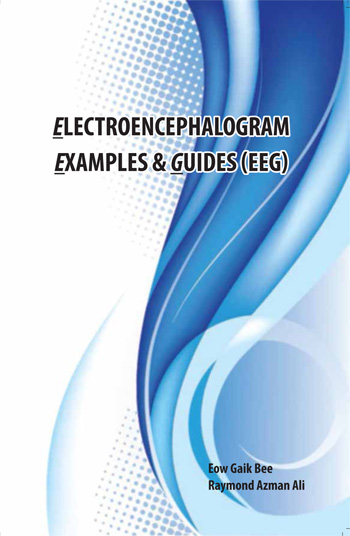
It is a privilege to introduce the book “Electroencephalogram Examples and Guides,” published in August 2013. The book provides a concise and comprehensive compilation of text and EEG recordings collected to shorten the learning curve toward competence in EEG interpretation.

This book will be valuable to a wide variety of readers. For specialists preparing for EEG certification examinations, such as those offered by the AsianEpilepsyAcademy – ASEAN Neurological Association (ASEPA-ASNA) or the American Board of Clinical Neurophysiology (ABCN), it could serve as an essential guide. For the practicing neurologist, it can serve as a useful quick reference. For beginners, including EEG technologists and neurology trainees, it can be an effective teaching resource.

Throughout the book, complex concepts are simplified without the sacrifice of fine details. Facts and comparisons are given in point forms and tables. Classifications of various EEG findings are presented in easy-to-understand algorithms. EEGs from common to rare conditions are presented in a stimulating quiz format. The index enables quick reference to EEG tracings from patients with different diagnoses.

Sections 1 and 2 deal with the basics of EEG, including indications, limitations and neurophysiologic principles. Section 3 provides actual EEG recordings, starting with normal findings, artifacts and benign variants and progressing through epileptiform and non-epileptiform abnormalities. Section 4 includes a systematic step-by-step approach to EEG interpretation, and Section 5 offers a quiz for readers to practice their EEG interpretation skills. Section 6 deals with EEG evaluation of syncope and blackout spells, while Section 7 deals with evaluation of coma and altered states of consciousness. Finally,  Section 8 provides an opportunity for self-assessment of the reader’s discernment of the various points in the book.

This innovative book is recommended for anyone who seeks to learn EEG in an effective and systematic way.  That the author also made the process pleasurable is a welcome benefit indeed.

Wylie is professor of the Lerner College of Medicine Epilepsy Center, Neurological Institute, Cleveland Clinic, Cleveland

Peripheral Nerve Injuries During World War I

Growing knowledge despite a lack of international cooperation.

Peter Koehler

Peter Koehler

This column on historical aspects of international relationships in the neurosciences usually deals with forms of international cooperation. Exactly 100 years ago, not only political relations collapsed; scientific relationships followed, even though some scientists hoped that their relations would remain above the cataclysm.

Living in neutral countries (Netherlands and Switzerland), at least two neuroscientists, Cornelis Winkler and Constantin von Monakow, hoped that “we neutral countries are … now in the first place obliged to continue to take care of values of humanity and culture and keep upright the international scientific relationships with all energy” (from their correspondence in November 1914). Only shortly after the beginning of World War I, however, they observed that artists and writers declared support to the German military actions, including Paul Ehrlich, Ernst Häckel, and neurologists Wilhelm Heinrich Erb and Hermann Oppenheim, who had sent back their scientific decorations they had once received from England. Monakow opined that this was an injudicious, unfortunate step, estranging them to international science “that has nothing to do with politics.”

Neurological knowledge, however, kept on growing, simultaneously, but now separately. A striking example was the knowledge of peripheral nerve injuries which had only gradually increased in times of peace, but now, sadly, much faster. During World War I, books on peripheral nerve injuries were published in England, France, Germany and the U.S. Probably the best known book on the subject was written by the French Jules Tinel (1879-1952), who published his Blessures des nerfs in 1916. His book became influential, and within a year, it was translated into English and published in New York.

Froment sign (from Presse Médical, Thursday, Oct. 21, 1915)

Froment sign (from Presse Médical, Thursday, Oct. 21, 1915)

On the French side, women published on the subject, as is witnessed in Chiriachitza Athanassio-Bénisty, a pupil of Pierre Marie (Paris). She wrote two books with the aim not only to publish the results of clinical experience, but also to improve the quality of examination of peripheral nerve lesions by less experienced physicians. It was translated by Edward Farquhar Buzzard, the English physician, working a.o. at the NationalHospital. From the English ranks, Arthur Henry Evans and James Purves-Stewart published their experiences, largely observations of injured soldiers, many of whom had fought at the various battles near Ypres, Belgium.

From the German side, neurologist/neurosurgeon Otfrid Foerster (1873-1941), who served as an advisory physician to the health office of the VI German Army on the western front in France, gathered abundant information on injuries to the peripheral nervous system and spinal cord. He published his findings in a multivolume handbook of medical experiences from World War I, but also in the supplement to Lewandowsky’s Handbuch der Neurologie, which was entirely devoted to war injuries of the peripheral nerves and spinal cord (single-authored altogether 1,152 pages). In the chapter on surgical therapy of peripheral nerve injuries, he mentioned 4,117 peripheral nerve injuries. Nearly 25 percent underwent surgical procedures, including nerve sutures, nerve transplantations and arm and leg plexus surgeries.

Froment sign (from Presse Médical, Thursday, Oct. 21, 1915).

Froment sign (from Presse Médical, Thursday, Oct. 21, 1915).

The U.S. entered World War I in 1917. After submarines sank seven U.S. merchant ships, U.S. President Woodrow Wilson went to Congress calling for a declaration of war on Germany. The U.S. Congress voted on April 6 to do so. Figures with respect to American peripheral nerve injury casualties during World War I were provided by several sources, including neurosurgeon Charles Harrison Frazier (1870-1936). Returning casualties with peripheral nerve injuries were treated in 12 peripheral nerve centers, usually located in general hospitals, where medical officers with experience in neurosurgery as well as consultant neurologists were working. Frazier provided the anatomic location of almost 2,400 peripheral nerve injuries. Byron Stookey (1887-1966) served with the British Royal Army Medical Corps (1915-1916) and the U.S. Army Medical Corps. In his Surgical and Mechanical Treatment of Peripheral Nerves (1922), he published relative frequencies of peripheral nerve injuries of 1,210 war casualties.

Of all nerve injuries described in the four countries (more than 10,000 in the various publications), radial nerve lesions were generally the most frequent. Partial lesions of the radial nerve were rare, in contrast to the frequency of partial injuries of the ulnar and median nerves. At least two eponyms are remembered from this dark period in the history of neurology. Working on different sides of the front, a French (Tinel) and a German (Paul Hoffmann:1884-1962) neurologist are remembered in one eponym, notably the Tinel-Hoffmann sign. It indicates radiating tingling sensations in the otherwise anesthetic skin area innervated by an injured nerve, upon light percussion of the area. The sign was considered to indicate the presence of new sensitive regenerating nerve fibers. Another French neurologist, Jules Froment (1878-1946), once a co-worker of Joseph Babinski, is remembered by his “signe de journal,” based on the fact that in ulnar nerve neuropathy, the action of the paralyzed adductor pollicis muscle is compensated for by the flexor pollicis longus muscle, innervated by the median nerve, resulting in flexion of the distal phalanx of the thumb.

Further reading

  1. Koehler PJ. Lessons from peripheral nerve injuries; causalgia in particular. In: Fine E (ed). History course syllabus. Lessons from war. AmericanAcademy of Neurology, San Diego, 2013.
  2. Koehler PJ, Lanska DJ. Mitchell’s influence on European studies of peripheral nerve injuries during WorldWarI. J Hist Neurosci 2004;13:326-35
Koehler is neurologist at Atrium Medical Centre, Heerlen, The Netherlands. Visit his website at www.neurohistory.nl

World Brain Day

By Mohammad Wasay, MD, FRCP, FAAN

Mohammad Wasay

Mohammad Wasay

There are many days related to neurological diseases being celebrated by professional organizations in collaboration with the World Health Organization (WHO), national organizations and local health ministries, including World Stroke Day, Epilepsy Day and Rabies Day. These days have proven to be extremely helpful in promoting public awareness and generating advocacy throughout the globe, including non-developed Asian and African countries. For example, the World Stroke Organization announced a global competition for public awareness and advocacy campaign focusing on World Stroke Day.

Brazil in 2012 and Sri Lanka a year later won the competition creating a huge impact at the national as well as regional levels. All of the days related to neurology are linked to neurological diseases. A few years ago, Vladimir Hachinski suggested celebrating a day for healthy brains. The human brain is so fascinating and is so closely linked to the health of whole human being that we should promote healthy brains. The future of this universe is linked to our brains so we should start a global campaign. The Public Awareness and Advocacy Committee came up with the slogan of “Our Brains-Our Future.”

The World Federation of Neurology was established on July 22. The committee proposed that July 22 should be recognized as “World Brain Day.” This proposal was announced at the Council of Delegates meeting in September at the WCN. The proposal was received with a warm welcome by delegates, Hachinsky, WFN president; Raad Shakir, WFN president- elect; Werner Hacke, WFN vice president; and other officials. The BOT meeting in February approved this proposal as an annual activity.

Our target audience is young brains throughout the world, and we want to promote healthy brain and brain health. Young students and minds are highly interested in knowing how the brain works and how  we can make it work better. Brain health is a huge topic covering many areas, including understanding of brain function, optimization of brain function, disease prevention, mental health and treatment of brain disorders.

We should target to approach one billion people around the globe to educate about brain in 2014. Most activities will focus on World Brain Day, but it is a year-long campaign. National societies should plan activities focusing on young school and college students. With the help of social and electronic media, the information could go to millions of people. All societies should share their plans and activities, and those organizations with highest impact public awareness activities should be awarded.  We should especially focus on Facebook and Twitter to connect with millions of people. Our young neurologists’ network on Facebook could be a great resource for this campaign. We should use multiple languages, especially focusing on Arabic, Chinese, French, German, Hindi and Spanish. We also could develop a five-minute promotional video with brief introduction of WFN in multiple languages. This video could be shared by millions through YouTube and Facebook. We have more than 100 member societies. If we are able to organize a few hundred programs on July 22 in all of those countries, it is bound to create an impact. Member societies could organize press conferences, media briefing sessions, lectures, seminars, conferences and poster competitions. Quiz competitions (Brain Quiz) have been successful and popular among school and college students.

Complexity of brain and neurological diseases often become a barrier for public awareness. “You should speak plain when you speak brain” was suggested by Keith Newton of the WFN. Our message should be simple and easily understandable for lay people. We could design a logo for this purpose, which may be a simple global message. WFN and local organizations could start a poster- or cartoon-designing competition to explain brain function and improve public awareness. Best posters, designs or cartoons could be awarded. We expect thousands of entries for this competition and some of these entries could become logos for our future campaigns.

There are many organizations working in this area, including the International Brain Council, International Brain Research Organization, the American Academy of Neurology, the International League Against Epilepsy and the World Stroke Organization. We should work with them for this common agenda. Strong liaison and lobbying with the WHO is important. If the WHO adopts this day in future, then this could be a great success for the WFN. If we are able to build a momentum around the globe in the coming years, we are sure it will become a WHO day in the future.

Wasay is chair of the Public Awareness and Advocacy Committee for the World Federation of Neurology.

JNS: Editor’s Update and Selected Articles

By John D. England, MD

John D. England

John D. England

The Journal of the Neurological Sciences (JNS) and its publisher, Elsevier, are working together to improve the journal and to make the submission process easier for authors.

A major complaint from authors concerns the necessity to format manuscripts to fit the idiosyncratic requirements of journals.  Since many high volume journals like JNS have a high rejection rate, authors frequently must reformat their manuscripts for submission to a different journal. This is not only a hassle, but a time-consuming process for authors.

In order to simplify the submission process for authors, JNS has eliminated strict requirements for reference formatting. As of now, we will accept manuscripts with no strict requirements for reference formatting. Any style of reference formatting will be accepted as long as the style is consistent.

If the manuscript is accepted for publication, then Elsevier will change the reference formatting to fit the style for JNS. As an extension of this process, in the near future JNS and Elsevier will begin accepting entire manuscripts without strict formatting or referencing requirements.

WFN_BreakingNewsElsevier already has introduced this feature, which is named “Your Paper Your Way (YPYW),” for several other journals in its portfolio. It has been so well received by authors that we will shortly begin offering this service to authors of manuscripts submitted to JNS.   Taken together, these new options for manuscript formatting flexibility should make it easier for authors to submit manuscripts to JNS.

As noted in the previous issue of World Neurology, we have begun an “Editor’s Selection” of articles from JNS.  Elsevier has agreed to allow free access to selected articles for members of the World Federation of Neurology.  The process for viewing these articles is now streamlined: Click on one of the two featured articles on the World Neurology Online page, or use the “See all free JNS articles” link below the featured articles. It will take you directly to the free article page on the JNS website.  In this issue, we share these two recent articles:

The Clinical and Pathological Phenotypes of Frontotemporal Dementia with C9ORF72 Mutations. Ying Liu and others from China have written a review on the phenotypes of frontotemporal dementia associated with C9ORF72 mutations.  The expanded hexanucleotide repeat (GGGGCC) in the chromosome 9 open reading frame 72 (C9ORF72) is now recognized as one of the major causes of hereditary frontotemporal dementia (FTD). It is also the most frequent genetic cause of the ALS/FTD complex.  However, the clinical and pathological phenotypes associated with C9ORF72 mutations appear increasingly diverse, and the mechanisms of disease are not known.  See Liu Y, et al.

Journal of the Neurological Sciences 335 (2013) 26-35.  An Association Between Benzodiazepine Use and Occurrence of Benign Brain Tumors. Tomor Harnod and others analyzed data from the National Health Insurance System of Taiwan to ascertain whether there is an association between long-term benzodiazepine use and the development of brain tumors. They identified 62,186 patients who had been prescribed benzodiazepines for at least two months between Jan. 1, 2000, and Dec. 31, 2009. These patients were compared with a matched non-benzodiazepine cohort of 62,050 patients. The incidence rate for benign brain tumors was 3.33 times higher in the benzodiazepine cohort compared to the non-benzodiazepine cohort with an adjusted hazard ratio (HR) of 3.15 (95% CI = 2.37-4.20).  Additionally, the adjusted HRs for benign brain tumors increased with benzodiazepine dose. Thus, in this cohort study, the authors found a significant association of benign brain tumors with long-term benzodiazepine use. There are many possible explanations for such an association, and the authors correctly avoid the conclusion of causation. But, benzodiazepines are a commonly prescribed medication, and further studies of this important topic are warranted. See Harnod T, et al.

Journal of the Neurological Sciences 336 (2014) 8-12.

England is editor-in-chief of the Journal of the Neurological Sciences.

Palliative Care as a Human Right: Where Does the Neurologist Stand?

By Raymond Voltz

Raymond Voltz

Raymond Voltz

Two recent international publications have put the spotlight on the situation of worldwide provision of palliative care. These are the “World Cancer Report,” published by the International Association of Research in Cancer (IARC) and the “Global Atlas of Palliative Care at the End of Life,” published by the WHO in collaboration with the World Wide Palliative Care Alliance. The “World Cancer Report” reminds us that the war on cancer has not been won and that still much remains to be done when treatment fails.

“The Global Atlas of Palliative Care at the End of Life” highlights the need for more palliative care: It estimates that 19.2 million people worldwide require palliative care with about 38 percent of patients dying from cardiovascular diseases, 34 percent of cancer and 10 percent of COPD. In the remainder, specific diseases such as multiple sclerosis (MS) and Parkinson’s are mentioned, and although less important on the global scale, patients with MMD/ALS. The atlas highlights the worldwide skewed distribution of palliative care services and access to palliative care depending on income and geographical regions worldwide. They conclude that palliative care is a human right, and that therefore each country should take improvement of palliative care up as a national strategy.

In some countries, it starts at the beginning: the access to opioids. A few weeks ago, a well-known Russian military committed suicide, stating in his final letter, that he did so because of an unbearable pain due to cancer and that he was not able to get opioids. Some countries are proud of their drug protection programs, which also means that they protect cancer patients from getting the right drugs at the right time. Here, a sensible political balance has to be reached. Opioids, of course, are important for the treatment of pain as well as the treatment of dyspnea. Provision of multiprofessional high-skilled palliative care follows those basic tenets.

So what does this mean for neurology? Cancer patients, cardiovascular patients and specific neurological diseases are treated within the responsibility of neurologists worldwide. As the atlas also states, their estimates only refer to patients at the end of their lives. The concept of early integration of palliative care has now fully reached the oncological world with the ASCO stating that all cancer patients should have access to palliative care. So, the need for palliative care is even greater than stated in the atlas, which the authors fully acknowledge. So where is the relevant interest in neurology? Do all neurologists worldwide know what palliative care structures already exist in their neighborhoods? Do they refer refer patients there early enough for common management of patients and families? Where is the topic of palliative care in the training of neurologists worldwide? Where are the neurologists who help establishing palliative care structures in their region? Where are the ones who advocate for a national palliative care strategy including all relevant patient groups? And finally, where is the interest of moving the field forward by valid research? Still, many neurologists think this in an unscientific topic not relevant to them, and that is a pity — mainly for many patients worldwide suffering not only from devastating neurological diseases but also from clear palliative care mismanagement by their neurologists.

Voltz is chair of the Department of Palliative Medicine at the University Hospital of Cologne, Germany.

International Conference on Freezing of Gait

Nir Giladi

Nir Giladi

By Mark Hallett, MD

Management of patients with Parkinson’s disease has progressed well. Levodopa and the dopamine agonists are effective, and when complications arise such as dyskinesias and fluctuations, deep brain stimulation (DBS) can be effective. However, another significant problem is now emerging. That is gait difficulties later in the course of Parkinson’s disease that are not responsive to either the dopaminergic therapy or DBS. Patients can look pretty good in many ways, but will have trouble relatively isolated to balance and gait.

One interesting feature of the gait difficulty is freezing of gait (FOG). With FOG, there is a failure to move forward despite intent to do so.  FOG can occur at gait onset or in the middle of walking, particularly in some settings such as walking through a doorway or turning. At times, there can be off-freezing that is dopa responsive; the problematic situation is on-freezing. FOG also occurs in other Parkinsonian conditions such as progressive supranuclear palsy.

There have been two prior workshops devoted to the topic, but recently there was an international conference Feb. 5-7, organized by Professors Nir Giladi and Jeff Hausdorff at the Dead Sea. The conference was sponsored by TelAvivUniversity, the TelAvivMedicalCenter and the International Parkinson and Movement Disorder Society. More than 160 attendees came from 20 countries.

Jeff Hausdorff

Jeff Hausdorff

Freezing also can occur with upper extremity movements or speech, but gait is more commonly affected and more debilitating. The topic is now under intense scrutiny. Gait is a complex movement requiring integrated activity of all parts of the brain and spinal cord with both balancing and stepping. This is likely why gait is more commonly affected than other movements.

There are a number of theories as to the etiology, and, as freezing is likely to be multifactorial, many of these may well be relevant in different patients. One type of problem is the deterioration of motor control capabilities, such as loss of the internal drive for movement, difficulties with multitasking and difficulties in carrying out automatic movements. Lack of gait symmetry is often correlated with freezing.

Another interesting motor control problem in Parkinson’s disease is the sequence effect, the progressive decline of movement amplitude in a sequence of what should be similar movements. Step length often gradually declines prior to a freeze, so this seems important at least in some circumstances. Cognitive problems, importantly loss of executive function, appear relevant. There is clearly a role of environment factors, including the path that needs to be traversed; the narrow doorway, for example.

There was considerable discussion of therapy. In the end, given the multiplicity of causes, therapy may have to be individualized.  Certainly, there should be care to optimize dopaminergic therapy. Physical therapy can play a role. Much discussion focused on the value of DBS of the pedunculopontine nucleus (PPN). The PPN sits in a complex region at the junction of the midbrain and the pons and appears to be a part of, or at least close to, the mesencephalic locomotor region. The literature is really still sparse, and it is not clear that the DBS electrodes are actually in the PPN in all patients, but some patients do appear improved.

Gait freezing remains an important clinical phenomenon, a fascinating set of problems for physiology and a challenge for treatment. Giladi and Hausdorff have been leaders in all aspects, and the attendees were grateful to them for organizing a useful conference that will set the direction of research in the next few years.

Africa-Canada International Behavioral Neurology Videoconference Rounds

Morris Freedman, MD, FRCPC, Tim Patterson, BA, Riadh Gouider, MD, Sandra E. Black MD, FRCPC, FAAN, FAHA, Cindy J. Grief, MD, FRCPC, MSc, and Peter Whitehouse

In front of screen in Canada (left to right) Ayman Selim, Carmela Tartaglia, Sandra Black, Arnold Noyek, Cindy Grief, Morris Freedman, Tim  Patterson. On the screen from Tunisia (foreground, left to right) Riadh Gouider, Mouna Ben Djebara and (background) Tunisian participants.

In front of screen in Canada (left to right) Ayman Selim, Carmela Tartaglia, Sandra Black, Arnold Noyek, Cindy Grief, Morris Freedman, Tim Patterson. On the screen from Tunisia (foreground, left to right) Riadh Gouider, Mouna Ben Djebara and (background) Tunisian participants.

In 2012, the World Federation of Neurology (WFN) eLearning Task Force of the WFN Education Committee proposed an expansion of the ground-breaking Canada-Tunisia videoconference as reported in the World Neurology Newsletter (Vol.27 – No.1-February 2012). This interactive and live videoconference was held in late May 2011, two weeks into the Arab Spring Uprising. It demonstrated both the WFN vision to connect international centers throughout the world and the determination of Tunisia to participate in a global educational videoconference at a time of great disruption to its society.

Building on the success of the Canada-Tunisia international rounds, the WFN awarded an educational grant to expand this initiative into an Africa-Canada series involving Tunisia, Morocco and Canada. The grant was in conjunction with the Peter A. Silverman Global eHealth Program, and the international rounds are under the auspices of the Canada International Scientific Exchange Program (CISEPO), Canadian Neurological Sciences Federation, Division of Neurology, University of Toronto and Razi Hospital La Manouba, Tunis.

Carmela Tartaglia and hosts Morris Freedman, Sandra Black and Riadh Gouider initiated the series Jan. 15, 2014, with a presentation titled, “An Uncommon Cause of a Common Problem.”Audience participants included sites from Canada and Tunisia. This event, and the videoconference series as a whole, serve as a prime example of the WFN’s goal to foster quality neurology and brain health worldwide. This educational format promises to be of great benefit, especially to those countries where educational resources in neurology are limited despite the quest for knowledge.

The series also demonstrates the universality of health care as a common language bridging societies together.

“These rounds have the potential to bring together health care professionals from across the world in a forum that transcends cultural and political differences that may otherwise pose barriers to dialogue,” said Freedman. In addition, “the rounds provide an opportunity to share the differences and challenges across national boundaries,” said Whitehouse.

Providing a Tunisian point of view, Gouider said, “The importance of the International Behavioral Neurology Rounds are in the content and also in the collaborative teaching methodology. It demonstrates that language and time differences between continents may be easily bypassed.”

The connectivity is accomplished through the Internet and bridging, with each of the receiving sites having the appropriate videoconferencing equipment. The videoconferences are monitored and evaluated under the successful University of Toronto Behavioral Neurology Rounds that have been videoconferenced to provincial and international sites over the last 10 years.

The launch of Africa-Canada international behavioral neurology rounds will act as a beacon for the success of future programming and hopefully as a stimulus for additional international projects involving disciplines in other areas of neurology using the medium of videoconferencing.

Freedman is with the Department of Medicine, Division of Neurology, Baycrest and University of Toronto, Toronto, and the Rotman Research Institute, Baycrest, Toronto. Patterson is with the Department of Telehealth, Baycrest, Toronto, and the Canada International Scientific Exchange Program. Gouider is with the Department of Neurology, Razi Hospital La Manouba 2010-Tunis-Tunisia. Black is Brill Chair in Neurology, University of Toronto, Sunnybrook Health Sciences Center, Toronto, and Department of Medicine, Division of Neurology, Sunnybrook Health Sciences Center and University of Toronto, Toronto. Grief is with the Department of Psychiatry, Baycrest and University of Toronto. Whitehouse is with the Department of Neurology, Case Western Reserve University, Cleveland, Ohio, and Department of Medicine, Division of Neurology, University of Toronto, Toronto.