European Board of Neurology Examination in Berlin in 2015

UEMS-EBN-logoThe European Board Examination in Neurology is a joint development of the UEMS Section of Neurology and the European Academy of Neurology. It is considered to be a tool for the assessment of European neurological education and to boost its European standards.

It is supervised by the examination committee of the UEMS/EBN and also observed by the EAN representing the European neurological scientific societies and the World Federation of Neurology.

The exam was held in 2009 for the first time, and since then 130 candidates have passed the exam. Beginning in 2015, the title “Fellow of the European Board of Neurology” will be conferred to European and non-European candidates.

The next UEMS/EBN examination will be organized one day prior to the 1st Congress of the European Academy of Neurology (EAN) on Friday, June 19, 2015, in Berlin, Germany. (http://www.eaneurology.org/)

UEMS-EBN-Exam-Istanbul-2014The European Board Examination in Neurology is a substantial step forward in the further harmonization and in the raising of the standards in European neurology. The cooperation with the scientific neurological societies is an important scientific input and a guarantee of continuous updates of the current knowledge of a European neurologist.

The European Examination in Neurology is a proof of excellence: Taking the examination shows the candidate’s commitment to lifelong learning. Even without legal recognition, this is known and recognized within the profession throughout Europe and the rest of the world, thus encouraging the mobility of specialists in neurology and giving an additional distinguishing mark to the individual candidate.

The deadline for application is the May 1, 2015. (http://www.uemsneuroboard.org/ebn/)

There is a reduced fee for candidates from low- and lower-middle income countries (see http://data.worldbank.org/about/country-and-lending-groups#Low_income) and for those who follow the early-bird registration procedure.

The examination consists of the following parts:

  • 80 MCQs (multiple choice questions)
  • 50 EMQs (extended matching question)
  • A short essay on a neurology-related public health or ethics-related topic to be orally discussed with the examiners.
  • A critical appraisal of a neurological topic to be discussed with the examiners.

Results of these four parts of the examination will be combined to one final mark.

We are happy to note that the number of participants taking the European Board Exam in Neurology is increasing year by year, and we aim to develop an exam that will be taken by all neurology trainees, particularly those who wish to extend their experience beyond the borders of their own country.

Any questions and comments can be sent to uems-sbn@medacad.org

Professor Dr. Jan Kuks: Chair of the examination committee: j.b.m.kuks@umcg.nl

Professor Dr. Wolfgang Grisold: UEMS/EBN past chair of the examination committee wolfgang.grisold@wienkav.at

Dr Walter Struhal: WFN website and social media, w.struhal@aesculapian.net

CONTACT address:

Mag.Gabrielle Lohner: uems-sbn@medacad.org

Section of Neurology –European Board of Neurology
c/o Vienna Medical Academy
Alser Strasse 4, 1090 Vienna
AUSTRIA
T (+43 1) 405 13 83 – 32
F (+43 1) 407 82 74
www.uems-neuroboard.org

Pediatric Neurology in Africa

Fellowship program builds skills for health practitionersAPFP_logo

By Jo Wilmshurst, MD

Doctors trained in the management of child neurology conditions are lacking in Africa1,2. Epilepsy is one of the major disease burdens in the continent and training in this area is even more scarce. EEG interpretation in children is very different to that for adults and grave errors can occur in patient management when misinterpretations occur.

The African Paediatric Fellowship Program (APFP) is a project developed by the Department of Paediatrics and Child Health at the Red Cross War Memorial Children’s Hospital, under the University of Cape Town in South Africa, to build skills capacity of health practitioners from Africa. The center is the largest dedicated children’s hospital in sub-Saharan Africa. Children are managed across primary to quaternary levels of care with the spectrum of diseases prevalent in Africa.

Figure 1. some of the 2014 apfp fellows attending the end-of-year discussion group. dr. kija, child neurology trainee from tanzania, is fourth from the left. represented in the group are doctors training in areas from pediatric urology to pediatric rheumatology from areas in africa inclusive of uganda, zambia, kenya, ghana, zimbabwe and malawi. the group remains as a cohesive support network and stay in contact after completion and following their return home.

Figure 1. Some of the 2014 APFP fellows attending the end-of-year discussion group. Dr. Kija, child neurology trainee from Tanzania, is fourth from the left. Represented in the group are doctors training in areas from pediatric urology to pediatric rheumatology from areas in Africa inclusive of Uganda, Zambia, Kenya, Ghana, Zimbabwe and Malawi. The group remains as a cohesive support network and stay in contact after completion and following their return home.

The APFP formed collaborations with tertiary centers across Africa and has assisted their identification of strategic training requirements based on their countries’ key health care needs. Structured training occurs at the pediatric units through the University of Cape Town. More than 65 specialists have completed, or are completing in 2015, the training program in diverse pediatric areas, referred from 33 centers in 12 different African countries. There has been a 98 percent retention rate of trainees returning to work in their home country since 2008. The program is evolving with training arms supporting nursing and ancillary services. The trainee becomes the trainer in his or her home center, and a key opinion leader equipped to lobby for changes to health care policy (Figure 1).

The grant provided by the WFN to support neurology training in 2013 has enabled the focused training for six general pediatricians from different centers in Nigeria, and three further trainees from Zimbabwe, who manage large caseloads of children with neurodisabilities and epilepsy. The University of Cape Town rolled out in 2015 a post-graduate diploma in “basic electrophysiology interpretation and the management of children with epilepsy.” This requires one-on-one training with a focus on areas relevant to the African context. The aim of the post-graduate diploma is to establish safe practice and not to train accredited epileptologists. In Africa, most child health practitioners who manage children with neurologic disorders must address the comprehensive needs of the child inclusive of the other health issues, such as co-infections, nutritional deficits, and social challenges (Figures 2 a, b).

At the current time in most African settings, it is not viable to work as an epileptologist without addressing these other health care issues (Figures 3 a, b). However as a result of more cost-effective neurophysiology equipment, and through equipment donations, there are an increasing number of EEG machines that are potentially being operated and interpreted by health practitioners with no pediatric training. This training program was devised out of the needs that this situation created. The audit of the preliminary findings of a pilot study on the impact of the training course while it was being established is in press. The audit confirmed that access to a basic handbook improved EEG interpretation skills, but that the optimal outcomes were seen in those doctors who had additional one-on-one training.

a.) Children attending the neurology clinic at Red Cross War Memorial Children's Hospital, enjoying a donation of new reading books. b.) One of the neurology patients occupied in puzzle play in the waiting area.

a.) Children attending the neurology clinic at Red Cross War Memorial Children’s Hospital, enjoying a donation of new reading books. b.) One of the neurology patients occupied in puzzle play in the waiting area.

Between 2013 and 2014, three doctors from Nigeria, Tanzania and Ghana entered the APFP for formal training to become accredited child neurologists. Tanzania has no accredited child neurologists and Ghana has two. These trainees, in addition to completing the full post-graduate clinical master’s degree in child neurology, also are completing research in areas relevant to the context they work in. The doctor from Tanzania is heading a prospective study to review the effects on bone mineral density in children on antiepileptic drugs in the African setting. Vitamin D supplementation is not part of standard care of these patients and it is hoped that the findings from this study will lead to data to support lobbying for this intervention to be part of standard practice. The doctor from Ghana will complete a study assessing the neurobehavioral influences on children from antiretroviral therapy. The doctor from Nigeria has completed a large prospective study assessing the efficacy of attaining sleep EEGs in children using melatonin.

In the next training cycle it is hoped that there will be funding to support applicants from Sierra Leone, Zimbabwe, Uganda, Kenya, Sudan and Zambia.

The training curriculum, while in line with international templates, also accommodates approaches novel to Africa, such as the neurological care for children with tuberculous meningitis, HIV, malaria and neurocysticercosis. The perinatal complication rates remain high in Africa with significant numbers of neonates suffering hypoxic ischaemic encephalopathy. Other neuroinsults are seen from the effects of central nervous system infections and motor vehicle accidents. The training must accommodate these areas in depth as well. The returning trainee must often function in all areas from social welfare to rehabilitation, the training is adjusted for this.

a.) Mothers attending the Queen Elizabeth Hospital, Blantyre, in Malawi, taken during an APFP site visit in 2013. This is the main teaching hospital in the country, which has one pediatric neurologist for the total population. b.) Child-care workshop for children with motor disabilities at the Child Rehabilitation Unit, Harare Hospital, Zimbabwe, taken during the APFP site visit to the referring units in the country in 2014.

a.) Mothers attending the Queen Elizabeth Hospital, Blantyre, in Malawi, taken during an APFP site visit in 2013. This is the main teaching hospital in the country, which has one pediatric neurologist for the total population. b.)
Child-care workshop for children with motor disabilities at the Child Rehabilitation Unit, Harare Hospital, Zimbabwe, taken during the APFP site visit to the referring units in the country in 2014.

Prevention and early intervention is one of the major aims for this project and all trainees in the program are facilitated in the knowledge gained during their training and assess its relevance to their home setting, how to introduce these skills to the optimal benefit to child health care and how these interventions can extend across all levels of health care—from primary to tertiary.

On the trainee’s return to their home center they maintain contact with their supervisor, and site visits are scheduled as needed to provide local input into service development and local training (Figure 3 a, b). Research collaborations also continue. These trainees are having a real impact in their home centers and are becoming voices in Africa lobbying to promote child health. One of the child neurology trainees who completed training in 2009 and returned to Kenya now sits on the national Kenyan pediatric body, assists selection of ongoing APFP trainees referred from the country, and is also on the Pediatric Commission for the International League Against Epilepsy. This trainee is part of a team developing its own subspecialty training program for East African doctors. This is viewed as a major future aim of the APFP. In order to grow and to fulfill the health care needs for the continent, more training sites are needed. It is important these remain within Africa with training relevant to the diseases of the region. There is much to learn from the approaches many innovative African centers undertake to cope with the challenges of scare resources.

While training experience in overseas centers offers obvious gains in skills development, the local relevance of the training may be questionable and the risk of the “brain drain” is high. A number of overseas specialists have opted to spend time working, training and lecturing in African centers. This is a superb way to assist skills development in African centers. Building on these relationships with regular visits develops sustained skills where often none existed before.

References

  1. Wilmshurst JM, Badoe E, Wammanda RD, Mallewa M, Kakooza-Mwesige A, Venter A, Newton CR. Child neurology services in Africa. J Child Neurol. 2011 Dec; 26(12):1555-63.
  2. Wilmshurst JM, Cross JH, Newton C, Kakooza AM, Wammanda RD, Mallewa M, Samia P, Venter A, Hirtz D, Chugani H. Children With Epilepsy in Africa: Recommendations From the International Child Neurology Association/African Child Neurology Association Workshop. J Child Neurol. 2013; 28 633-644
Dr. Wilmshurst is the head of paediatric neurology, Red Cross War Memorial Children’s Hospital, University of Cape Town, South Africa, and the director of the APFP.

Neurology Fellowship: UC San Francisco/Yale University

55844195A neurology fellowship is offered by the Memory and Aging Center at the University of California, San Francisco, and the Neurology Department at Yale University (position can be filled at either location) through the NeuroHIV Cure Consortium, which operates numerous neurological research studies in acute HIV infection and cure strategies in Thailand and Africa. The Consortium is a research collaboration directed by Victor Valcour, MD, PhD (UCSF), Serena Spudich, MD (Yale University), and Jintanat Ananworanich, MD, PhD (U.S. Military HIV Research Program).

The NIH-funded work gives a neurologist opportunities to conduct research. The fellow will provide at least six months of neurological expertise at research sites in Thailand, including neurological evaluation of subjects and lumbar punctures, while developing a personal research portfolio. The incumbent will be expected to actively participate in manuscript preparation and analyses for publication as lead author, and apply for awards and other funding to support continued involvement.

The Consortium leaders have extensive experience as career and research mentors and will provide guidance and assistance in data analysis and preparation of presentations and manuscripts, as well as in independent fellowship and grant applications. The position is for one year, with opportunities for extension. The fellowship may be combined with a neurobehavioral fellowship at UCSF.

For more information: www.inhcc.net/opportunities.html.

Eye Movement Disorders in Clinical Practice by Dr. Shirley H. Wray

Wray SH (2014) Eye Movement Disorders in Clinical Practice. New York NY: Oxford University Press.

By Steven L. Galetta, MD

Eye Movement Disorders in Clinical Practice is written by a legend in the field of neuro-ophthalmology, Dr. Shirley H. Wray. The book is a wonderful compendium of the common and challenging disorders of ocular motility. Chapters are organized topographically beginning with the cortical control of eye movements, followed by chapters on the eyelids, the extraocular muscles and nerves, disorders of horizontal and vertical gaze, dizziness, the cerebellum and ocular oscillations.

Each chapter begins with a generous discussion of the relevant anatomy and physiology of a particular area of the brain. The key anatomical points are then summarized in a box to distill the critical information that is about to be given clinical relevance in the remainder of the chapter. Chapters in this book take some of the most complicated neuroanatomy and make it come to life with beautiful figures, clear discussions and key references. After the anatomy and physiology are reviewed within each chapter, the reader is then taken to cases where the history and examination are dissected, with an emphasis on how signs and symptoms are localized and correlated. Each case has a great video attached to it. The videos are of outstanding quality and clearly demonstrate the ocular motility disturbance. In fact, many cases are the best examples that I ever seen of the eye movement disorder. One minor criticism would be that the videos often do not point out the exact findings as you are taken through the standard neuro-ophthalmological examination. Nonetheless, such findings are summarized in the text and the reader is provided ample guidance on what to look for.

From the video section, the reader is taken back to the text for further discussion of localization, differential diagnosis, testing and treatment of the problem, emphasizing the importance of neuro-ophthalmological signs in managing the patient. The learning experience is then supplemented by special explanatory notes; these break down the problem and often provide some historical context to the findings.

The beauty of this book is its redundancy; the reader is never left to struggle with a single description of the neuro-ophthalmological topic. This is a great feature for both the novice and the expert and makes each case even more memorable.

It was a real pleasure to read this book. There were many moments when I could not put it down because it was fun to read such an organized and a logical approach to eyelid and ocular motility disorders. The chapters on the cortical control of eye movements and the cerebellum are true masterpieces; they take some of the most complicated anatomy and disorders of ocular motility and make them easy to understand. Another feature that makes this book so special is the simple way in which the examination is explained, and how symptoms are emphasized as a guiding factor in what the examiner should look for. I often found myself taking notes on the side, and trying to remember all the gems being offered.

Eye Movement Disorders in Clinical Practice is a keeper! I would recommend it to neurology and ophthalmology trainees and senior clinicians alike.

Dr. Galetta, is professor and chair of the Department of Neurology, New York University School of Medicine.

New Society Works to Improve Quality, Establish Standards

Guenther Deutschl

Guenther Deutschl

By David B. Vodušek and Guenther Deutschl

The European Academy of Neurology (EAN) was founded on June 3, 2014, in Istanbul by joint efforts of the two parent societies at their (first and last) Joint Congress of European Neurology, the congress of the European Federation of Neurological Societies (EFNS) and the European Neurological Society (ENS)1. (See also Grisold W, WFN Newsletter http://www.worldneurologyonline.com/article/ens-efns-ean/).

The society is established according to Austrian law, the main goal being to promote “excellence in neurology in Europe.” EAN is a new and vigorous society, which is taking over the good program, functions and initiatives from both parent societies along with the previous EFNS head office in the prestigious Museum District in Vienna, a fine centrally situated “Pentagon” of European neurology, assuring frictionless functioning of the diverse EAN organs.

The bulk of the new group of governing officers, the EAN Board, was elected in June by the hybrid Assembly of Delegates from 45 National European Societies (previously constituting EFNS) and 45 delegates from individual members (elected by the ENS in spring 2013). Prof. Günther Deuschl from Kiel, Germany was elected as president, and the other members of the EAN Board are:

  • Vice President: Prof Franz Fazekas, Graz, Austria
  • Secretary General: Prof Didier Leys, Lille, France
  • Treasurer: Prof Marianne de Visser, Amsterdam, The Netherlands
  • Chair Liaison Committee: Prof David B. VoduÅ¡ek, Ljubljana, Slovenia
  • Chair Scientific Committee: Prof Antonio Federico, Siena, Italy
  • Member at large: Prof Per Soelberg Sørensen, Copenhagen, Denmark

The Board has taken up the reins immediately, and after a call for applications, Dr. Hannah Cock from London, UK, was chosen as chair of the Education Committee, and Prof. Paul Boon from Gent, Belgium, as Chair of the Congress Programme committee. All committees were replenished with committee members by October 2014, with some members continuing from the previously active EFNS/ENS Committees of same name, and a strong representation of new names from all over Europe.

David B. Vodusek

David B. Vodusek

Europe needs strong leadership in the field of neurology. A recent analysis has found the annual costs for neurological diseases in Europe to be  greater than 300.000 Mio €2, and this will increase in the years to come as in Europe the population above 65 years as a fraction of those below 65 years will rapidly increase from less than 25 percent in 2015 to more than 52 percent in 2060 3. The threat of increasing the burden and the costs of disease is the stimulus for politicians to make plans to find solutions, and a chance for EAN to step in and help. The European Commission and the European Parliament have developed programs to support research and development, and over time the amount of money has been steadily increased. The term for the next program “Horizon 2020”4 has begun. The amount of money for Horizon 2020 is almost 80 billion €. Of this money 8 billion € are reserved for health research. EAN is ready to help the 19,000-plus European neurologists to meet the challenges of the burden of neurological diseases and to increase their research efforts to fight them.

EAN is aware of the different national schools of neurology, which have also been exported in the last two centuries to many countries worldwide. These different cultures of practicing neurology will be respected and integrated in EAN. We consider such diversities of neurological cultures to be a strength of European neurology. On the other hand, we move forward together to improve quality of care within all countries, and also to establish minimal standards that can objectively be defined on the basis of sound scientific evidence. Thus, the member societies can use them to negotiate for appropriate financial resources in their respective countries.

It is EAN’s purpose to bring European neurologists together. Berlin 2015 will be our first congress, the program is ready and we believe it will aptly demonstrate the richness and quality of European neurology. Participants from all over the world are most welcome, of course! We will hold our EAN congress annually and all new developments in the field of science and healthcare development as relates to neurology will be covered during these congresses. From 2016 in Copenhagen on, each congress will be dedicated to a certain theme. The scientists of Europe and from all over the world will present new insights and we will recruit our best teachers to have the latest knowledge transferred into clinical practice in the teaching courses.

EAN is reinforcing ties to member societies and is paying much attention to increase individual membership, striving to involve neurologists as individual contributors to developing neurology. EAN has already established closer links to the European Association of Young Neurologists and Trainees (EAYNT), and will make sure that the future generations of neurologists feel represented within the society.

EAN is aware of the important cooperation with UEMS, the representative body of all European specialists at the level of European Union, and is cooperating with its Section of Neurology. The main aims of UEMS are harmonization of postgraduate education, ensuring quality control of education, and delivering care to patients all over Europe. One of the great past achievements of the cooperation between the professional  body (Section of Neurology, UEMS) and the scientific society (at that time EFNS and ENS) has been  the organization of the European board examination in neurology, which EAN will continue to support.

EAN has also kept the ties previously established by EFNS to the leading voice of neurological patients in Europe, the European Federation of Neurological (patients’) Associations (EFNA), and is already planning coordinated actions aimed at improving patient care across Europe.

Looking back at the short interval since the establishment of EAN much has already been achieved in the past months, thanks to the good preparatory efforts of the parent societies in the past and the ambitious team leading the European Academy of Neurology.

References:

  1. Bassetti C L, R Hughes. The European Academy of neurology is founded: a fundamental step linking the glorious past with our future challenges. Eur J Neurol. 2014 Jun;21(6):809-813
  2. Olesen J, Gustavsson A, Svensson M, Wittchen HU, Jonsson B. The economic cost of brain disorders in Europe. Eur J Neurol. 2012 Jan;19(1):155-62
  3. http://epp.eurostat.ec.europa.eu/statistics_explained/index.php/.
  4. COUNCIL DECISION of 3 December 2013 establishing the specific programme implementing Horizon 2020 – the Framework Programme for Research and Innovation (2014-2020) and repealing Decisions 2006/971/EC.

Congress Offers Inspiration, Motivation for Beginning Neurologist

By Dr. Pamela Noella Correia

I am a young neurologist from India and have just finished my training. I have a keen interest in the field of cerebrovascular disease. It was possible for me to attend the World Stroke Congress in Istanbul in October 2014, due to the World Federation of Neurology Junior Travelling Fellowship. The conference was held at the Halic Congress Centre, very scenically located at the Beyoglu, “The Golden Horn.”

I presented my abstract “Correlation of prothrombotic markers with genetic markers of hypercoagulability in ischemic stroke” under the genetics subsection at the Congress. I also took teaching courses on acute stroke treatment and uncommon causes of stroke. The lectures on translational stroke research and global stroke policies offered a good retrospective. The session on challenges in acute stroke trials also addressed some very important issues that are relevant in the implementation of any trial.

Participation in the WSC presented a unique opportunity to discuss my research in stroke genetics with many delegates from different nationalities from around the world. Such conferences help us to perceive that the world community must stay united across frontiers in search of solutions to problems that we all have: implementation in acute stroke care and improving stroke awareness globally. At the cusp of the matter though, is to address the issues of timeliness of action, intervention and holistic treatment thereby paving the way toward a fuller life after stroke.

It was very encouraging to listen to the results of the MR CLEAN trial (Multicenter Randomized Clinical trial of Endovascular treatment for Acute ischemic stroke in the Netherlands), which were announced on the last day of the congress, and gave new hope in the field of endovascular stroke treatment.

They say that education is never complete without traveling, and so I managed to squeeze in some time into my itinerary to soak up a fair glimpse of the city of Istanbul. The Minia Turk, which was located very close to the congress venue, is a nice park displaying many historical Turkish locations in a miniature form—with cryptic details to encapsulate the significance—so as to flip through a moment in this cradle of civilization. I could traverse the memory lanes of time past: of monuments like Hagia Sofiya and the magnificent Blue Mosque. In the evenings we savored a few delicious Turkish specialties in Taksim.

Overall it was an enthralling experience for me—getting an impetus at the right time in my career and providing me with a better world view of the field of stroke neurology.

As inhabitants of this planet we all have a shared responsibility in the welfare of our fellow beings; as medical professionals, in true Hippocratic traditions, we have a commitment to alleviate the rigors of suffering humanity anywhere and everywhere with our combined resources, knowledge and skills. In this sense I am very grateful to the World Federation of Neurology for their initiatives, graciousness and support.

Rabat Center Intern Trains in Neurophysiology

By Mohamed Albakaye, MD

Left to right: W. Grisold, K. Al Zemmouri, M. Albakaye, C. Hicham, and El Alaoui M at the Maghrebian Congress of Neurology in Agadir, Morocco, November 2014.

Left to right: W. Grisold, K. Al Zemmouri, M. Albakaye, C. Hicham, and El Alaoui M at the Maghrebian Congress of Neurology in Agadir, Morocco, November 2014.

I was delighted to be selected for the first WFN training program at Rabat Center. The program is for 10 months of clinical neurophysiology training, from September 2014 to June 2015, in the Department of Neurology at the Mohammed V University, Rabat, Morocco.

Before starting my training, I was given a precise program regarding my training in electromyography and electroencephalography. In September, I began my training in electromyography full time. I examine patients admitted for EMG and then review the EMG examination under the supervision of a teacher.

I also attend the multidisciplinary consultation on myopathies with Prof. Nazha Birouk, who has taught me diagnostic approaches and treatment guidelines for these diseases.

I also participate in various weekly meetings organized in the Hà´spital des Spécialités: the neurophysiological conference,  where cases involving neuromuscular disease or epilepsy are discussed, and the conference on general neurology, where I presented my first case—epilepsy associated with cerebral cavernoma.

In December, with the support of the Moroccan Society of Neurology, I had the opportunity to participate to the Maghrebian Congress of Neurology in Agadir, where for three days I attended high-level scientific conferences and teaching courses.

I thank Prof. Mustapha El Alaoui Faris, Prof. Rida Ouazzani, and all the neurophysiological team for the welcome and mentoring that they have provided for me. I also thank the WFN for supporting my training. I hope that many African neurologists will have the opportunity to enjoy this high-quality scientific training in Rabat.

Dr. Albakaye is a resident in the final year of training in neurology at the University Hospital Mohammed VI, Marrakech, Morocco. He is the first awardee of WFN for neurophysiology training at Rabat Center.

Clinical Neurophysiology and Neurorehabilitation 2014

Russian meeting’s attendance doubles from previous year

By Vladislav Voitenkov, MD, Ph

A large scientific event was held by the Scientific Research Institute of Children’s Infections of Federal Medical-Biological Agency of Russia this November. Clinical Neurophysiology and Neurorehabilitation 2014 (November 25-26) took place in Mosckovskye Vorota Congress Center in St. Petersburg. This event attracted 330 registered participants. Both the attendance and the scale of the scientific program were significantly larger than that of previous year, which attracted 170 registered delegates. This may highlight the growing interest of the neurophysiologists and neurorehabilitation specialists in the event.

The scientific program was organized around specific themes and methods in neurophysiology and neurorehabilitation, which were covered by the plenary lectures and seven symposia. Plenary lectures included such themes as diagnostic and therapeutic approaches in paraneoplastic syndromes (presented by Prof. W. Grisold, Ludwig Boltzmann Institute for NeuroOncology, Vienna) and the role and place of classic neurophysiologic methods in modern medicine (presented by Prof. V. Gnezditskiy, Scientific Center of Neurology of Russian Academy of Sciences, Moscow).

Themes of the symposia were scientific and clinical aspects of electromyography (EMG), electroencephalography (EEG), diagnostic and therapeutic magnetic brain stimulation (TMS) and evoked potentials (EP), neurorehabilitation techniques in children and adults, neuroorthopedics, clinical aspects of functional state of CNS in children with systemic diseases, neurophysiologic monitoring, and ultrasonography of the brain. Ninety-three speakers presented their data on topics listed above. Symposia included talks from leading Russian and overseas speakers, and presentations from early-career researchers whose material had significant impact in their field.

Speakers for the conference were selected from a wide a geographical spread, with no domination of a single institution in any of the symposia. There were four master-classes: EMG, TMS, micropolarization and taping techniques.

There were 143 abstract submissions from authors from Russia and abroad. The distribution across themes is comparable to the distributions of symposia.

Delegates came from more than 100 locations. About 290 delegates were from Russia, and 40 came from locations outside it: Belorussia, Kazakhstan, Ukraine, Hungary, Austria and Germany. This is the first time that so many of international delegates have attended a conference on this topic in Russia. It should be noted that some of the “local” delegates came from far away parts of the country: Far East (Vladivostok) and Siberia (Irkutsk, Krasnoyarsk, etc).

Feedback from the delegates and speakers about organization of the event was quite positive. The planning process for the next event is underway; we are keen to repeat what was done right, and eager to make it even better. The third conference will take place in St. Petersburg at the end of November 2015.

Dr. Voitenkov is Executive Secretary of the Conference, Scientific and Research Institute of Children’s Infections Federal Medical-Biological Agency of Russia.

Apparent Death and Coma in the 18th Century

Curious Practices Arise from Fear of Being Buried Alive

Figure 1. Title page of the History and Memories of the Society for the Rescue of Drowned Persons (1780).

Figure 1. Title page of the History and Memories of the Society for the Rescue of Drowned Persons (1780).

by Peter J. Koehler

Coma has been a phenomenon of interest for physicians as well as lay people through the ages and was associated chiefly with stroke (“apoplexy”) and trauma1. One chapter in the history of coma has two extraordinary perspectives, notably coma following drowning and the fear of being buried alive, which played a role particularly during the late 18th century.

Drowning Rescue and Resuscitation

A considerable number of books on comatose persons, usually drowning victims, often referred to in the titles as “apparently dead,” appeared during the 18th century. These books were published when the first societies for the resuscitation of drowning people had been established. It is of no surprise that the first of these societies was founded in the Netherlands (1767), notably the Amsterdam Society for the Rescue of Drowning Persons (Maatschappij tot Redding van Drenkelingen). Due to the many canals, drowning was a frequent event in Holland. The society paid premiums for saved drowning persons and thus in the 1780 publication (Fig. 1), it is reported that “73 premiums had been distributed to good and indefatigable surgeons and other persons” in the years 1778-9. The lifesavers could choose between six gold ducats or a gold medal.

Figure 2. English translation of the Amsterdam Society by Thomas Cogan (1773).

Figure 2. English translation of the Amsterdam Society by Thomas Cogan (1773).

Drowning persons were supposed to be brought inside a house, airways inspected, wet clothes removed, warmed up by rubbing with woollen clothes, and administered tobacco smoke fumigation by rectum. Following this warming up, bleeding could be applied from the arm or neck, but not too superfluous. Only if signs of swallowing acts were observed, not earlier, some hard liquor could be poured down in the mouth and the rapid spirit of ammonia salt kept under the nose. If this did not work, the drowning person should be laid in a preheated bed, accompanied by a naked person who provided natural heat. The book contains short histories of failed resuscitations and longer cases histories of successful ones.

A case history (March 27, 1778, 10;30 a.m., Noordwaddingsveen): a 5-year old boy, Jan van Someren, was missed for half an hour and found in the water, apparently dead, by his parents Cornelis van Someren and Aagje Joosten Robberts. A surgeon, Pieter de Nick, was sent for, the child brought inside and warmed up. The usual resuscitation methods were applied and only after a prolonged period (one hour) the blue lips disappeared and he began to cry. He was laid in a warm bed with another person and after some time he started to speak. He recovered completely the next day and Pieter de Nick received the gold medal.

Figure 3. American Society (Boston, 1788).

Figure 3. American Society (Boston, 1788).

The Dutch example was soon followed by several other countries. In 1774, the English society was founded by physicians William Hawes and Thomas Cogan, the latter becoming interested after a visit to Amsterdam (Fig. 2). An American society was founded in 1787, notably The Institution of the Humane Society of the Commonwealth of Massachusetts (Fig. 3)2-5. Although John Hunter (1776) suggested cessation of respiration was the primary cause of death and cardiac arrest secondary, and also wrote about ventilation, it would be more than a century before it was routinely applied (see also6).

Buried Alive

Next to coma in drowning persons, there was another aspect of apparently dead, notably a great fear for being buried alive, a.o. appearing from the titles of the publications, for instance, the book The uncertainty of the signs of death, and the danger of precipitate interments and dissections … with proper directions, both for preventing such accidents, and repairing the misfortunes brought upon the constitution by them. The book contains  chapter titles such as: “A woman, falling into a syncope, occasioned by a violent fit of passion, suppos’d to be dead, and put into a coffin” and “Precautions to be us’d in order to recover those who have been drown’d or buried alive” (Figs. 4 and 5)7. Another example is the French Lettres sur la certitude des signes de la mort. O๠l’on rassure les Citoyens de la crainte d’àªtres enterrés vivans (Fig. 6)8.

Figure 4. The Uncertainty of the Sign of Death (1746).

Figure 4. The Uncertainty of the Sign of Death (1746).

The English physician John Fothergill suggested that in some situations it might be profitable to “distend the lungs with air,” in particular in “sudden Deaths from some invisible Cause; Apoplexies, Fits of various Kinds, as Hysterics, Syncope’s, and many other Disorders, wherein, without any obvious Prae-indisposition, Persons in a Moment sink down and expire” (Fig. 7)9. Next to artificial ventilation, the use of electric shock, not unexpected in this age of medical electricity, was recommended10.

Figure 5. Fear of being buried alive (1746).

Figure 5. Fear of being buried alive (1746).

The fear of being buried alive led to curious practices including the one advised by the English lawyer and philosopher Jeremy Bentham to nail a wooden pin through the brain or heart for the prevention of interment of apparently dead (“require that a spike of appointed length, kept for the purpose, be run either through the heart, or into the brain, through the secket of the eye”) (Fig. 8)11. The Danish-born French anatomist Jacob Winslow wrote a thesis about the uncertainties of the signs of death (translated and augmented by Jacques-Jean Bruhier), which contains stories of persons, who were buried almost too early. Huston was critical about Winslow’s and Bruhier’s “fabulous stories of recovery”12. Winslow himself would have escaped a premature burial two times and concluded that putrefaction is the only real sign of death13. He referred to the well-known (but controversial) case, autopsied by the 16th century physician and anatomist Andreas Vesalius, who appeared to be alive, after which Vesalius was prosecuted for murder12. The king of Spain changed the sentence into a voyage to the Holy Land.

Figure 6. French book on Certainty of Signs of Death by Louis (1752).

Figure 6. French book on Certainty of Signs of Death by Louis (1752).

Later, even Charles Dickens was concerned about the apparent dead, as can be read in a contribution in his weekly journal, in which he warned against prematurely buried persons while still alive14. Coma, in drowning persons as well as the fear of being buried in such condition was an issue among physicians as well as lay persons for centuries in many countries.

Dr. Koehler is neurologist at Atrium Medical Centre, Heerlen, The Netherlands. Visit his website at www.neurohistory.nl.

This article was adapted from a section of Koehler PJ. The history of coma. In: Boes CJ (ed.). The History of Certain Disorders of the Nervous System. American Academy of Neurology, Philadelphia, 2014.

References

    1. Koehler PJ, Wijdicks EFM. Historical study of coma: looking back through medical and neurological texts. Brain 2008;131:877-889
    2. Historie en gedenkschriften van de maatschappij tot redding van drenkelingen. Amsterdam, Meijer, 1768.
    3. Johnson A. A short account of a society at Amsterdam instituted in the year 1767 for the recovery of drowned persons with observations shewing that the utility and advantage that would accrue tot Great Britain from a similar institution etc. London, John Nource et al., 1778.

      Figure 7. John Fothergill's publication (1744).

      Figure 7. John Fothergill’s publication (1744).

    4. Hawes W. The transactions of the Royal Human Society. London, Nichols, 1796.
    5. The Institution of the Humane Society of the Commonwealth of Massachusetts: With the Rules for Regulating Said Society, and the Methods of Treatment to be Used with Persons Apparently Dead: With a Number of Recent Cases Proving the Happy Effects Thereof. Boston, 1788.
    6. Payne JP. On the resuscitation of the apparently dead. Ann R Coll Surg Engl. 1969;45:98–107
    7. The uncertainty of the signs of death, and the danger of precipitate interments and dissections…with proper directions, both for preventing such accidents, and repairing the misfortunes brought upon the constitution by them. London, Cooper, 1746.
    8. Louis M. Lettres sur la certitude des signes de la mort. O๠l’on rassure les Citoyens de la crainte d’àªtres enterrés vivans. Paris, Lambert, 1752.
    9. Fothergill J. Observations on a Case Published in the last Volume of the Medical Essays, &c. “of recovering a Man dead in Appearance, by distending the Lungs with Air. Printed at Edinburgh, 1744” in The Works of John Fothergill, M. D . . . London, 1784. [Ed. J. C. Lettsom]

      Figure 8. Jeremy Bentham, English philosopher and jurist.

      Figure 8. Jeremy Bentham, English philosopher and jurist.

    10. Kite, C. An Essay on the Recovery of the Apparently Dead. London, Dilly, 1788.
    11. Bentham J. Works vol. 6. Edinburgh, Tait, 1843, p.571.
    12. Huston KG. Resuscitation. An historical perspective. Wood Library – Museum, Park Ridge, Illinois, 1976, p.2.
    13. Winslow JB. Dissertation sur l’incertitude des signes de la mort et l’abus des enterremens, & embaumens précipités. Transl and commented by Bruhier JJ. Paris, Morel et al. 1742.
    14. Dickens, C. “Apparent Death” in All the Year Round, New Series, Vol.II, No. 31 (Saturday, July 3, 1869), pp. 109-114.


ASAPP Combats Global Epidemic of Stroke

Organization Conducts Screenings in Uganda, India

Chin_World Neurology_01
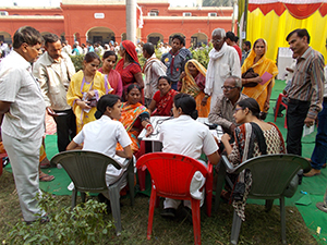
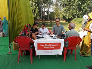
Stroke is the third leading cause of premature death and disability worldwide. The burden of stroke is growing in low and middle-income countries due to many factors including population growth and aging, urbanization, unhealthy diets, physical inactivity and smoking. More importantly, these demographic and epidemiologic factors are driving a rise in the prevalence of high blood pressure, the leading independent risk factor for both ischemic and hemorrhagic stroke. In many less developed countries, particularly in rural areas, awareness of high blood pressure is extremely low and screening services are non-existent. On the other hand, treatment for high blood pressure is widely available and relatively inexpensive.

Figures 1. and 2. Dr. Jerome Chin and volunteers at an ASAPP project site in India in November 2014.

Figures 1. and 2. Dr. Jerome Chin and volunteers at an ASAPP project site in India in November 2014.

Since 2010, Dr. Jerome Chin, a neurologist in the U.S., has been volunteering for two months annually as an attending physician on the neurology ward at Mulago Hospital, the national referral hospital of Uganda in the capital Kampala. The neurology ward admits more than 50 acute stroke patients monthly, the majority with severe previously undiagnosed high blood pressure. In October 2011, Dr. Chin founded the Alliance for Stroke Awareness and Prevention Project (ASAPP) in Kampala to reduce the incidence of stroke in Uganda. ASAPP volunteers, who are mostly medical and other health professions students, provide free community-based screening and counseling for high blood pressure every week at places of religious worship. Individuals with elevated blood pressures are advised to make dietary and lifestyle changes and are referred for medical treatment if indicated. In December 2012, Dr. Chin visited the neurology ward at the All India Institute of Medical Sciences (AIIMS) in the capital Delhi. Similar to Mulago Hospital in Uganda, the majority of patients admitted to AIIMS with acute stroke have severe previously undiagnosed or untreated high blood pressure. In December 2013, Dr. Chin launched ASAPP in India.

ASAPP currently supports six project sites in Uganda and three projects sites in India. In addition, ASAPP is partnering with the Uganda Ministry of Health and other organizations including Rotary International and Impact India Foundation to provide free high blood pressure screening and counseling at special health camps and events. ASAPP project sites have provided more than 55,000 free screenings for high blood pressure since 2011. In the next few years, ASAPP plans to launch additional project sites in Uganda and India and expand to Nepal and other less developed countries. ASAPP is a U.S. tax-exempt non-profit charitable organization. For more information, visit www.asapp.org.