Applied Research Group in Neurological Infections Launches

By Kiran Thakur, MD, and Sarosh Katrak, MD, DM, FRCP(E)

Neurological infections continue to ravage populations in developing and developed countries. In many regions, central nervous system (CNS) opportunistic infections due to AIDS and tropical diseases remain a major contributor to morbidity and mortality.

In resource-rich settings, where new immunomodulatory medications are being frequently used, CNS infections are being increasingly recognized. There are a growing number of emerging and re-emerging neurotropic infectious diseases, and proper diagnosis and management often require neurologic expertise that may not be available in certain global regions.

Despite significant scientific advances in the field, the burden of undiagnosed neurological infections remains unacceptably high. Major research gaps exist in our understanding of the pathogenesis and cost-effective diagnostics, as well as the CNS penetration and optimal treatment schedules of many neurological infections.

As co-chairs of the newly established applied research group in neurological infections, we are excited to enhance the education, training and research in neurological infections to the global neurology community. The World Federation of Neurology is well positioned to make a major impact in this field through its representation of more than 100 countries, many of which have neurologists with expertise in neurological infections.

We hope to engage experts in neurological infections in collaborative research and educational projects and those interested in improving their knowledge of neurological infectious diseases. We encourage those interested in participating to become members and participate in the educational and research endeavors of our group.

Research Goals

  • Surveillance of emerging and re-emerging neurological infections
  • Continued vigilance in understanding the burden of CNS opportunistic infection risk in patients on immunomodulatory therapies
  • Surveillance of undiagnosed infectious diseases in the global community
  • Enhanced pathogen discovery testing globally, with increased access to advanced testing in resource-limited settings
  • Drug-development trials, including studies on CNS penetration of medications for neurological infections and setting drug-dosing and treatment protocols specific for CNS infections

Training and Educational Goals

  • Develop and enhance educational sessions on neurological infections at the World Congress of Neurology meetings
  • Training sessions in neuroinfectious diseases for general practitioners and neurologists
  • Develop training modules on neurological infections with a focus on acute meningitis/encephalitis, chronic meningitis, opportunistic infections in immunosuppressed patients, viral encephalitis and tropical neurology
Kiran Thakur, MD, is an assistant professor in the department of neurology at Columbia University College of Physicians and Surgeons, New York, and Sarosh Katrak, MD, DM, FRCP(E), is director of the department of neurology at Jaslok Hospital and Research Centre, in Mumbai, India.

AFNA, AKUH Team Up for Training, Telemedicine

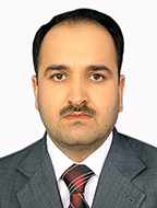
By Esmatullah Hamed, MD

Esmatullah Hamed

Esmatullah Hamed

Trained neurologists are lacking in Afghanistan. Neurological disorders are one of the major disease burdens in this part of the world, and training in this area is scarce. EEG interpretation both in children and adults is a specialized job, and grave errors can occur in patient management when misinterpretations occur.

The Afghan Neurological Association (AFNA) was established to promote the development of clinical neurology and neurological science throughout the country and to promote friendship among neurologists in Afghanistan and the region.

The AFNA formed collaborations with Aga Khan University Hospital (AKUH) Karachi for assessment and identification of strategic training requirements based on the country’s key health care needs. The program is evolving with training arms supporting residents and physicians.

The program not only focuses on the training aspect of physicians, but it also reaches patients in the far parts of Afghanistan with the (FMIC) Medical Institute for Children conducting telemedicine neurology clinics. This effort gives patients access to a modern health facility and expert neurologists in Kabul. To get expert opinions, telemedicine clinics also are conducted with AKUH Karachi.

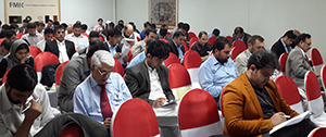
Regular conferences and workshops are major components of the AFNA, AKU collaboration.

Regular conferences and workshops are major components of the AFNA, AKU collaboration.

One of the major components of training and sharing knowledge is through regular conferences and workshops. A major function of such meetings of neurology is to facilitate the sharing of knowledge and to help develop ongoing working relationships that can lead to many advances for all. Although publications and electronic communications provide essential ways to communicate, an international meeting offers unparalleled access to peers whose workplaces and problems are far from home, but may be extremely informative.

Both information sharing and clinical and research collaboration become real possibilities. Clinical collaboration today often takes the form of setting up periodic videoconferences, supplementing important opportunities to visit each another.

In this regard, the second Neurology Certificate Course was conducted via video conference June 6-9 in Kabul in collaboration with the FMIC, the Afghan Neurological Association and Aga Khan University in Karachi.

AFNA, AKUH meetings of neurology facilitate knowledge sharing and help develop ongoing working relationships.

AFNA, AKUH meetings of neurology facilitate knowledge sharing and help develop ongoing working relationships.

Seventeen speakers from neurology, neurosurgery, psychiatry and radiology departments participated, among which 14 were from the Aga Khan University and three speakers from Kabul. The World Federation of Neurology and the Asian Ocean Association of Neurology provided funding for this course.

The lectures covered a broad spectrum of neurological disorders, including stroke, central nervous system infections, headaches, epilepsy, neurological Investigations, coma and brain death.

Seventy residents and physicians participated in the conference from different parts of Afghanistan. The levels of interest were clear by the high level of engagement during the each question-and-answer period at the end of each session, participant feedback after the course revealed that the participants highly appreciated the course and called on planners for more frequent courses.

Esmatullah Hamed, MD, is president of the Afghan Neurological Association and consultant neurologist at the French Medical Institute for Children, Kabul, Afghanistan.

The Hansen-Neisser Controversy Concerning the Discovery of Mycobacterium Leprae

By Douglas J. Lanska

Douglas J. Lanska

Douglas J. Lanska

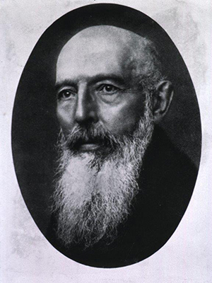
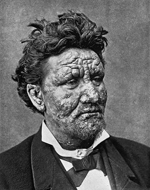
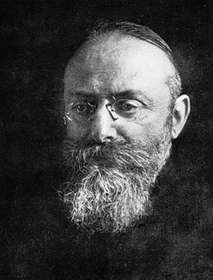
In the late 1860s, on the basis of his clinical and anatomical studies, Norwegian physician Gerhard Armauer Hansen (1841-1912) concluded preliminarily that leprosy was a distinct disease with a specific cause, and not simply a degenerative condition with multiple potential etiologies. (See Figures 1 and 2.) His subsequent epidemiological studies in Norway found no association between the occurrence of leprosy in various districts and general mortality rates, provided evidence that leprosy was contagious rather than hereditary, and demonstrated that isolation of cases produced a decline in incidence.

In 1873, Hansen discovered rod-shaped bodies — Mycobacterium Leprae, sometimes called Hansen’s bacillus — in leprous nodules, although he did not clearly identify them as bacteria. He described these in a report to the Medical Society of Christiania (now Oslo) and in his main treatise in 1874, with a shorter English version in 1875:

Figure 1. Gerhard Armaner Hansen. Photograph by J.F. Lehman, Munich, 1912. Public domain. Courtesy of the U.S. National Library of Medicine.

Figure 1. Gerhard Armaner Hansen. Photograph by J.F. Lehman, Munich, 1912. Public domain. Courtesy of the U.S. National Library of Medicine.

“While leprosy may be … indirectly proved to be a specific disease by demonstrating its contagiousness, it would, of course, be the best if a direct proof could be given. I will briefly mention what seems to indicate, that such proof is, perhaps, attainable. There are to be found in every leprous tubercle extirpated from a living individual — and I have examined a great number of them — small staff-like bodies, much resembling bacteria, lying within the cells; not in all, but in many of them. Though unable to discover any difference between these bodies and true bacteria, I will not venture to declare them to be actually identical. Further, while it seems evident that these low forms of organic life [i.e., bacteria] engender some of the most acute infectious diseases, the attributing of the origin of such a chronic disease as leprosy to the apparently same matter must, of course, be attended with still greater doubts. It is worthy of notice, however, that the large brown elements found in all leprous proliferations in advanced stages … bear a striking likeness to bacteria in certain stages of development … .”

Figure 2. A 24-year-old Norwegian man with lepromatous leprosy. From Leloir H. Traité pratique et théorique de la lè
pre. Paris: A. Delahaye et Lecrosnier. 1886. This figure was later reprinted by Hansen in his monograph (1895). Public domain. Courtesy of the Bibliothè
que nationale de France.

Figure 2. A 24-year-old Norwegian man with lepromatous leprosy. From Leloir H. Traité pratique et théorique de la lè
pre. Paris: A. Delahaye et Lecrosnier. 1886. This figure was later reprinted by Hansen in his monograph (1895). Public domain. Courtesy of the Bibliothè
que nationale de France.

Hansen tried unsuccessfully to stain his preparations. In 1879, when Hansen was visited by Albert Neisser (1855-1916), a young colleague from the laboratory of German physician and pioneering microbiologist Robert Koch (1843-1910) in Breslau, Hansen, encouraged him to try to stain the bacteria. (See Figure 3.) Shortly after Neisser returned to Breslau, he succeeded in staining the bacteria, and then promptly announced his findings, suggested that these bacteria were indeed the infectious agent of leprosy, and claimed priority for the discovery.

Hansen replied quickly and tried to assert his own priority, and by 1880 he had also succeeded in staining the bacteria. (See Figure 4.)

Figure 3. Albert Neisser. Public domain. Courtesy of the U.S. National Library of Medicine.

Figure 3. Albert Neisser. Public domain. Courtesy of the U.S. National Library of Medicine.

“It was not my intention to make any of my investigations on this subject public at present, but as not only Dr. Edlund to whom in the preceding year I showed preparations, and mentioned that I considered leprosy a parasitic disease, in his little work on ‘Leprosy’ speaks of its precise origin as something that he has discovered in the form of “micrococci,” by also Dr. Neisser, of Breslau, who passed some portion of this summer in Bergen has just published the result of his investigations of those preparations that he made while here, and as these results also point out that in general, the preparations are filled with ‘bacilli’ which he supposes to be peculiar to leprosy, and as its ‘contagium’— I feel myself called upon to announce what I have attained to, up to the present time, in my researches after the same ‘contagium,’ and, this, partly to assert my priority with reference to this discovery, and partly in order to advance those details in research which I omitted to announce on account of the still uncertain result in my report to the Medical Society of Christiania [Oslo], 1874, concerning my investigations into the etiology of leprosy.”

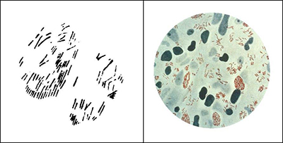
Figure 4. Left: Hansen's drawing (1880) of “brown elements colored with methyl violet, from a tubercle treated with osmic acid.” From Hansen (1880). Right: A photomicrograph of Mycobacterium Leprae (small red rods), taken from a leprosy skin lesion. Public domain. Courtesy of the U.S. Centers for Disease Control and Prevention, Public Health Image Library (PHIL) #2123.

Figure 4. Left: Hansen’s drawing (1880) of “brown elements colored with methyl violet, from a tubercle treated with osmic acid.” From Hansen (1880). Right: A photomicrograph of Mycobacterium Leprae (small red rods), taken from a leprosy skin lesion. Public domain. Courtesy of the U.S. Centers for Disease Control and Prevention, Public Health Image Library (PHIL) #2123.

Although an international consensus generally favored Hansen in this priority dispute as the discoverer of Mycobacterium Leprae, neither Hansen nor Neisser succeeded in fulfilling Henle’s postulates — the criteria to establish a causative relationship between a microbe and a disease propounded in 1840 by German physician, pathologist and anatomist Jakob Henle (1809-1885):

  1. The microbe occurs in every case of the disease under circumstances that account for the pathological changes and clinical course of the disease;
  2. The microbe occurs in no other disease as a nonpathogenic parasite; and
  3. After being isolated and grown in pure culture in an artificial medium, it can induce the disease in an experimental host. These criteria were later augmented by Koch and subsequently known as the Henle-Koch postulates.

Neither Hansen nor Neisser demonstrated that the bacteria observed in cases of leprosy were specific to that disease, nor was Hansen able to transmit the disease to animals or humans using leprous material from patients. Hence, at the time of the Hansen-Neisser controversy, it remained unproven that leprosy is infectious, despite even an unethical attempt by Hansen to transmit leprosy to a patient without informed consent. Furthermore, to this day Mycobacterium Leprae has not been grown in pure culture in an artificial medium.

References:

  1. Evans AS. Causation and Disease: The Henle-Koch Postulates Revisited. Yale J.
  2. Biol Med. 1976;49(2):175-195.
  3. Fite GL, Wade HW. The Contribution of Neisser to the Establishment of the Hansen Bacillus as the Etiologic Agent of Leprosy and the So-called Hansen-Neisser Controversy. Int J Lepr. 1955;23:418-428.
  4. Hansen GA. Spedalskhedens Arsager (causes of leprosy). Translation by Pierre Pallamary. Intl J Leprosy 1955;23:307-309.
  5. Hansen GA. On the Etiology of Leprosy. Br Foreign Medico-chirurgical Rev 1875;55:459-489.
  6. Hansen A. The Bacillus of Leprosy. Q J Microscopical Sci 1880;20:92-102.
  7. Hansen GA, Looft C. Leprosy: In Its Clinical & Pathological Aspects. Translated by Norman Walker. Bristol: John Wright & Co., 1895.
  8. Irgens LM. The Discovery of Mycobacteriom Leprae: A Medical Achievement in the Light of Evolving Scientific Meathods. Am J Dermatopathol 1984;6(4):337-343.
  9. Vogelsang TM. The Hansen-Neisser Controversy, 1879-1880. Int J Lepr. 1963;31:74-80.
  10. Vogelsang TM. Gerhard Henrik Armauer Hansen: 1841-1912: The Discoverer of the Leprosy Bacillus. His Life and his Work. Int J Lepr 1978;46:257-332.
  11. Peter J Koehler is the editor of this history column. He is neurologist at Atrium Medical Centre, Heerlen, The Netherlands. Visit his website at www.neurohistory.nl.


International Coursework

INTERNATIONAL DIPLOMA IN MENTAL HEALTH, HUMAN RIGHTS AND LAW

The International Diploma on Mental Health, Human Rights and Law is currently accepting applications for the academic year 2015-16. The diploma, now in its eighth year, is a collaboration between the World Health Organization and the ILS Law College in Pune, India. The course builds the capacity of students to advocate for human rights and to influence national legislative and policy and service reform in line with the U.N. Convention on the Rights of Persons with Disabilities and other key international human rights standards. It is a one-year diploma and includes two residential sessions and distance learning.

Students to date comprise health and mental health professionals, lawyers, mental health service users/survivors, government officials, social workers, human rights defenders, families and careers. The course is taught be a faculty of renowned international experts in the area.

More information about the diploma is also available at www.cmhlp.org/diploma.

The prospectus and application forms are available at http://cmhlp.org/applications-and-fees/download-prospectus-and-forms.

In addition, the Open Society Institute (OSI) will provide funding for two students from Central and Eastern Europe/former Soviet Union to participate in the course. These are fully funded fellowships and include tuition fees, travel, accommodations and living expenses for the residential sessions. If you are interested and qualify for this grant, please state on your application that you wish to be considered for the OSI fellowships.

INTERNATIONAL MASTER IN MENTAL HEALTH POLICY AND SERVICES

The International Master in Mental Health Policy and Services, an international course promoted by the NOVA University of Lisbon in collaboration with WHO, is currently accepting applications for the academic year 2015-2017. The main scope of the Master Degree in International Mental Health Policy and Services (MHPS) is to build capacity of mental health professionals to lead and contribute to conceiving, formulating, implementing and evaluating:

  • national mental health policy
  • national mental health legislation
  • mental health services and care delivery

The course will start Monday, Oct. 12, 2015, with a two-week residential session, at the Faculdade de Ciàªncias Médicas campus in Lisbon. A second two-week residential session will take place April 4 in Lisbon. Between the two residential sessions, the students will participate in e-learning teaching activities under the orientation of supervisors.

The second year of the course will be dedicated to the development of a project and the elaboration of the dissertation, under the orientation of a supervisor. Additional information about the course can be found at the following site: www.fcm.unl.pt.

Neurosonology in Egypt

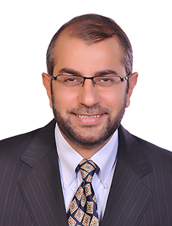
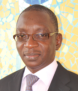
By Prof. Foad Abd-Allah

headshot_Foad

Foad Abd-Allah

The Cairo University Neurosonology Unit (CUNU) is a distinguishable, highly specialized center for sonographic assessment of the nervous system. The unit was founded in 2006, and, since then, it has become a hub for large volume sonology service provision, education and research activities. Team members in the unit are highly trained and practice the state-of-the-art of neurosonology.

The unit was established following the post-doctoral scientific visiting fellowships of Dr. Foad Abd-Allah, founder of the unit, first with Prof. David Russell at Rikshospitalet, the National Teaching Hospital in Oslo, Norway, and then with Prof. Manfrad Kaps at Gissen University, Germany, during the 2004-2005 academic year. Thereafter, the department procured a color duplex ultrasound machine. Coupled with the interest of senior doctors in the department and the enthusiasm of young neurologists, training in neurosonology was started. Currently, the unit possesses four pieces of equipment, with well-trained operators working within two lab facilities. Most of the members of CUNU are certified by the Intersociety Commission for Certification in Neurosonology, the Neurosonology Research Group of the World Federation of Neurology (NSRG) and European Society of Neurosonology and Cerebral Hemodynamic.

CUNU provides high-quality, high- impact services in a timely fashion for patients with neurological disorders. More than 1,200 cases are examined every year by its team. The mainstay of the service is the neurovascular assessment of stroke patients to tackle steno-occlusive disease of cerebral vasculature. Additionally, follow up of patients with subarachnoid hemorrhage for vasospasm as well as cerebral circulatory arrest in brain death is another important task. Recently, nerve muscle neurosonology was introduced. The diagnostic possibilities of neuro-ultrasound have not yet been exhausted. Anyone interested in neurosonology is offered a comprehensive initiation into this fascinating diagnostic tool.

Foad-TeamThe neurosonology team members created a training program, titled “The Neurosonology Professional Diploma.” The course is designed in five comprehensive modules to be presented annually from October to June, and provides candidates with basic theory and practical skills in commonly applied neurosonology techniques. The NSRG of the World Federation of Neurology reviewed the program and certified it as an outstanding high standard teaching program. Many candidates already expressed their interest in it. Full details of the program can be found at www.medicine.cu.edu.eg/cunu/index.html.
The Neurosonology Annual Workshop was launched in 2008, in collaboration with Prof. Manfred Kaps from Giessen, Germany, and since then it has become a landmark event in the annual neurology conference in Egypt. Every year, a new topic is presented in this conference with more than 100 attendees; some of whom have received training in the CUNU lab and eventually have gone on to establish their own laboratories in their respective departments.

CUNU supports candidates for their master’s and PhD theses. Since 2006, team members have performed more than 20 projects for both master’s and doctoral degrees. In addition, many post-doctoral research projects have been completed and even published in peer reviewed journals. The main research interest of our group is the role of ultrasound in cerebrovascular and neurocritical care.

Last month, the WFN Educational Board visited the department of Neurology at Cairo University for accreditation as a training center for English-speaking countries in Africa. The visiting board was impressed by the department as a whole, and many positive comments were received with special attention to the neurosonology unit, recognizing it as “exceptional.”

Abd-Allah is professor of neurology and stroke medicine and head of the neurosonology unit at Cairo-University, Egypt. He is also an executive member of the Neurosonology Research Group of WFN and the WSO board of directors for Middle East and North Africa.

Meeting of Headache and Pain Management

By Gallo Diop

Gallo Diop

Gallo Diop

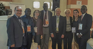
The first Turkish-African Meeting of Headache and Pain Management was held May 2-6 in Istanbul, Turkey. It was convened by the Turkish Headache Society and organized by Prof. Hayrunnisa Bolay from Gazi University, Ankara. There was major support from the Turkish International Cooperation Agency (TIKA). It was held under the auspices of and with support from the International Headache Society (IHS). In attendence was Alan Rapoport, president; three other board members of the IHS (Hayrunnisa Bolay, Peter Goadsby, Andy Charles); Dimos Mitsikostas, president of the European Headache Federation; Zaza Katsarava, vice president of the European Headache Federation; and Aksel Siva, president of Turkish Headache Society. This scientific neuro-event was attended by almost 70 senior and young neurologists from different African countries (Botsawana, Djibouti, Egypt, Ethiopia, Kenya, Morocco, Nigeria, Senegal, Sudan), Germany, Georgia, Greece, Denmark, U.K., U.S., and the hosting country Turkey, represented by headache experts from universities of Istanbul and of Gazi (Ankara). The key highlights were:

  • To promote the cooperation and interactions among headache specialists from IHS, headache societies across African countries and Turkey
  • Aim to educate young leaders and train clinicians to alleviate pain and increase quality of life of people with headache disorders in Pan Africa
  • To learn more about the current conditions and requirements for headache in Africa
  • To increase skills for and knowledge about headache and pain management
  • Leading international faculty to teach about headache and pain
  • Particularly useful for young neurologists, algologists, senior residents and other medical doctors who care for patients with headache and painful conditions
  • Membership to IHS will be provided to participants without charge if they are from a country listed as one of the 100 poorest countries in the world.

The meeting covered a wide range of important topics related to headache and pain medicine. The first part of the event ran on the website and involved the theoretical reading of basic papers and live international webinars. The second part of the meeting brought together more than a dozen world-renowned headache and pain experts to teach and mentor the junior physicians from Africa and Turkey. Teaching lectures, interactive sessions, case-based learning and practical interventional courses took place in Istanbul during four days. Professors assistant, professors and residents from African universities took part in lectures, cases presentations and discussions.

Day 1

Day 1 was the opening with welcome statements from Bolay, trustee of IHS and convener of the meeting; Prof. Najib Kissani, neurologists at the of University of Marakesh, Morocco; Rapoport, president of International Headache Society from the University of California, Los Angeles; and Siva, president of Turkish Headache Society, from Istanbul. It was followed by lectures.

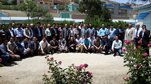
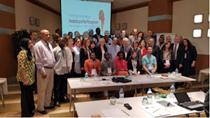
Attendees of the first Turkish African Headache and Pain Meeting with IHS staff.

Attendees of the first Turkish African Headache and Pain Meeting with IHS staff.

Yohannes W. Woldeamanuel, trained in neurology in Ethiopia (2009-2013), is an IHS scholarship awardee and post-doctoral fellow at the Stanford Headache Program. He reported on ‘’Burden of Headache in Africa and Emerging Challenges.” Woldeamanuel emphasized that headache was the 13th cause of YLDs in 2010; migraine represents 15 percent of Africa’s DALYs. In Africa, it is noted a great number of seconday-type headaches from infectious disorders (WHO, 2011, Atlas of Headache Disorders and Resources in the World; Woldeamanuel, 2014, J. Neur. Sci., 342). He suggested collecting more population data with incidence and prevalence rates, prospective research to increase awareness about pain and headache, and more research about traditional management.

Prof. Kissani reported on ‘’Headache in Morroco.” Even if the ratio is better than many African
countries, there is still great lack of specialists for taking care of pain. Prevalence of migraine in Morocco is estimated to 13 percent. Eighteen percent of patients suffering from headache have at least visited one or many healers (Kissani et al, 2009). Results of research supported by ‘’Lifting the Burden: The Global Campaign to Reduce the Burden of Headache Worldwide” Initiative revealed that 85 percent of the 3,600 interviewed people reported more than one headache last year. About 22.5 percent of them suffered from chronic headaches: tension type (48 percent) and migraine (26 percent). Fifty percent used modern medications (55 percent paracetamol; 10 percent aspirin; cost: $22/month). (Some use of combinations also are reported). Ten percent reported consulting traditional healers.

Hellen Kariuki, professor of physiology at Nairobi University, reported on ‘’Natural Traditional Methods to Overcome Pain.” She reported about the dramatic and rapid change of Africans’ lifestyle. Plants are rich sources of pain management around the world. She described some plants that are used by local tribes as painkillers. She discussed how to benefit from these findings and local oportunities to concretely improve management of pain in developing countries.

Rachid Bezad, professor of gynecolgy (Morocco), discussed ‘’The Contribution of Morocco and Africa to Medicine” and described the health system in Morocco and the opportunities of training and cooperation offered by his country.

Days 2-4

Days 2-4 were dedicated to various lectures, cases presentations and practical courses (such as group learning with patients suffering from headaches and practical training in invasive analgesia techniques, peripheral nerve blocks, trigger point injection and acupuncture). Various thematic topics were developed during exciting lectures: classification and evaluation of headache; how to investigate headache patients in restructed resource-setting; choice of treatment options; migraine headache and its mechanisms and management; chronic daily headache and its neurobiology and differential diagnosis; history taking of headaches and pain by specialists; secondary headaches; headaches attributed to infections; women and headache; headache in children and adolescents; chronic pain disorders; and invasive treatment in headache and pain.

From left to right, Aksel Siva, Mustafa, Dr. Ozge, Pr Ndiaye; Alan Rapoport, Hayrunnisa Bolay, Dr. Uluduz and Gallo Diop.

From left to right, Aksel Siva, Mustafa, Dr. Ozge, Pr Ndiaye; Alan Rapoport, Hayrunnisa Bolay, Dr. Uluduz and Gallo Diop.

The meeting was successful in all aspects: organization, scientifc program, and social and friendship environment.

This educational meeting was an excellent opportunity to learn about the diagnosis and management of headache and pain disorders, new developments in the science of headache medicine and the care of headache sufferers.

Through the meeting, everybody has learned more about the current conditions in and requirements for headache in Africa. The meeting will promote the cooperation and interactions among headache specialists from the IHS, various headache societies across African countries, Turkey and worldwide.

It was a contributive additional action for the vision of World Federation of Neurology to promote, via its Africa Initiative, training and exchanges as a leveraging opportunity for trainees and specialists continuing professional development in Africa. This meeting comes also as an additional contribution of Turkish Neurology Society in regard to this aim, because this country is already offering (over three years) two scholarships per year, for a month short-term training in neurology departments of Turkish universities for young African neurologists.

Next plans will aim to educate young leaders and increase skills for and knowledge about headache and pain management to alleviate pain and increase quality of life of people with headache disorders throughout Africa. A short- and long-term plan of action has been discussed and will increase the implication of Turkey, IHS and other specialties to fulfill training needs in Africa, in addition to their various contributions during the Regional Teaching Courses organized by European Academy of Neurology and WFN in Africa for 8 years. For more information, visit www.tahpm.org.


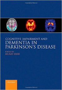
Cognitive Impairment and Parkinson’s Disease Dementia, Second Edition

DemCoverThe medical book publishing industry is challenged nowadays to turn out products quickly and efficiently, lest the rapid dissemination of today’s scientific advances through the Internet render the content of a book out of date on arrival. The second edition of Murat Emre’s “Cognitive Impairment and Dementia in Parkinson’s Disease” has avoided this fate, in large part because of the organizational skill of the editor and his recruitment of the same authoritative thought leaders that contributed to the first edition in 2010. Hence, this continuity of authorship has allowed for a seamless update of the topics covered before.
The co-authors of each of the 22 well- written chapters have handled their assignments with balanced attention to recent discoveries and to the potential for practical application. There is also a healthy regard for the concept that all roads of investigation ultimately lead back to the patient, whose struggle to cope with the twin burdens of progressive motor and cognitive impairment in Parkinson’s disease (PD) has not been significantly helped by a true breakthrough in treatment since the introduction of levodopa in the 1960s. The failure to develop more effective symptomatic or game-changing, disease-modifying therapies has been one of the great disappointments of modern-day clinical research despite years of valiant effort.

HurtigAs Dr. Emre observes in his elegant introduction, cognitive impairment as an essential feature of PD was mostly unrecognized by James Parkinson in his essay on the Shaking Palsy (1817) because lack of treatment doomed its victims to a severe physical disability and a shortened lifespan. The stark reality of cognitive impairment in advanced PD became apparent only after the remarkable benefit of levodopa enabled people with PD to function better physically and thereby live longer. Well-designed, long-term cohort studies in the early part of this century revealed not only the shocking news that 70-80 percent of people with PD would develop dementia as they aged and progressed, but also that subtle cognitive abnormalities, particularly in executive function, were prevalent in a sizeable minority at early stages of the disease.
This book thoughtfully reviews the important developments in clinical and basic research of the last two decades, and it highlights new and refined information that has emerged in the five years since the first edition was published. References are up to date as of 2013 with a sprinkling of 2014, the year of publication. Early chapters on epidemiology, natural clinical history and neuropsychological assessment are comprehensive and succinct. The differential diagnosis of dementia in the setting of parkinsonism can be difficult, but the neuropsychological profile of cognitive dysfunction in PD, showing the typical executive, visuo-spatial, attention and memory deficits of PD sets it apart generally from Alzheimer’s disease, wherein disturbances of memory and language are the classic hallmark findings.

The histopathology of PD is the focus of several chapters, which emphasize the near unanimity of opinion that misfolded alpha-synuclein is the key molecular abnormality and the main component of the signature intracytoplasmic inclusion Lewy body. The clinical and pathological continuum of Parkinson’s disease dementia (PDD) and dementia with Lewy bodies (DLB) is also effectively explained, including elements of the debate over the contribution of the plaques and tangles of Alzheimer disease (AD) as a minority component of PDD/DLB pathology. Current evidence suggests (without being close to consensus) that DLB, which is defined arbitrarily as dementia occurring within a year of the onset of parkinsonism, has a pathological substrate of alpha-synuclein and Alzheimer plaque (sans tangles), whereas the substrate for PDD — dementia beginning more than a year after the onset of parkinsonism — is more likely to be alpha-synclein alone. Another chapter covers the neurochemistry of PD and PDD and the central relevance of the well-known deficiencies of dopamine and acetylcholine as they relate to the evolution of dementia.

The ferment in biomarker research — a relatively new phenomenon with great promise for predicting cognitive outcomes in the preclinical and early clinical stages of PD and for identifying subgroups for targeting in clinical trials of new therapies — is included in several chapters. Also since the first edition, official criteria for Parkinson dementia and mild cognitive impairment (MCI) in PD have been established under the auspices of the International Parkinson and Movement Disorder Society, using the model of the Petersen criteria for pre-Alzheimer MCI. Several chapters on recent findings in brain imaging are well executed, although it could be argued that all imaging should have been condensed into a single chapter. One of the hottest areas of research in the basic science of PD is the rapidly expanding field of Parkinson genetics. The chapter on the genetic basis of PDD is excellent and comprehensive.

Ian McKieth and Murat Emre join forces in the pentultimate chapter to discuss management of PDD and DLB, and the result is a masterful presentation of a humane message about the time-honored clinical fundamentals of taking a good history, listening empathically and caring long term for the patient and family.

Finally, the closing brief chapter by John Hardy is a sobering statement of how far we still must go before truly meaningful treatment comes into view. He acknowledges the substantial “incremental” progress in molecular biology and genetics of the last decade. The identification of numerous potentially causative somatic and mitochondrial genetic loci in some patients clears some of the fog around pathogenesis, although these advances apply only to a small fraction of the much larger universe of patients with PD, for whom genetic markers are nowhere to be found. Moreover, it is far from clear which pathogenetic pathways are influenced by alterations in the genome or, perhaps more important, how the growing number of these changes interact to produce disease and which ones are the most critical in the pathogenetic cascade. In short, the goal of a cure is “still beyond the near horizon.”

“Cognitive Impairment and Dementia in Parkinson’s Disease” is a solid achievement. The authors have created an informative and useful compendium of the universally accepted wisdom as well as the latest more controversial advances in the field. It is easy for an armchair reviewer to find fault with even the best publications, but there are a few minor shortcomings here. First, the list of contributors should have included the specific disciplines of each person in addition to their institutional affiliations. Second, some of the references at the end of each chapter were duplicated or unrelated to the citations in the text. Third, redundancy across chapters was generally appropriate for emphasis of the most important concepts and facts, but more careful editing could have minimized useless redundancy. Fourth, the importance of impaired olfaction in preclinical PD and the voluminous body of research devoted to it was all but ignored; olfaction received only a brief paragraph in one of the early chapters and no attention was paid in the chapter on biomarkers. And fifth, there was no mention of the so-called “prion hypothesis” that has been invoked in the last several years to explain how alpha-synuclein pathology spreads rostro-caudally throughout the brain as the disease progresses and leads to the development of dementia. These small quibbles hardly detract from the many assets of this fine contribution to the growing shelf of literature on one of the most pressing problems in clinical neurology.

The Diamond and the Rose

By Richard Peatfield

Richard Peatfield

Richard Peatfield

During the last 40 years, the world of headache has been blessed with two remarkable men: Dr. Seymour Diamond and Dr. Frank Clifford Rose. They have both recently published autobiographies. Rose’s autobiography is, of course, posthumous following his death in 2012, while Seymour, just the elder, is still enjoying his retirement at the age of 89.

Although living and working in different environments on different continents, Diamond and Rose have striking similarities over and above their lively personalities. They were born within 16 months of one another of Jewish immigrant stock. Rose’s parents settled in the East End of London after getting married and having two children in Romania. Rose was the youngest of seven surviving siblings.

Diamond’s parents, by contrast, had arrived in Chicago as children from Slovakia and from the Ukraine. He was the youngest of their four children. Both had that combination of inherent talent and industry that enabled them to move out of their original backgrounds.

I think it is fair to say that Rose’s path was advantaged by the grammar school and the university system of his time in Britain, while Diamond grew up in the challenging environment of wartime America. Never is this more clear than in their early clinical training. Rose was able to do all of his jobs within London, whereas Diamond tells the tale of his in-laws driving him, his new wife and all of their possessions from Chicago to Arkansas and then to Ohio every year or so.

Both spent the bulk of their careers as practicing physicians with interests in headache, though Diamond was never a board-certified neurologist as he had been accredited in family medicine and had not done a residency program in neurology.

Both set up dedicated migraine clinics. Rose’s clinic is dedicated to Princess Margaret, while Diamond’s bears his own name. Both wrote and edited a large numbers of books and conference proceedings.

Both were superb administrators and used their talents in a wider field. Rose was the more international, playing a major role in the evolution of the International Headache Society and serving as secretary treasurer general of the World Federation of Neurology.

Diamond, in contrast, devoted much of his energy to the inpatient and outpatient facility he established in Chicago and developed a nationwide reputation as a physician “who cared.” He was one of the first to try tricyclic antidepressants in headache patients and was the leading light in many of the trials of drugs that are currently part of the every physician’s drug armamentarium.

Diamond played a major role in the evolution of the American Association for the Study of Headache and was its chief executive for many years. His autobiography makes it clear that his relationship with the neurological establishment was often fraught. Nevertheless, it is a tribute to his diplomatic skills that the American Headache Society (as it is now called) covers such a broad spectrum of practitioners from neurologists and pediatricians to physiotherapists, psychologists and other colleagues. He also initiated the influential patients’ own National Migraine (now Headache) Foundation.

Rose, in contrast, was more of a neurological polymath, with interests not only in headache but also in motor neurone disease, Parkinson’s disease and stroke. He was perhaps more dependent on his junior colleagues on both the clinical and academic fronts. Diamond’s concentration on one symptom attracted devoted patients from all over the continent. Both were renowned for their approachability.

Diamond’s autobiography was written in conjunction with a medical journalist, in the third person in somewhat more popular style, with many fascinating personal details, including his prowess at Bridge and his innocent dealings with the local mafia. Rose wrote his in the first person, and his voice and characteristic energy come through.

They were both devoted to their wives and children. Diamond and his wife, Elaine, had three girls, of whom one now runs the eponymous Clinic in Chicago after his retirement. Rose and his wife, Angela, had three boys, all established in careers away from medicine.

Both autobiographies give fascinating insights into medical training during and immediately after World War II, as well as into the evolution of the clinical and academic world of headache on each side of the Atlantic. Both are fitting tributes to two outstanding men.

The Headache Godfather: The Story of Dr. Seymour Diamond and How He Revolutionized the Treatment of Headaches By Seymour Diamond and Charlie Morey. Skyhorse Publishing, Inc., New York 2015 ISBN-10: 1629145386 $24.95

Autobiography: By Any Other Name By Clifford Rose Privately printed 2014 [Copies may be available for £12 + p&p. Expressions of interest should be made to r.peatfieldimperial.ac.uk]

Myalgic Encephalomyelitis/Chronic Fatigue Syndrome: A New Name & Diagnostic Criteria

Maggie McNulty

Maggie McNulty

In March 2015, a report from the Institute of Medicine (IOM) was published in the Journal of the American Medical Association to redefine the illness known as Myalgic Encephalomyelitis/Chronic Fatigue Syndrome (ME/CFS).

Over the years, clinicians and researchers have developed different diagnostic criteria for ME and CFS; however, the two terms describe conditions with similar symptoms. In the World Health Organization’s “International Classification of Diseases,” 10th Revision, both ME and CFS are coded the same and classified as disorders of the nervous system (ICD G93.3). The term “benign myalgic encephalomyelitis” was first used in the 1950s in London when describing an outbreak in patients who experienced a variety of symptoms, including “malaise, tender lymph nodes, sore throat, pain and signs of encephalomyelitis.”

The cause was never found, but it appeared infectious in etiology, and the term “benign myalgic encephalomyelitis” was used to reflect “the absent mortality, the severe muscular pains, the evidence of parenchymal damage to the nervous system and the presumed inflammatory nature of the disorder.” Then in 1970, two psychiatrists reviewed reports of 15 of these outbreaks and concluded that the outbreaks “were psychosocial phenomena” caused by mass hysteria or altered medical perception of the community.

However, the idea that the condition was psychogenic in origin was refuted by Dr. Melvin Ramsay. In 1986, he was the first to publish diagnostic criteria for ME and, at this point, the term “benign” was dropped as the disease often was severely disabling for those patients afflicted. Around the same time, in the mid-1980s, there were two outbreaks of an illness that resembled mononucleosis characterized by “chronic or recurrent debilitating fatigue and various combinations of other symptoms, including sore throat, lymph node pain and tenderness, headache, myalgia and arthralgias.” This illness was at first linked to the Epstein-Barr virus (EBV); however, further research ruled out this as the cause, and, in 1988, the term “chronic fatigue syndrome” was coined by the Centers for Disease Control and Prevention (CDC).

ME/CFS is characterized by symptoms of profound fatigue, cognitive dysfunction, sleep abnormalities, autonomic manifestations, pain and other symptoms that are worsened by any type of exertion. The syndrome affects women more than men with an average age of onset of 33 years with a wide range of distribution ranging from 10 to 77 years old.

ME/CFS is a common disorder that is currently estimated to affect 836,000 to 2.5 million Americans. Despite the prevalence of this disorder, less than one-third of medical school curricula and only 40 percent of medical textbooks include information regarding this syndrome. This likely contributes to delays in diagnosis time for these patients (e.g., 29 percent of patients report symptoms for >5 years prior to receiving a diagnosis), and it is estimated that 84 to 91 percent of people with this condition have not yet been diagnosed.

Table1There is significant economic burden associated with this condition as one quarter of patients are bed- or house-bound at some time during their illnesses. ME/CFS patients have been found to be more functionally impaired than patients with other disabling illnesses, such as diabetes mellitus, congestive heart failure, hypertension, depression, multiple sclerosis and end-stage renal disease. Unemployment rates range from 35 to 69 percent in these patients. ME/CFS patients have loss of productivity and high medical costs that lead to an estimated economic burden of $17 billion to $24 billion yearly.
In general, ME/CFS is a condition that is poorly accepted as its pathophysiological mechanisms are poorly understood, and many contest the characteristics needed to make a diagnosis. There continue to be many misconceptions regarding ME/CFS, including that it is a psychogenic illness. Many times, patient symptoms are met with skepticism or even dismissal. After diagnosis, many people with ME/CFS report being subject to hostile attitudes from their health care providers. The cause of ME/CFS is currently unknown; however, symptoms may be triggered by different infections or other prodromal events, including “immunization, anesthetics, physical trauma, exposure to environmental pollutants, chemicals and heavy metals and, rarely, blood transfusions.”

The IOM was asked by the Department of Health & Human Services, the National Institutes of Health, the Agency for Health Care Research & Quality, the CDC, the Food & Drug Administration and the Social Security Administration to convene an expert panel to review the evidence basis for ME/CFS. This was a comprehensive committee of 15 members that convened in September 2013 and took into account data from patients, clinicians and researchers while also reviewing almost 1,000 public comments. This was followed by a comprehensive literature review to help identify new diagnostic criteria to be used by clinicians. Based upon their work, a new name was recommended to replace ME/CFS. Previous studies have shown that the term “chronic fatigue syndrome” can negatively affect patients and medical professionals’ perceptions of the illness and trivialize the seriousness of the disease. “Myalgic encephalomyelitis” is also inappropriate as there is no evidence of encephalomyelitis in these patients, and myalgia is not a core symptom of this disease. The new name that has been recommended for use is systemic exertion intolerance disease (SEID); this new name is felt to encompass the central characteristic of the disease: the fact that exertion of any kind can negatively affect patients in multiple different organ systems.

A new set of diagnostic criteria was also developed by the group with the intent to ease the process of making a diagnosis of ME/CFS (SEID) and hopefully, decrease the time to make a diagnosis for many patients. The new diagnostic criteria that were developed by the IOM committee are detailed in Table 1. The core features include: fatigue and impairment, post-exertional malaise (PEM) and unrefreshing sleep. All of these features need to be present for one to be diagnosed with ME/CFS (SEID).

Fatigue as defined in the dictionary is “weariness from bodily or mental exertion.” Sufficient evidence has been found that fatigue is profound in ME/CFS (SEID). Dramatic examples of reports of fatigue in patients with ME/CFS (SEID) include feeling “too exhausted to change clothes more than every 7-10 days” and experiencing “exhaustion to the point that speaking is not possible.” More commonly, patients report fatigue as “exhaustion, weakness, a lack of energy, feeling drained and an inability to stand for even a few minutes.” The fatigue must be associated with a significant reduction or impairment in the ability to engage in pre-illness levels of occupational, educational, social or personal activities. This degree of fatigue must also persist for more than six months.

Another core symptom, post-exertional malaise (PEM), is found consistently in patients with ME/CFS (SEID) and felt to help distinguish it from other conditions. A patient’s symptoms worsen after exposure to physical or cognitive stressors that were previously well tolerated prior to onset of the disease. Descriptions provided by patients after an exertional task include “crash,” “exhaustion,” “flare-up,” “collapse,” “debility” or “setback.” PEM may occur within 30 minutes of an exertional task or be delayed up to seven days after an exertional task with the duration of PEM lasting hours to months. In studies comparing ME/CFS (SEID) patients with healthy controls, 86 percent of patients report minimum exercise makes them tired compared to 7 percent of controls. Additionally, 85 percent of ME/CFS (SEID) patients report that they feel drained after mild activity compared to 2 percent of healthy controls.

The third and final core symptom is unrefreshing sleep for which 92 percent of ME/CFS (SEID) patients report compared to 16 percent of controls. The typical sleep-related symptoms described by ME/CFS (SEID) patients include difficulty falling asleep, frequent or sustained awakenings, early-morning awakenings and nonrestorative or unrefreshing sleep (persistent sleepiness despite adequate duration of sleep). Primary sleep disorders such as sleep-disordered breathing, restless legs syndrome and narcolepsy should be considered and evaluated for if clinically indicated as treatment of these disorders can be effective in reducing or relieving symptoms of unrefreshing sleep. However, a polysomnogram is not required to diagnose ME/CFS (SEID). Currently, there is no strong evidence to identify ME/CFS (SEID)-specific sleep pathology despite some studies revealing differences in sleep architecture in a subset of ME/CFS (SEID) patients compared to healthy controls.

In addition, there are two supportive criteria included in the new definition, of which one of two is required to be present to meet a diagnosis of ME/CFS (SEID). These include cognitive impairment and/or orthostatic intolerance. Common features of cognitive impairment that are seen in ME/CFS (SEID) include complaints of problems remembering, difficulty expressing thoughts, difficulty paying attention, slowness of thought, absent-mindedness and difficulty understanding. It is suggested that slowed information processing plays a central role in the cognitive impairment associated with ME/CFS (SEID). This deficit can lead to significant disability that results in loss of employment as well as functional capacity in social environments. Neuropsychological testing has found that patients with ME/CFS (SEID) display deficits in working memory compared with healthy controls with reduced verbal and visual memory being the most consistent finding. The second feature, orthostatic intolerance, refers to worsening of symptoms upon assuming and maintaining an upright position. Symptoms are typically improved but may not be alleviated by lying down or elevating their feet. There is sufficient evidence that indicates a high prevalence of orthostatic intolerance in patients with ME/CFS (SEID) based on bedside orthostatic vital signs, tilt table testing or by patient reported worsening of symptoms with standing in day-to-day life.

The committee also described additional frequent findings found in ME/CFS (SEID) patients, which include pain, immune impairment, infection and miscellaneous symptoms. Pain was found to be common in patients who had ME/CFS (SEID) with complaints of headaches, myalgias and arthralgias being most common. There was sufficient evidence found that supported the finding of immune dysfunction in these patients with data revealing poor NK cell cytotoxicity that correlated with illness severity in ME/CFS (SEID). This finding, however, is not specific to ME/CFS (SEID). Additionally, there was evidence that ME/CFS (SEID) can occur after infection with EBV, but there was not sufficient evidence to conclude that all cases of ME/CFS (SEID) are caused by EBV. Less frequent symptoms that were found in patients with ME/CFS (SEID) include gastrointestinal impairments, genitourinary impairments, sore throat, painful or tender axillary/cervical lymph nodes and sensitivity to external stimuli (e.g., foods, drugs, chemicals).

In summary, ME/CFS (SEID) is a serious, chronic, complex and systemic disease that often significantly limits the day-to-day activities of those affected. It is characterized by a prolonged, significant decrease in function; fatigue; post-exertional malaise; unrefreshing sleep; difficulties with information processing, especially under time pressure; and orthostatic intolerance. A thorough history, physical examination and targeted evaluation are necessary and can be sufficient to make a diagnosis. Despite the high prevalence of this condition with associated high economic burden, little research has been conducted to study the etiology, pathophysiology and effective treatment of this disease. Moving forward, it will be vital to distinguish this disease against other complex fatiguing disorders as the majority of previous research has compared ME/CFS (SEID) patients to healthy controls. Additional research into ME/CFS (SEID) is essential for further progress to be made, and the term “chronic fatigue syndrome” should no longer be used due to the associated stigma, which often precludes patients from receiving appropriate care. It is critical that we do our part to help stop the stigma associated with this condition and provide optimal care of these patients.

Maggie L. McNulty is an assistant professor at Rush University Medical Center, Department of Neurological Sciences.

HIV Infection Is a New Target for Stroke Prevention

Infection is an independent risk factor for both ischemic and hemorrhagic stroke

By Jerome H. Chin, MD, PhD, MPH

Jerome H. Chin, MD, PhD, MPH

Jerome H. Chin, MD, PhD, MPH

Stroke is the third-leading cause of premature death globally as measured in years of life lost1.  Demographic and epidemiologic changes, including population growth and aging, urbanization and unhealthy diets are driving a rise in the incidence of stroke in low- and middle-income countries (LMIC)2. Due to inadequate primary health care services to screen for and treat the most common stroke risk factors, particularly hypertension and diabetes, the age-standardized incidence rates of stroke in LMIC exceed those in high-income countries2. In addition, strokes due to atrial fibrillation and rheumatic heart disease contribute a significant share of the stroke burden in LMIC as a result of both diagnostic and therapeutic resource limitations for these conditions.

Figure. HIV-associated stroke: (a) middle cerebral artery infarct; 25-year-old HIV-infected male, CD4+ cell count = 42 cells/mm3; (b) bilateral basal ganglia infarcts; 25-year-old HIV-infected female, unknown CD4+ cell count.

Figure. HIV-associated stroke: (a) middle cerebral artery infarct; 25-year-old HIV-infected male, CD4+ cell count = 42 cells/mm3; (b) bilateral basal ganglia infarcts; 25-year-old HIV-infected female, unknown CD4+ cell count.

HIV infection has recently emerged as an independent risk factor for both ischemic and hemorrhagic stroke3-8. Two cohort studies conducted in the United States reported an elevated risk of ischemic stroke in HIV-infected individuals compared to non-HIV-infected individuals4,5. Lower CD4+ cell counts or higher HIV RNA levels were associated with an increased risk of stroke. Cohort studies from Canada and the United States have reported an association of HIV infection with an increased risk of intracerebral hemorrhage6,7. A community-based case-control study performed in Tanzania found HIV infection to be an independent risk factor for stroke (ischemic and hemorrhagic combined) with an adjusted odds ratio of 5·618. Only 40 percent of the 200 stroke cases had a CT scan. The pathogenic mechanisms responsible for the increased risk of stroke associated with HIV infection are multiple and may include chronic inflammation and immune activation leading to endothelial dysfunction and subsequent vasculopathy3,9. Both small-vessel (e.g. lacunar) and large-vessel strokes are observed in HIV-infected individuals (figure) in addition to intracerebral hemorrhages.

Thirty-five million people are living with HIV worldwide10. The population-attributable risk of stroke due to HIV infection will depend on the prevalence of HIV infection in a particular region or country. Given the higher prevalence of HIV infection in sub-Saharan Africa compared to other regions, a substantial number of incident strokes in sub-Saharan Africa may be the result of HIV infection. However, reliable figures for stroke incidence, mortality and comorbidities are difficult to obtain in most countries of sub-Saharan Africa due to inadequate stroke surveillance and vital registration data. The above-mentioned epidemiologic studies provide support for the inclusion of HIV antibody testing in the diagnostic evaluation of patients with acute stroke in all regions of the world. Furthermore, stroke prevention should now be considered another potential benefit of the early initiation of antiretroviral therapy in HIV-infected individuals through both a reduction in HIV-associated vasculopathy as well as through the prevention of HIV transmission to their uninfected partners.

Dr. Chin is president of the Alliance for Stroke Awareness and Prevention Project (ASAPP).

References

  1. GBD 2013 Mortality and Causes of Death Collaborators. Global, regional, and national age–sex specific all-cause and cause-specific mortality for 240 causes of death, 1990–2013: a systematic analysis for the Global Burden of Disease Study 2013. Lancet. 2015;385:117-171.
  2. Krishnamurthi RV, Feigin VL, Forouzanfar MH, et al. Global and regional burden of first-ever ischaemic and haemorrhagic stroke during 1990–2010: findings from the Global Burden of Disease Study 2010. Lancet Glob Health. 2013;1(5):e259-e281. doi:10.1016/S2214-109X(13)70089-5.
  3. Cole JW, Chin JH. HIV infection: a new risk factor for intracerebral hemorrhage? Neurology. 2014;83:1690-1.
  4. Marcus JL, Leyden WA, Chao CR, et al. HIV
    infection and incidence of ischemic stroke, AIDS. 2014;28:1911–1919.
  5. Chow FC, Regan S, Feske S, Meigs JB, Grinspoon SK, Triant VA. Comparison of ischemic stroke incidence in HIV-infected and non-HIV-infected patients in a U.S. health care system. J Acquir Immune Defic Syndr. 2012;60:351-358.
  6. Chow FC, He WS, Bacchetti P, et al. Elevated rates of intracerebral hemorrhage in individuals from a U.S. clinical care HIV cohort. Neurology. 2014;83:1705–1711.
  7. Durand M, Sheehy O, Baril JG, LeLorier J, Tremblay CL. Risk of spontaneous intracranial hemorrhage in HIV infected individuals: a population-based cohort study. J Stroke Cerebrovasc Dis. 2013;22:e34–e41.
  8. Walker RW, Jusabani A, Aris E, et al. Stroke risk factors in an incident population in urban and rural Tanzania: a prospective, community-based, case-control study. Lancet Glob Health. 2013;1:e282–e288.
  9. Benjamin LA, Bryer A, Emsley HCA, Khoo S, Solomon T, Connor MD. HIV infection and stroke: current perspectives and future directions. Lancet Neurol. 2012;11:878–90.
  10. UNAIDS, The Gap Report, available at: http://www.unaids.org/en/resources/campaigns/2014/2014gapreport/gapreport/ Accessed January 25,  2015.