Call for Applications for 2017 WFN Junior Traveling Fellowships

By Steven L. Lewis, MD,
and Wolfgang Grisold, MD

This year, the WFN is again able to offer Junior Traveling Fellowships for young neurologists from countries classified by the World Bank as Low or Lower Middle Income, to attend approved international meetings.

Applications for 2017 would be most welcome.

There will be 30 awards; applicants should be neurologists in training or early in their careers, have an MD degree or equivalent medical degree, hold a post not above that of associate professor, and be no older than 45 years of age.

Candidates are asked to apply online via www.wfneurology.org/junior-travelling-fellowship-grants-form.

You will be asked to provide:

  • The name and dates of the meeting for which you wish to register
  • A CV and bibliography
  • A letter of recommendation from the Head of your Department
  • An estimate of expenses, to a maximum of £1,000, no excess will be granted

It is expected that applicants participate actively in the meeting (e.g. presentation, poster) that they attend. The submission of an abstract is encouraged. A copy of the abstract also should be included. Preference will be given to candidates who have not previously received an award and to candidates who intend to attend the 2017 World Congress of Neurology in Kyoto, Japan.

Applications must be received at the WFN office no later than Wednesday, March 15. All applications will be reviewed by the education committee, and the awards will be announced as soon as possible thereafter.
Seven New African Professors of Neurology Announced

By Gallo Diop, MD

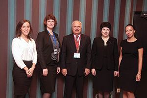
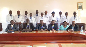
Seven new neurology professors and four psychiatry professors in Africa were chosen from the candidates for the positions. Photo by Gallo Diop, MD.

Seven new neurology professors and four psychiatry professors in Africa were chosen by juries from French-speaking universities. Photo by Gallo Diop, MD.

From Nov. 7 to 15, 2016, the 18th biannual African contest for selecting new university professors was organized in Dakar, Senegal. After a tough competition, the assistant professors are upgraded to the grade of professor by different juries whose members come from all French-speaking universities and, depending on the needs, from other countries such as Belgium, France, and Canada. All the Francophone universities of the African Council for Higher Education (www.lecames.org), use this exam for enriching their faculty. They submit the candidacy of their assistant professors several years after they have received their doctorates. All the specialties of medical, pharmaceutical, dental, and veterinary sciences are involved.

This year, there were 172 successful candidates out of a total of 201 candidates from 24 universities. For neurology, seven candidates competed, and all succeeded. The awardees, in order of merit, are: Dieudonne Gnonlonfoun (Benin), Yacouba Mapoure (Cameroon), Paul Ossou (Congo), Thierry Adoukonou (Benin), Komi Assogba (Togo), Fode Cisse (Guinea), and Moustapha Sarr (Senegal).

This is a great achievement for local neurology training because six of them were entirely trained for four years, 15 years ago, at the University of Dakar, and one (Dr. Adoukonou) from the University of Abidjan (Ivory Coast). They also benefitted from one to two years of complementary focused training in France. 

Gallo Diop, MD is a professor of neurology at the University Hospital of Dakar, Senegal, and co-opted trustee of the WFN.

International Conference Tackles Air Pollution, Emerging Medical Issues

By Peter Spencer and Jacques Reis

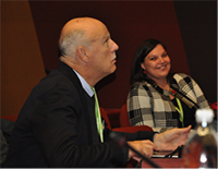
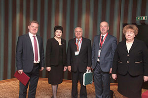
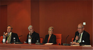
Speaking at the welcome at the Council of Europe were (from left) Professor Christman of the Faculty of Medecine, Professor de Seze of the Department of Neurology, Dr. Mengus of the COE and Professor Sandner of the University of Strasbourg.

Speaking at the welcome at the Council of Europe were (from left) Professor Christman of the Faculty of Medecine, Professor de Seze of the Department of Neurology, Dr. Mengus of the COE and Professor Sandner of the University of Strasbourg.

Air pollution is an increasing concern for humanity. The subject urgently needs more attention from medical scientists and society alike. It has long been recognized as a major health threat with multiple impacts, increasing mortality, and morbidity. Pulmonary and cardiovascular diseases are well known outcomes. Brain impacts are an emergent challenge, as are related epigenetic changes and developmental issues.

For the first time, a conference tackled these issues in an inter-professional and translational approach. Basic researchers, epidemiologists, public health specialists, and neurologists assessed current knowledge and identified key gaps that mandate further research.

The conference, which took place in a pleasant Christmas atmosphere in Strasbourg, Germany, was organized by the Club de Neurologie de l’Environnement, NEUREX, and the Environmental Neurology Applied Research Group of the World Federation of Neurology

Prof. Grisold and Mrs. Reis, a local organizer, at the International Conference.

Prof. Grisold and Mrs. Reis, a local organizer, at the International Conference.

While compelling data are lacking, there is growing concern that continuous exposure to heavily polluted air might perturb brain development, increase the risk of ischemic stroke in adults, and even contribute to the development of neuro-degenerative and behavioral disorders.

The historical correlation between childhood exposure to lead from gasoline, population IQ attainment, and violent behavior during adulthood, coupled with experimental evidence of persistent epigenetic changes in the brains of lead-exposed primates, serves as an early warning of the dire potential effects of neuropollution.

That fine particles suspended in breathable air enter the brain is certain, whether directly via the sensory innervation of the olfactory epithelium or through pulmonary transfer to the circulation, the lymphatic system or other peripheral route. How the brain responds to neuropollution is the subject of debate. Is the particulate garbage removed without incident by normal physiological processes? Does its presence trigger a neuroinflammatory response? Is this a short-term event or can this lead to temporary or persistent brain cell dysfunction and damage? Can this initiate or exacerbate a neurodegenerative process related to amyotrophic lateral sclerosis, Parkinson’s disease or Alzheimer’s disease?

Attendees enjoy the Alsatian atmosphere.

Attendees enjoy the Alsatian atmosphere.

Given the extraordinary levels of ambient air pollution in many population centers around the globe, the ever-increasing trend of human urbanization, the rapid projected growth of some of the most populous cities in Africa and Asia, and the growing evidence for adverse health consequences of neuropollution, the Strasbourg conferees urged immediate action to curb air pollution and greater investment in research to establish the health effects and their underlying mechanisms.

The organizers thank La Région Grand Est, the Council of Europe, and the municipality of Oberhausbergen for their kind support. 

Peter Spencer, PhD, FANA, FRC, is a professor of neurology at the School of Medicine and senior scientist at the Oregon Institute of Occupational Health Sciences, both at the Oregon Health & Science University, Portland. Jacques Reis, MD, is with University Hospitals, Strasbourg, France, and is chair of the WFN Environmental Neurology Research Group.

The Profile of Neurology Has to Grow for Greater Recognition

By Raad Shakir, MD

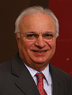
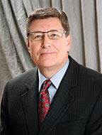
Raad Shakir, MD

Raad Shakir, MD

With the publication of Global Burden of Disease (Lancet Oct. 8, 2016), it is now clear that brain health is at the forefront of diseases leading to morbidity and mortality. Moreover, if we combine neurological, mental, developmental, and substance-use disorders (NMDS), then the group is the leading cause of mortality and morbidity, surpassing cancer and cardiac disease.

These facts, in my opinion, have not trickled to the policymakers, let alone the public. If this does not happen, then desperately required funding and support would not follow soon. Neurologists have to be more proactive in “promoting” the discipline across the world. In many parts of the world, our numbers are so small that the sheer number of patients overwhelms us, and our specialty is not recognized as a priority by governments. At times, we have to explain our discipline to the lay public, as the neurology profession is not instantly recognizable. In the eyes of many, being a heart specialist is easy to comprehend while a neurologist may not be. We may have to use the word “brain” more liberally, and stating that we are a “brain specialist” could be an option, which may perhaps be more recognizable. Although this does not cover those of us who look after the disorders of the peripheral nervous system, in my opinion, the issue of recognition of our specialty is much more important.

In this context, we have to understand that many other interest groups and those responsible for statistics come into play. As glaring examples, I will mention two. The first and most obvious is the issue of cerebrovascular diseases. Sixty years ago, International Classification of Diseases (ICD) 7 decided that anatomically all strokes are diseases of vessels, and classified them under cardiovascular diseases! This served the interests of some groups, but not those who suffer a stroke or those who look after them. This will hopefully change with the World Health Organization (WHO) modernizing its International Classification of Diseases next year. This has led to the lumping together of the two most prevalent causes of death and disability, i.e. heart attacks and strokes. Both cardiologists and neurologists have clearly stated their opinion to the WHO that the two have different physiology, etiology, pathology, epidemiology, and clinical outcomes, and fall under different types of physicians. Both recommended rectifying this erroneous state back in 2011. We are now working closely with the WHO statisticians to remedy this anomaly while at the same time keeping statistical continuity when previous data are compared to new ones.

The second issue in the Global Burden of Disease is that it had to utilize diagnoses based on ICD10 that are flawed and rather outdated, and therefore attribution is totally dependent on the original etiology. Let me explain. When including statistics on epilepsy as an example, the Global Burden of Disease only included primary generalized seizure disorders and not any other forms of epilepsy! This means that perinatal injuries as well as all post-traumatic epilepsy in general are classed under injuries, post-meningitis/encephalitis epilepsy under infections, and so on. This may sound bizarre, but this is how the situation is in the world we live in. Moreover, neurologists deal with complications of general medical and toxic conditions, but the burden lies outside neurology. All neurological complications of diabetes, alcohol abuse, and toxic etiologies, as examples, are classed as due to the original issue, however remote. This leaves us with a major deficiency in our statistics and makes the majority of our work attributed to some other specialty. 

One can understand the dilemma of statisticians producing classifications and the major pressures they experience.  However, this rather rigid procedure may “simplify” various matters but it does not serve the accurate reporting of specialty-specific complications. Moreover, it will not help epidemiologists who are interested in ensuring that clinicians seeing such complications report and categorize them accurately.

Dementias are the leading cause of death and disability, and the incidence is increasing exponentially. The matter is beyond the health finances of most countries. Again, neurology is short-changed here. I do not think that it is recognized by many health providers that dementia is an organic disease of the brain. However, traditionally our psychiatrist colleagues deal with many aspects of the condition, and hence it is a joint endeavor between both disciplines. This is yet another reason why talking about brain health is in many ways a better description of all the disorders that we deal with on a daily basis.

When this logic is followed, there is no doubt that brain disorders are by far the leading cause of mortality and morbidity. We, as those responsible for looking after millions of affected individuals, must make our voices heard and be advocates for our patients. Some of these issues may sound obvious to some practicing in the more developed world, but it certainly is not the case in most parts of the world. Even if we look at the more developed sections of the world, we will find a huge discrepancy between various geographic locations, sometimes within the same country. Care provision is certainly not uniform, and it is our duty to inform the public of what is available and feasible in their localities so that the correct provisions can be made.

Globally, the number of neurologists is woefully low. It is most important to emphasize that our numbers are increasing, but so is the need for our services. This puts a huge pressure on us to perform efficiently with meager resources. Training is the way ahead, and perhaps as important is for us to emphasize that it is not enough to have neurological training programs without the support of the most essential specialties for proper service provision. Along with neurologists, health care providers will need neurosurgeons, neurophysiologists, neuroradiologists, reliable laboratory services, physiotherapists, geneticists, psychiatrists, immunologists, and nurse specialists. I could go on, but the point is made that we cannot work in isolation. When addressing these facts to governments, we have to clearly state the benefits of such integrated systems, and only then can neurological health provisions make inroads into the death and disabilities related to brain diseases.

I have so far stayed away from research, which is the cornerstone of our progress. This is performed now at varying levels in all parts of the world. Encouraging collaboration between regions and institutions is a core responsibility of the WFN, which we diligently pursue.

The profile of neurology has to increase, and our role in global health is being more recognized. The road is long, but the obstacles are not insurmountable. Most of this is in our hands. 

A Brief Review of the Historical and Modern Aspects of Saint Petersburg Neurology and its Influences

Prof. Sergey Lobzin and Natalie Zinserling, North-Western State Medical University

Neurologists of Saint Petersburg and Northwestern Federal District

Neurologists of Saint Petersburg and Northwestern Federal District

Saint Petersburg neurology has a great and deeply rooted history. In 1835, Prof. Shipulinsky was the first scientist who taught neurology in Saint Petersburg. Later, separately from psychiatry, neurology was developed by Prof. I. Mierzejewski (1835-1908). He worked with K. Westphal and R. Virchow in Berlin, Gudden, Leidesdorf, and Meinert in Vienna, and Broca, Ranvier, Claude Bernard, Vulpian, and Charcot in Paris.

The History of Saint Petersburg neurology was always associated with European neurological societies. Prof. Mierzejewski was a corresponding member of the Anatomical Society of Paris, an active member of the Society of Anthropology of Paris, a corresponding member of the Paris Biological Society (1876), an honorary member of Medico-Psychological Association of Great Britain, a member of American Neurology Society in New York (1882), an honorary member of the New York Academy of Anthropology (1888), and a corresponding member of the Paris Society of Medicine (1897). Prof. Mierzejewski made a significant contribution to the study of amyotrophic lateral sclerosis, and, together with V. Betz, described giant pyramidal neurons in the fifth layer of the cerebral cortex and progressive facial hemiatrophy (1883), and was a teacher of the outstanding scientist V. Bekhterev.

(From left) Kseniya S. Krylova, manager of the Department for International Affairs, and Natalya V. Zinserling, head of the Department for International Affairs and assistant professor in the Department of Neurology, both of North-Western State Medical University; Raad Shakir, MD,WFN president; Prof. Alla B. Guekht, Scientific and Practical Centre of Neuropsychiatry, named after Z.P. Solovyov; and Alyona D. Kubina, manager of the Department for International Affairs, North-Western State Medical University.

(From left) Kseniya S. Krylova, manager of the Department for International Affairs, and Natalya V. Zinserling, head of the Department for International Affairs and assistant professor in the Department of Neurology, both of North-Western State Medical University; Raad Shakir, MD,WFN president; Prof. Alla B. Guekht, Scientific and Practical Centre of Neuropsychiatry, named after Z.P. Solovyov; and Alyona D. Kubina, manager of the Department for International Affairs, North-Western State Medical University.

Prof. Vladimir Bekhterev (1857-1927) was an exceptional psychiatrist and neurologist who learned from notable scientists such as I. Mierzejewski, Paul Flechsig, and Jean M. Charcot. Prof. Bekhterev was a founder of the neurological school and author of “Pathways of the Spinal Cord and Brain” (1896). About 90 of his students became professors, and 40 of his students headed various psychoneurological institutions set up by Prof. Bekhterev. He described a specific nosological form of “spinal stiffness” known as ankylosing spondylitis (morbus Bekchterev). He and his student, L. Puusepp (the world’s first professor of neurosurgery), opened Russia’s first clinic of nervous diseases with a neurosurgical department (1897). He set up the world’s first psychoneurological institute (1907), which developed in the North-Western State Medical University, named after I.I. Mechnikov. Moreover, he created 12 scientific journals and edited them himself. Prof. Bekhterev established several departments of neurology in educational institutions and initiated the foundation of the world’s first neurosurgical institute (1918) and brain institute. He made a number of eminent discoveries in the field of neuroanatomy and neurophysiology, describing tens of neurological symptoms and syndromes. German anatomist F. Kopsch wrote, “There are only two persons who know the brain anatomy perfectly—God and Bechterev.” He brilliantly mastered hypnosis, using it in his medical practice.

Among Prof. Bekhterev’s students were two future neurologists: Michael Astvatsaturov, author of the evolutionary-biogenetic research method in neurology and founder of a nervous disease clinic (with 180-bed capacity) at the Peter the Great Hospital (now known as NWSMU, named after I.I. Mechnikov); and Alexander Triumphov, author of topical diagnosis of nervous system diseases. Prof. Alexander Panov was the first in the world who described tick-borne encephalitis (1935).

(From left) Prof. Alexey V. Silin, vice rector for Research and Innovations, North-Western State Medical University; Prof. Alla B. Guekht, Scientific and Practical Centre of Neuropsychiatry, named after Z.P. Solovyov; Raad Shakir, MD, WFN president; Prof. Sergey V. Lobzin, Northwestern State Medical University; and Olga A.Kasantseva, vice governor of Saint Petersburg.

(From left) Prof. Alexey V. Silin, vice rector for Research and Innovations, North-Western
State Medical University; Prof. Alla B. Guekht, Scientific and Practical Centre of
Neuropsychiatry, named after Z.P. Solovyov; Raad Shakir, MD, WFN president; Prof. Sergey V.
Lobzin, Northwestern State Medical University; and Olga A.Kasantseva, vice governor of Saint Petersburg.

Nowadays, Saint Petersburg neurology continues to develop closely with foreign colleagues, exemplified by the annual conference Davidenkov Readings. This conference is organized by NWSMU and its Department of Neurology, named after S.N. Davidenkov. Academician Davidenkov is one of the founders of Russian neurogenetics.

The first Davidenkov Readings were held in 1998 as a small intra-city event. However, in 2016 the number of registered attendees exceeded 1,000 participants.

On Sept. 29-30, 2016, the conference was held under the auspices of the World Federation of Neurology (WFN), and was opened by its president, Raad Shakir, MD. He delivered a speech on the challenges of global neurology education, then noted the considerable contribution of Russian scientists. Dr. Shakir highlighted the importance of further integration of Russian neurology in WFN educational programs and fruitful cooperation in clinical neurology.

Alla B. Guekht, honored doctor of Russia, delivered a commencement speech on behalf of the Russian Scientific Society. Her presentation focused on changes in the sphere of neurology education programs in Russia, as well as participation with the WFN on educational programs and collaboration prospects with European colleagues.

Within the conference was the Russian-Norwegian seminar on multiple sclerosis and migraine, nine breakout sessions on topical areas of modern neurology, and workshops on pain relief.

Prof. Lars Bø, director of the Norwegian Multiple Sclerosis Competence Centre in Norway, presented a lecture dedicated to multiple sclerosis and advances in immunomodulatory treatment, and noted the frequency of demyelinating diseases in Norway. Prof. Bø shared his experience in cell technology usage in multiple sclerosis treatment, and problems and advantages in this sphere.

Marte Helene Bjørk, MD, of the University of Bergen, Norway, gave a report on headache and cerebrovascular disease. NWSMU and the University of Bergen are partners and conduct joint educational and training programs in the field of neurology.

Summing up the results of the conference, Dr. Shakir was impressed by the conference organization and the contribution of classics of Russian neurology, and discussed topics and visited with guests from various regions of Russia and foreign countries. He expressed confidence in strengthening of relationships between the Russian Federation and the WFN.

The Neurological Society of Saint Petersburg is keen on cooperating with foreign colleagues. Please contact the University Department of International Affairs at interndept@szgmu.ru

Editor’s Update and Selected Articles From the Journal of the Neurological Sciences

John D. England

John D. England, MD

By John D. England, MD

Within the first few months of 2017, the Journal of the Neurological Sciences will begin using EVISE, which is Elsevier’s new web-based editorial system. EVISE will replace the older system, the Elsevier Editorial System (EES). For more than a decade, the journal has been using EES to submit and manage manuscripts, but EES has gradually become outdated. We are hopeful that EVISE will provide an improved author and reviewer experience by enhancing editorial speed and increasing quality content. Since all of the Elsevier journals will eventually use EVISE, authors and reviewers will be able to access all of the Elsevier journals using a single sign-on. For authors, the submission process will be simplified, and proofing of articles will be faster. For reviewers, automated reference-linking to the bibliographic record will be available via CrossRef. Once deployment is accomplished, the journal’s EVISE page can be accessed via the “Submit Your Paper” link on the left of the journal’s home page. We expect that the new system will help both authors and reviewers streamline their tasks. 

In our ongoing attempt to inform readers of important and interesting new developments in the journal, the editorial staff has selected two new “free-access” articles for our readership. The new selected articles provide a theoretical and experimental basis of a possible new treatment for carbon monoxide (CO) poisoning using methane-rich saline (MRS).

1. Methane has been reported to exert a protective role in ischemia-reperfusion injury of liver and heart via anti-oxidative, anti-inflammatory, and anti-apoptotic activities. As these same processes are thought to be responsible for brain injury associated with CO poisoning, Meihua Shen and colleagues studied the effects of MRS in the brains of rats that underwent experimental CO poisoning. The MRS was administered intraperitoneally after CO poisoning. MRS was able to protect the brain from CO injury as indicated by decreased neuronal injury. Measures of lipid peroxidation, DNA oxidative stress, and inflammatory factors were all decreased, suggesting that the neuroprotective effects of MRS are mediated by its anti-oxidative and anti-inflammatory activities.

Meihua Shen, et al., Neuroprotective effects of methane-rich saline on experimental acute carbon monoxide toxicity, J. Neurol. Sci. 369 (2016) 361-367. researchgate.net/publication/308017578_Neuroprotective_effects_of_methane-rich_saline_on_experimental_acute_carbon_monoxide_toxicity

2. In an accompanying editorial, Seref Kerem Corbacioglu comments on the potential significance of using MRS as a potential new treatment option in CO poisoning. He points out that CO poisoning is one of the most important causes of poisoning-related death and neurological damage in the world. At present, the only treatment for patients with CO poisoning is hyperbaric oxygen therapy, and this treatment remains somewhat controversial. Therefore, a safe and potentially better option to prevent the brain injury from CO toxicity is needed. Whether methane treatment in one form or another
will satisfy this need is an open question. The answer will require additional studies in animals and humans. The preliminary data would seem to indicate that such studies should proceed.

Seref Kerem Corbacioglu, New potential-treatment options in brain injury due to acute carbon monoxide poisoning in the future, J. Neurol. Sci. 369 (2016) 292-293. jns-journal.com/article/S0022-510X(16)30547-0/references

Report from the XLVII Congress of the Italian Society of Neurology

By Bruno Giometto

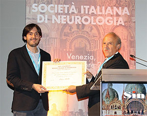
The Past President of Italian Neurological Society, Prof. Quattrone (right) presents an award to Dr. Luca Bello for his paper “DMD Genotypes and Loss of Ambulation in the CIRNG Duchenne Natural History Study,” published in Neurology.

The Past President of Italian Neurological Society, Prof. Quattrone (right) presents an award to Dr. Luca Bello for his paper “DMD Genotypes and Loss of Ambulation in the CIRNG Duchenne Natural History Study,” published in Neurology.

This year, the Italian neurological community gathered in Venice for the XLVII Congress of the Italian Society of Neurology (SIN). Over 1,500 delegates took part in four days of intense updating divided into three plenary sessions, 10 refresher courses, and 21 workshops, in addition to teaching conferences and sponsored symposia. Venice has always been a leader in the treatment of diseases of the nervous system, and the appeal of the illustrious city has given the national neurological community added visibility in scientific, care provision, and educational terms.

At the inaugural ceremony, held at the Cinema Palace in Venice, 10 young researchers were awarded for first authorship in high-impact medical journals, while Dr. Luigi Bertinato gave a very interesting keynote lecture on “Controlling the Plague in the Serenissima Republic of Venice and Its Relevance to the Present Day.” He described how the public health system actually started in Venice in 1500 when, to curb the spread of the plague, which was decimating the city’s population, the first quarantine station was set up to isolate the sick, and ships arriving from the East (the Silk Route) were subject to rigorous control. (Read Dr. Bertinato’s speech.)

In the following days, all neurological conditions were covered, focusing on the most innovative aspects of the field and on translation into clinical practice. Special emphasis was paid to recently available therapeutic resources and care pathways, with a view to modifying the development of many neurological diseases.

More than 1 million Italians suffer from dementia, 600,000 of whom have Alzheimer’s disease. Every year, 120,000 Italians are affected by stroke, and 930,000 manifest residual disability related to the pathology. Finally, 90,000 patients have multiple sclerosis, 200,000 have Parkinson’s disease, and 500,000 are affected by epilepsy. In Italy, there has been a significant increase in the elderly population, implying a heavy burden in terms of care for age-related pathologies.

Particular attention was devoted to identifying the most appropriate models for neurological patient care at both the hospital and outpatient levels. Neurological diseases require highly specialized expertise and a multidisciplinary approach to management of the most advanced stages of the diseases. •

Bruno Giometto, MD, is the director of the Second Clinic of the Department of Neurology in Padova and a member of the Board of Arbitrators of the Italian Society of Neurology.

Main Topics at the XLVII Congress of the Italian Society of Neurology

  • Headache
  • Dementia and Aging
  • Movement Disorders
  • Pain
  • Epilepsy
  • Cerebrovascular diseases
  • Degenerative diseases
  • Motor Neuron diseases
  • Neuromuscular diseases
  • Neuroepidemiology
  • Clinical Neurophysiology
  • Clinical Neurophysiology and Sleep
  • Neurogenetics
  • Neuroimaging
  • Neuroimunology
  • Neuroinfectious diseases
  • Clinical Neuropsychology
  • Basic Neuroscience
  • Neuro-Oncology
  • Neurotraumatology
  • Neurological Rehabilitation
  • Multiple Sclerosis
  • Sleep
  • History of Neurology

Brain Stones

By Peter Koehler

“Cutting the stone” is the title of a well known painting (1494)  by Hieronymus Bosch (c.1450-1516), who died 400 years ago. The painting is displayed at the Prado Museum in Madrid. Another name by which the painting is known reads, “The extraction of the stone of madness or the cure of folly.” An inscription on the painting says (in old-Dutch), “Meester snijt die keie ras, Mijne name is Lubbert Das,” which in English would read, “Master, cut away the stone soon, my name is Lubbert Das.” In Dutch literature of the day, the name was given to a comical character. The theme was well known at the time, and the scene depicted by several other painters, including Pieter Brueghel (senior), Peter Huys, and Jan Steen. The idea of a stone in the brain made me think of a discussion taking place in medical literature over a century later.

The Seat of the Soul  and the Pineal Gland

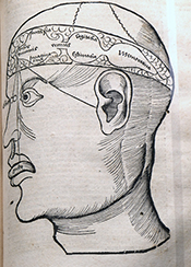
Figure 1. From Gregor Reisch (Margarita Philosophica cum Additionibus Novis, 1517) depicting the localization of the functions of the soul: sensorium commune and imaginatio in the first cell (lateral ventricles), cogitatio (reasoning) in the middle, and memory in the posterior cell (our fourth ventricle).

Figure 1. From Gregor Reisch (Margarita Philosophica cum Additionibus Novis, 1517) depicting the localization of the functions of the soul: sensorium commune and imaginatio in the first cell (lateral ventricles), cogitatio (reasoning) in the middle, and memory in the posterior cell (our fourth ventricle).

Looking at medical literature of the 17th century (and at CT scans nowadays), we know that calcifications within the skull really exist. An issue from this period that is of particular interest with this respect deals with the seat of the soul. Through the ages, the seat of the soul has been localized in various structures. In the ancient Greek period, mental functions, including memory, imagination, and thinking, were discerned. The concept of sensorium commune referred to the part of the mind that brings together the impressions from all five senses. It goes back to Aristotle, who localized the sensorium commune (the seat of the soul) in the heart, whereas Hippocrates and Galen localized it in the brain. For many ages, the soul, the immaterial part of human being, was localized in the cerebral ventricles or cells (Figure 1), in which model the lateral ventricles were considered one cell. In the first cell, imagination was localized; in the second, thinking or reasoning was localized; and in the third cell (our fourth ventricle), memory was localized.

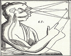
Figure 2: From Descartes’ posthumously published Traité de l’homme (1664).

Figure 2: From Descartes’ posthumously published Traité de l’homme (1664).

The localization of the soul changed in the 17th century, among others by René Descartes (1596-1650), who, as is well known, made a clear distinction between body and mind, and localized the soul in the pineal gland. In a rational way, he became convinced that sensory information from the body should convene in one place, an unpaired organ that he usually called “la petite glande,” the small gland. This is the place where he localized the sensorium commune, imagination, as well as thinking. It was the seat of the soul, he believed, situated between the beginning of the aquaduct of Sylvius that connects the (present) third and fourth ventricles. An important argument for Descartes, who stayed in the Netherlands for a large part of his life (1628-1649), was that this would be the place where the two images from each eye come together and form one image. The pineal gland is the only unpaired organ where this could take place and, moreover, it lies in the extension of the optic tracts. (Figure 2.)

A Stone in the Pineal Gland

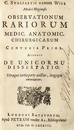
Figure 3: Title page of Stalpart van der Wiel’s Observationum Rariorum (Latin edition of 1687).

Figure 3: Title page of Stalpart van der Wiel’s Observationum Rariorum (Latin edition of 1687).

Not everyone was convinced of this localization. One of the physicians, who opined differently, was the Dutch Cornelis Stalpart van der Wiel (1620-1702). He had studied at the University of Franeker (no longer existent), where he also wrote his thesis and practiced in the city of The Hague, where his father had been mayor. In 1682, he published his experiences of 40 years of medical practice in the book Hondert seldzame aanmerkingen, so in de Genees- als Heel- en Snijkonst [Hundred rare comments, in medicine as well as surgery and dissection; also published in Latin, Figure 3, and French]. He wished to pass on his experiences to his son Pieter, who would succeed him six years later. The 12th chapter is titled “Steen in de Pijn-appel-klier en Saadtvaten gevonden” [Stone found in the pineapple gland and seed vessels]. The text reads “When the body of a certain woman was opened by my brother the physician Johan Stalpart van der Wiel, here at the dissecting room some years ago, I have seen that a small stone, the size of a hempseed, was found in the pineapple gland…” Being aware of Descartes’ opinion, he concluded “that aforementioned gland cannot be the seat of the soul, nor that the most important faculties or acting powers (as has been made up by some) have their origin here.” Moreover, Stalpart van der Wiel argued that the gland “in animals, who seem to have no imagination, no memory, nor any remarkable powers of the soul” was found to be fairly large.

Indeed, it had been a well-known fact that the pineal gland could easily be missed at autopsies of human beings. Galen was aware of the gland, but most probably did his research in cattle. At a public anatomy lesson in Leiden (1637), Professor Adriaen van Valkenburg (Falcoburgius, 1581-1650) admitted, if requested, to René Descartes, who attended the lesson, that he had never found the gland in human beings.

Brain Stones

Stalpart van der Wiel’s case description is followed by an extensive discussion of contemporary literature, referring to his Dutch compatriots Reinier de Graaf, Job van Meekeren, Justus Schraderus, and the Danish Thomas Bartholinus. Stones had been found in several body parts as had been noted by many physicians. Several of them had written that these could be found in various places within the head. Stalpart referred, for example, to the German physician Johannes Schenckius (in Lithogenesia) and the French Isaac Cattierius (Cattier, physician to the king), who, in his Observationes medicinalis (1653), published a case of a stone growing attached to and under the dura mater. The list of references provided by Stalpart van der Wiel is quite large. A certain Kentmannus (lib. de Lapid in corpori inventis or book on stones found in the body) relates of a stone found in the brain with the form and size of a mulberry. Furthermore, the Dutch Theodor Kerckring (observ. 35) is referred to, who had found a stone the size of a hazelnut in the right ventricle.

Figure 5: Theodor Kerckring by Jurgen Ovens (1660).

Figure 5: Theodor Kerckring by Jurgen Ovens (1660).

Although I was unable to check all references by Stalpart van der Wiel, the latter book by Kerckring could easily be found. Theodor Kerckring (1638-1693; Figure 5), often working with a microscope made by his former schoolmate (at Van den Enden’s Amsterdam Latin school) Baruch Spinoza, became well known because of his description of the circular folds of the intestine. In his Spicilegium Anatomicum (Figure 6) of 1670 there is indeed a chapter named “Lapis in cerebro repertus” [Stone found in the brain]. It is about the 14-year old boy Johannes Nicolai Westphalus, who died June 8, 1667. At autopsy, hydrocephalus was found, probably caused by a white calcification of the right ventricle (Figure 7a and 7b).

Another interesting case referred to by Stalpart van der Wiel is that of a stone the size of a Turkish bean, which was found at the origin of the optic nerves and that had caused headache and blindness (referring to Zodiaco Med. Gall. anni 1, fol. 81).

Concluding Remarks

Figure 7a and 7b: From Kerckring’s observation XXXV on p. 76.

Figure 7a and 7b: From Kerckring’s observation XXXV on p. 76.

Although the scene on Bosch’s painting — in fact, not a stone but a flower appears from the skull — obviously is a critical depiction of credulity of common people on one side and quackery on the other. Several other interpretations have been given, but brain stones, as we know today, are not only a fantasy, and a touch of truth is present. In a recent paper, Celzo et al. (Insights Imaging 2013;4: 625–635) provide a pictorial review of brain stones with modern illustrations. They distinguish intra- and extra-axial stones. The first comprise stones of various origin, including neoplastic (oligodendroglioma), vascular (arteriovenous malformations), infectious (toxoplasmosis, rubella, etc.), congenital (Sturge-Weber syndrome) and endocrine/metabolic (Fahr’s syndrome) origin. The extra-axial stones find their origin in tumors (meningiomas) and exaggerated physiological calcifications. The authors conclude that “brain stones are more common than previously thought.” Although Stalpart van der Wiel was already aware of this, he would have been interested to read this paper and to realize that it is even more frequent than he thought.

Latest Developments of the European Board Examination in Neurology

Dr. Michael Ackerl

The European Union of Medical Specialists (UEMS) — European Board of Neurology (EBN) autumn meeting took place in Venice on Oct. 22, 2016. The main topic was the further development of the European Board Examination, which, in the last year, was under the lead of Prof. Jan Kuks of the Netherlands, who managed it very professionally. Nevertheless, new collaborations to improve the examination were, and are, in discussion.

At the Venice meeting, the board members decided unanimously to collaborate with the Swedish company Orzone for the next three years, which already helps many other sections of the UEMS in managing their exams. This guarantees that the principles of our exam stay unchanged (open- and closed-book questions, oral part). There were also negotiations with the Royal College of Physicians in the last year, and these negotiations will be continued for possible further collaborations. The board members also decided to create an examination task force in common with EAN and have a more intensive collaboration with EAN in training issues.

The next examination will take place in Amsterdam on June 23, during the EAN congress. Candidates from all over the world are welcome. Do not hesitate to contact us through our website.

Dr. Michael Ackerl is the Austrian delegate of the EBN.

Mediterranean Routes and the Bulwarks of Plague Control During the “Serenissima Republic in Venice”

By Luigi Bertinato, Venice, Italy

plague_mainNavigation and trade have always been the cornerstones of multiple cultural relationships with civilizations of the Mediterranean and the Far East countries. Indeed, the caravans that transported the rarest and most precious wares by sea to the Venetian warehouses arrived from as far as Constantinople and Alessandria.

The patricians of the Serenissima Venetian Republic, as Niccolò Machiavelli pointed out in The Prince, were the only noblemen in the world who thought themselves to be above the humility of practical work. Trading was for these people not only a source of financial gain, but an art form.

To navigate and trade successfully, it was important to gather information on the routes, the coastlines, the islands, and on geographical and climatic details, as well as to understand markets, rates of exchange, money, weights and the differing customs of the people located in other parts of the world. It was important to know how to communicate using conventional sign systems and gestures marked by innate pragmatism reminiscent of those civilizations that converged along the coasts of the huge Mediterranean basin.

From 1348, a dark foreboding began to make its way into the Mediterranean: the plague. Even today, the plague has its natural breeding ground in Kurdistan, in Assyria, and in Central Asia, where it is endemic. It would spread to the convoys that followed the lines of the caravan routes, thereby reaching the ports of the Eastern Mediterranean. The contagion would spread via two main vehicles: a rodent, the rattus-rattus, and the Xenopsilla Cheopis, a flea that lodged itself in the surface hair of the rodent, and then pass on to humans. In the dark, dank corners of the ship’s hold, the rats not only represented a serious threat for the goods and wares, but, more importantly, for the people who would come into contact with them.

Due to its constant contact with the East, Venice would find itself in the frontline. Venice could not give up its trading links with the East on which it based its whole wealth. Therefore, it had to adopt measures for the protection of public health. The result was the invention of the quarantine, or lazzaretto.

The Birth of the Venetian Lazzaretti
From 1348 in this globalized ante litteram world raged the spectre of the plague, which spread along the Mediterranean routes, threatening the survival of peoples in its wake.

While science at the time groped helplessly in the dark for solutions, it was clear to many states that the lethalness, contagiousness and sheer virulence of the plague constituted such an enormous threat that it was necessary to take recourse to collective measures.

On Aug. 28, 1423, during the umpteenth attack of the plague, the Venetian Senate instituted isolation facilities where the plague victims would be housed. In these buildings, there would be medical and nursing personnel at hand, paid by the state. After much deliberation, the choice of location fell upon the Island of Santa Maria di Nazareth, which carried the name of the pre-existing convent, vulgarized thereafter into the term “Nazaretum,” then into “Lazaretum,” and finally into “Lazzaretto.” This name was then adopted by similar types of isolation facilities modeled on the Venetian Lazzaretto, which sprang up elsewhere in the western world.

In 1423, the Senate underlined the importance of information as an indispensable premise for effective and rapid isolation, and the success of prevention. Each and every Venetian — from the captains of large vessels to the sailors on deck, to the citizens themselves — was involved in the fight against the spreading of infection and was encouraged to collect every piece of news, when it surfaced, on cases of the plague in the city and on board the ships, so that affected persons and goods could be isolated immediately.

However, certainly the most original model was that of the quarantine lazzaretto, founded in 1468, which would take in the recovered plague victims for a period of 40 days before they were allowed to return to the city. The lazzaretto, called “Nuovo” or “New” to distinguish it from the pre-existing site named “Vecchio” or “Old,” was used as a quarantine for persons and goods arriving from infected countries. Isolation and purification measures were implemented, requiring constant investment of economic resources as well as the slowing down of the traffic of goods and persons. The priority was to spend on prevention rather than risk economic and demographic ruin as a result of widespread epidemics.

The system of the Venetian lazzaretti was managed, in the context of a wider national and international health care strategy, by the Public Health Care Magistracy of the Venetian Republic (instituted in 1486), thereby creating the model for all Mediterranean ports, which intended to compete with the “Serenissima” Venetian Republic.

Mass Communication on Preventing Measures
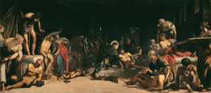
It was no easy task to force plague victims and those suspected of carrying the plague virus to be hospitalized in the lazzaretti, which during the 15th Century were tainted by scandal and condemned by their priors for the maltreatment of patients. The health care information campaign and the sensitization of the people to isolation practices reached a peak as a result of the veneration of Saint Rocco who, through his example, inspired people of that time to accept marginalization and quarantining as a means of preventing the spread of disease in populations.

Saint Rocco embodied and, at the same time, exorcised the fears and obsessions which beset the Christian West since 1348, in whom, as a beggar and wayfarer, people saw the image of Christ in need of help. Following the disappearance of the plague in Europe, the fear remained that there could be individuals still around who could spread the infection and cause further deaths. Saint Rocco represented the pilgrim, the traveller who was conscious of the danger of his movements, and the possibility of constituting a vehicle for the spread of infection.

To dissuade people from violating laws on compulsory isolation for plague victims and to discourage all infringements of quarantine norms and of the expurgation of goods and passengers, the Public Health Magistracy adopted exemplary measures such as capital punishment before the seat of the Magistracy at St. Mark’s.

During the plague of 1576, since there were numerous victims and deaths in Venice as a result of coming into contact with the infection raging through the city, the number of citizens who had to be subjected to quarantining procedures in Lazzaretto Novo was high. The small island could not accept all these people, which meant that the sick had to be housed in thousands of ships moored off the island, crammed next to one another, thereby increasing the size of the territory and increasing the ability of the island to take on infected people. At a short distance away from the island, a boat equipped with a gallows served as a warning to those who thought of escaping isolation, and other armed boats patrolled the surrounding waters to tighten the security even further. The proclamation enforces the ban on those coming from infected areas (or suspected areas) when trying to enter the city without undergoing a period of quarantine.

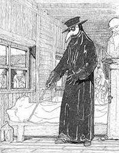
plague-2A plague doctor (see the photo at the right),was a medical physician who treated victims of the plague. They were specifically hired by the Senate of Venice that had many plague victims in times of epidemics.

The plague doctors wore a special costume. The garments were invented by Charles de L’Orme in 1619; they were first used in Paris, but later adapted in Venice with a special mask with a prominent nose. The protective suit consisted of a heavy, waxed fabric overcoat, a mask with glass eye openings and a beak-shaped nose, typically stuffed with herbs, straw, and spices. Plague doctors would also commonly carry a cane to examine and direct patients without the need to make direct contact.

This was thought to protect the doctor from miasma.

The Great Quarantine Warehouses and the Rules of Expurgation
In the 17th century, the health care cordon around the city was optimized, thereby preventing any ship from approaching Venice without being subjected to the necessary controls by the Public Health Care Magistracy. Armed boats patrolled the lagoon to check that nothing was unloaded from the ships arriving from foreign waters. The captains of these ships were obliged to go before the health care office and avoid contact with anyone else, and then to fill out a detailed report about the trip. The Magistrate’s scribe had to collect the details of the captain’s report, and check health certificates and licenses issued by port authorities in the ports visited by the ship in question. These documents had to prove the ship had set sail from ports that were not infected by disease. In case of false declaration, the captain would risk the death penalty. It was quite common that seafarers who had set sail from plague-ridden ports would destroy the documents, thereby asking for licenses to be issued by other ports free from infection. This was a futile trick because the Republic would plant informers and spies all around the Mediterranean area.

In order to gain access to the Venetian ports of the Mediterranean, it was compulsory to exhibit a bill of health. The bill of health or the health license (passport or passpartout) certified the health of individuals and the safety and hygiene levels of goods that were shipped by providing detailed information on ports of call and transit ports used by a particular vessel.

This documentation was issued by the Chancery of the Venetian consulates, which was at the head of the markets in the numerous near eastern ports where ships would berth. At the end of a journey, the licenses were returned to their owners indicating all the ports entered by the vessel.

This is the reason why such documents have been preserved in the consular archives or in the archives of the Venetian ambassador (“bailo”) who resided in Constantinople, and was in charge of the consulates stationed in the Near Eastern Mediterranean.

The Health Care Magistracy of the Serenissima Republic and International Health Care Bulletins
From the date of its establishment in 1486, the health care magistracy had founded preventative policies based on a systematic identification of epidemics in all the countries that traded with Venice, which included the publication and distribution of news concerning the latest developments, through a network of health care offices, diplomatic representatives and similar agencies abroad.

In 1770, a total of 6,990 copies of health decrees rolled off the ducal press, carrying warnings of the dangers posed by the outbreak of plague in specific areas, with relevant dates and bearing the supposed office’s address. All this went to show what a fine system of networks had been adopted in efforts to reach even the most distant localities, and stop the spread via unexpected and lesser known avenues, and avoid the suspension of ties with infected zones.

Infected war vessels as well as those under suspicion of being so were duly reported so that ports could deny them access or send them on to areas equipped with infected lazzaretti. Near-escapes and cases of having resolved the epidemic onboard ships were also made known using the same system of publication and news bulletins.

The Serenissima’s loss of domination over the Adriatic, known for centuries as the “Venetian Gulf,” was having to come to terms in the meantime with yet another business rival, that of Ancona.

Despite the loss of her military and commercial supremacy, the old Republic did continue through its health authorities to control the dissemination of information about epidemics and to spread health warnings via its public announcements, many of which in fact fell on the ears not only of friendly but less than friendly ports.

Poveglia: The Very Last of the Lazzaretti
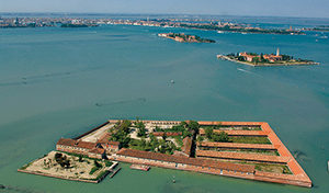
Over the course of the 17th century, the New Lazzaretto faced a slow and irreversible degradation, made no easier by the surrounding lagoon areas falling prey to swamplands. This forced the Senate into taking into consideration two alternative solutions: either that of undertaking drastic restorations, or building a third lazzaretto on another island. In 1782, the island Poveglia was carefully selected for its proximity to the mouth of the port of Malamocco, with its deep and navigable canals, which had long been in use for the placement of ships in quarantine.

Victims of the plague were isolated on islands to keep the disease from spreading to Venice.

Victims of the plague were isolated on islands to keep the disease from spreading to Venice.

This would have afforded impressive savings on the costs of transporting the goods and passengers to the islands, thus cutting the costs of peate, towing, transfers, and overtime for the health administrators who had the task of keeping their eye on such operations. The project however, had to be set aside, due to a lack of funds, and it was only the emergency created in 1793 by the arrival of an infected Tartan that prompted having to equip the island with quarantine facilities. This necessitated the building of two wooden control centers, one for infected persons and the other for the guards, who in patrolling the surrounding waters with armed boats managed to contain the spread of infection.

After the fall of the Republic, Poveglia continued to be used as an emergency quarantine base for crews afflicted by the plague and, after 1803, yellow fever. The following year, the Austrian government decided that suspected ships coming from Spain were better turned away from the Austrian lazaretti and should instead be sent to the Venetian reception points, which were far better equipped by their isolated position to deal with them. In 1805, plans were being considered for the creation of a large lazzaretto at Poveglia, an idea also taken into consideration by the successive Kingdom of Italy in 1808, but the major obstacle remained in the fact that the island was already used by the military.

In 1814, while the plague continued to break out in many ports of the Levante, Poveglia was finally handed over to the Health authorities, functioning from then on as an isolation center for all Austrian sea ports, and managing the infection to a high degree of success.

Any historical reflection on the role of the Mediterranean Lazzaretti demonstrates that the West had already expressed a common intention regarding health research, many centuries prior to the birth of the European community anticipating the rules adopted today by the World Health Organization, within the so called “International Health Regulation.” 

Luigi Bertinato, MD, is public policy adviser in the areas of international public health policies and currently Health Care Manager at the University of Verona,Italy