By Mark Hallett, MD, NINDS, Bethesda
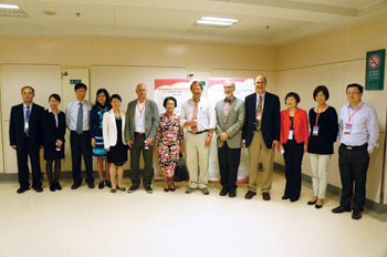
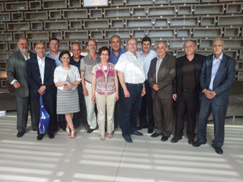
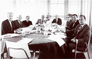
Faculty at Changsha meeting.
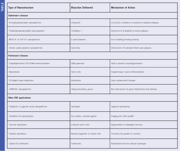
The paroxysmal dyskinesias are rare familial disorders, but very dramatic. There are three main types: paroxysmal kinesigenic dyskinesia (PKD), paroxysmal non-kinesigenic dyskinesia (PNKD) and paroxysmal exertional dyskinesia (PED).
PKD is typically precipitated by a quick movement, PNKD is precipitated by stressors such as coffee, tea and alcohol, and PED is produced after long periods of exercise. In the past few years, the main mutations responsible for all three have been determined. The gene for PNKD is myofibrillogenesis regulator 1, now called PNKD gene, and the gene from PED is the SLC2A1 gene that leads to GLUT-1 deficiency. Only recently has the gene for PKD been found to be PRRT2. As is often the case, when a gene has been found, some surprises emerge.
On Oct. 24, 2013, the Xiangya Hospital of Central South University in Changsha, China, held its Fifth Xiangya International Congress on the Clinical and Basic Research of Neurodegenerative Disorders focused on PRRT2 related diseases. The basic phenotype of typical PKD cases had been refined by Louis Ptacek’s group in 2004, and this certainly helped in narrowing the gene search. Already at that time, it became clear that there was a significant overlap between PKD and infantile convulsions. At about the same time, two years ago, Bei-Sha Tang’s group from CentralSouthUniversity and Ptacek’s group from University of California, San Francisco, identified PRRT2 as the relevant gene. Tang and his group as well as Ptacek came to the meeting.
Clinically, the PRRT2 mutation brings PKD and Benign Familial Infantile Convulsions (BFIC) together. Lu Shen, also from XiangyaHospital, discussed the clinical aspects of the BFIC cases. Another significant phenotype is hemiplegic migraine, as discussed by Pierre Szepetowski from Marseille. Zhi-Ying Wu from FudanHospital, Shanghai, widened the spectrum further by pointing out that PRRT2 mutations also have been seen in some cases of paroxysmal torticollis, episodic ataxia, childhood-absence epilepsy, febrile seizures, and both surprisingly and confusingly, in cases of PED and PNKD. Hence, while there is a most typical phenotype of PRRT2, it appears to be able to cause a variety of paroxysmal disorders, mostly in young persons.
Qing Liu from PekingUnionHospital, Beijing, speaking for Li-ying Cui, reported on SPECT neuroimaging in ictal attacks of PKD. There have only been a few cases and findings are not completely concordant, but it appears that there is hypermetabolism of the basal ganglia or thalamus during an attack. This confirms the general suspicion that the basal ganglia are the site of origin of PKD, but the nature of the abnormal activity is still unclear. There was discussion as to whether this might be a subcortical seizure, but clearly more data would be needed to determine this.
Ptacek led the discussion about the basic cellular mechanism of PRRT2. It is a novel protein and its role is not yet clear, but it interacts with SNAP-25. SNAP-25 is a SNARE protein that plays a critical role in synaptic release mechanisms, and is well known by neurologists as the target for botulinum toxin type A. He speculated that PKD might be a type of synaptopathy, a new general mechanism for paroxysmal disease, distinct from channelopathies, which cause other types of paroxysmal disorders. He noted that the PNKD protein also plays a role in exocytosis at the synapse.
Thus, PRRT2 mutations can lead to a variety of paroxysmal diseases that at the meeting were referred to as the PRRT2-related paroxysmal diseases (PRPDs). Taken all together, this class is not as rare as it might first appear. Moreover, knowledge of the gene function is leading to a new general mechanism for paroxysmal disorders.
The meeting, organized by Hong Jiang of Xiangya Hospital, provided a worthwhile current synthesis of this field, which certainly will have more surprises coming.
 The World Federation of Neurology (WFN) is a huge and complex structure, representing neurologists worldwide. To achieve its aim, many international neurologists collaborate and work on WFN projects, represent the organization as officers or serve as editors or authors for WFN media. These initiatives play an important role in advocating the interests of neurologists on a global scale.
 Bringing worldwide science and patient care closer together is a strong objective of the WFN. At www.wfneurology.org, you will find details on the World Brain Alliance, an umbrella group of international neurological organizations. WFN applied research groups organize scientific projects and educational activities in neurology subspecialties, and publish their activities on the website on an annual basis.