Edward J. Fine, MD
By Edward J. Fine, MD
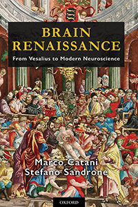
In Brain Renaissance From Vesalius to Modern Neuroscience, authors Marco Catani and Stefano Sandrone have written chapters based on translations from Andreas Vesalius’ book on the brain from his De Humani Corporis Fabrica, followed by commentaries. The book is published by Notting Hill Editions, 2016.
The first chapter adroitly reviews the history of dissection of human bodies and Galen’s hegemony prior to Andreas Vesalius’ first edition of his seminal and revolutionary anatomical textbook of 1543.
Chapter 2 recounts Andreas Vesalius’ illustrious lineage in a family of physicians and pharmacists who attended royalty. Andreas Vesalius was born on Dec. 31, 1514, in Sablon, a neighborhood of Brussels. His father was the royal apothecary (pharmacist) to Charles V of Spain. At age 15, Vesalius entered the Castle College in Leuven, which promoted the philosophy of humanism. At age 18, Vesalius left Leuven to study medicine in Paris.
Chapter 3 recalls Vesalius’ medical education in Paris and his encounter with profound differences between what he saw in human cadavers and Galen’s incorrect descriptions. Galen’s texts of anatomy were the canon from the time of the Roman Empire to publication of Vesalius’ De Humani Corporis Fabrica. Vesalius left Paris abruptly without graduating, due to war between Henry II of France and Charles V of Spain, patron of Vesalius’ father.
The fourth and fifth chapters highlight Vesalius graduating from Leuven Medical School, traveling to and joining the medical school at Padua, Italy, renowned for its eminence in teaching anatomy from human dissection. Through dissection, Vesalius found so many more errors in Galen’s descriptions of human anatomy that Vesalius concluded Galen had dissected monkeys and other animals rather than humans. Chapters 6, 7, and 8 deal with production of the Fabrica, a volume of nearly 700 pages of which the seventh book deals with the brain, making 200 woodcuts used to print illustrations, and sources for backgrounds in anatomical illustrations. Vesalius’ friendship with Contarini, the podesta (chief magistrate) of Padua, provided corpses of recently freshly executed criminals for dissection.
Disgusted by nefarious intrigues of jealous surgeons and physicians at the Court of Phillip II in Spain, Vesalius escaped to the Holy Land after contacting colleagues who would petition his return to the chair of anatomy and surgery at Padua. The authors cite reliable accounts that Vesalius studied medicinal herbs there. He died attempting to return to Padua. His death on the island of Zante was attributed to exhaustion from scurvy, or illness caused by a prolonged voyage in the tempestuous Mediterranean Sea.
Chapter 13 defines Vesalius’ concept of the brain as the source or sensation and voluntary movement. Vesalius was versed in comparative anatomy: “… there is no difference at all in the structure of the brain in the parts that I have dissected in the sheep, goat, monkey … when compared with the human brain.” He recognized that man’s intelligence was due to “… the human brain larger proportionally to his body but also larger than all of the animals … .” Vesalius stridently denied the presence of the rete mirabilie in the base of the human skull, a structure Galen insisted was present in humans. This structure is part of bovine skulls. Vesalius also decried Galenic beliefs about ventricular function.
In Chapters 14 and 15, Vesalius denotes dura and pia as hard and soft membranes surrounding the brain. He reminds readers of similarities and differences between these structures and pericardium and epicardium. Vesalius presciently mentions that the surface of the thin membrane (pia) is covered with aqueous liquid (cerebrospinal fluid) and that this membrane “provides a defensive wall for the brain against collisions with the skull.”
In Chapter 16, Vesalius describes the corpus callosum, brainstem, and cerebellum with emphasis on the position and external surface of the cerebellum. Vesalius rebuked Galen for not describing accurately the morphology of the cerebellum: “Oh, Galen, you have been deluded without good reason by things of little importance and sometimes also by your apes.” Vesalius comments on the mid-position of the corpus callosum — that it is a band of fibers connecting the hemispheres. The commentary begins with the statement that Galen and Vesalius assigned a mechanical function to the corpus callosum to keep the cortex from collapsing into the ventricles.
In the commentary, we learn that some anatomists succeeding Vesalius assigned absurd roles to the corpus callosum such as the seat of the soul. Spurzheim and Gall proposed that the corpus callosum “contributed to producing action and reciprocal reaction between the hemispheres.” We read that persons born with agenesis of the corpus callosum may be nearly normal or be developmentally impaired. Catani and Stefano Sandrone accurately summarized Gazzaniga and Sperry’s research on the functions of persons who underwent transection of the corpus callosum for control of epilepsy. Catani and Sandroni inform us that the anatomical fornix was linked to arches in Rome where prostitutes met their customers. They recounted the discovery of connections of the fornix to mammillary bodies by Felix Vicq d’Azyr in 1786. These authors advanced the idea that Papez, who is credited for “discovering” a circuit for emotion in 1937, was actually scooped by Paul Broca in 1878. Moreover, Christfried Jakob’s circuit for emotions closely resembled James Papez’s 1937 diagram. John Fulton and Jacobsen observed chimps were less aggressive after they had resected portions of their frontal lobes. Antonio Moniz, assisted by the neurosurgeon Almeida Lima, injected pure ethanol into frontal lobes of chronic schizophrenics. They were convinced they had improved the patients’ behavior. They were visited by Walter Freeman, an ambitious neurologist, who later performed 3,000 trans-orbital leucotomies that partially controplled violent behavior, but caused profound inability to plan and initiate activities. Catani and Sandrone summarized the tragic case of H.M. who suffered anterograde amnesia after bilateral removal of his hippocampi in an attempt to control partial complex epilepsy.
In the chapter on the pineal gland, Vesalius localizes this structure to the base of the third ventricle and its function as a gland. Sandrone and Catani summarized knowledge of pineal function from Descartes’ opinion as the seat of the soul to current knowledge of its role in secretion of melatonin. Connections between the photoreceptors in the eye to the suprachiasmatic nucleus in the hypothalamus and then to the pineal gland control increase melatonin release from the pineal gland at night and suppress its secretion during daylight. I was amused to learn that farmers expose hens to excessive artificial light to suppress melatonin release and increase egg production.
Although the titles of chapters on Testes and Buttocks of the Brain and Sex on the Hills may strike readers as risqué, the content is about the colliculi and the dependent pineal gland resembling these organs. Catani and Sandrone explain lucidly how the expansion of occipital lobes replaced the superior colliculi as the principal structure for vision in the ascent of evolution from birds to man. They mention the role of the inferior colliculus in the auditory pathway from medulla to the primary auditory cortex.
In Chapter 22 on the cerebellar processes, Vesalius lambasted Galen for his fanciful and fallacious concept of the function of the cerebellum as a valve to control the flow of the animal spirits to the spinal cord and then to the nerves. Vesalius vehemently objected to the misnomers of Galen used to describe folds of the cerebellar cortex as vincula or chains.
Commentary following Vesalius on the cerebellum contains a plethora of interesting facts, including that the elephant has the largest cerebellum in absolute and relative size. We learn that Costanzo Varoli removed the brain from the skull, inverted it, and described the pons (Latin for bridge). Thomas Willis in 1664 discovered its three peduncles. Albrecht von Haller corrected Willis’ statement that the cerebellum controlled involuntary movements of the heart and lungs by locating these functions in the brainstem. Luigi Rolando used electrodes supplied with current from Volta’s electrical pile to stimulate regions of the brains of pigs. He found muscular movements were strongest when he stimulated the cerebellum. Pierre Flourens in 1824 correctly assigned coordination of movement to the cerebellum and movement to the spinal cord. Catani and Sandrone end their commentary tersely, describing cerebellar cellular anatomy and Golgi’s vicious defense of the syncytial theory of brain architecture in his Nobel lecture in 1906.
Vesalius described the infundibulum as the structure that collects and drains cerebral phlegm. He believed drainage from the infundibulum out of the cranium occurred through spaces surrounding arteries, veins, and nerves. The commentary describes the connections between the hypothalamus and pituitary, and the regulatory effects of the hypothalamus on thyroid, adrenal, and ovarian or testicular hormones. The authors cited the amazing case of the pituitary giantess Aama Bataillard, whose autopsy revealed a greatly enlarged pituitary. They summarized extensive research on the influence of pituitary extracts on growth and maturation in Chapter 24.
Subsequent chapters deal with the variations of the blood vessels of the brain and Vesalius’ precise description of structures within the eye. Vesalius’ closing chapter deals with methods on how to remove and dissect the brain.
Chapter 31 contains useful chronological tables of advances in microscopy, electrophysiology neuroanatomy, and neuroimaging. A subsequent chapter tersely comments on phrenology, discovery of animal electricity, and the action potential.
The following chapters provided more information on advances made in staining brain tissue by Golgi and Cajal, emergence of the neuronal doctrine, and Cajal’s declaration of proof of dynamic polarization based on Cajal’s microscopic observations. Staining techniques that differentiated regions of human brain led to construction of elaborate maps by Campbell and Brodman.
Chapter 37 omits the discoveries of Richard Caton who first recorded electrical potentials recorded from cerebral cortices of moving mammals (electrocorticography) in response to light and tactile stimuli. No comments are offered regarding Hans Berger’s meticulous descriptions of normal and abnormal EEG activity in awake and sleep, after trauma, and during seizures. The authors’ final chapter summarizes the emerging science of functional MRI imaging of connections between regions of brain during specific tasks.
This book has some minor but detracting errors and omissions. The authors did not mention the Papal edict that blocked human dissection for four centuries and how the Medici family’s Pope ended this prohibition. This information would explain the resurgence of human cadaver dissection in the Renaissance. In the section about limbic system connections to the prefrontal cortex, these authors should have mentioned that Ignaz Moniz received the Nobel Prize for discovery of the harmful prefrontal leucotomy, but not for initiating the highly beneficial technique of cerebral angiography. In some areas, the commentary seems haphazard and sometimes omits important material.
These authors’ commentary on the cerebellum omits anatomical studies of James S. Risien Russell, who discovered the uncinate fasciculus, known as the hook bundle of Russell. That chapter overlooks clinical observations of American authors Charles K. Mills and Theodore Weisenberg regarding lesions in the anterior versus posterior vermis that cause loss of tone and respectively falling forward or backward. No mention was made of Babinski’s discovery of dysdiadochokinesis in 1902 or Granger Stewart and Gordon Holmes’ publication in Brain in 1904 on loss of check or “rebound” in patients with cerebellar hemispheric lesions. This chapter minimizes Gordon Holmes’ meticulous and exhaustive observations of 40 World War I soldiers as they recovered from gunshot wounds to their cerebella. Holmes described initial hypotonia of limbs ipsilateral to destruction of the cerebellar hemisphere, later deviation of gait toward the side of the cerebellar hemisphere. The figure showing extreme hypotonia of a soldier is the sole illustration from Holmes’ opus. The authors could have included Holmes’ graphic recordings of disturbed movements induced by cerebellar hemispheric lesions.
The commentary would be improved by comment on the Galvani versus Volta controversy, the landmark publication of The Functions of the Brain by David Ferrier in 1876, based on ablation and electrical stimulation of cerebral cortex of primates and other mammals. Elaborating on discoveries of Caton and Berger, cited only in the chronological tables, would have added much.
Typographical errors mildly mar the book. In pages 40 and 41, the erroneous date 1664 appears three times instead of 1564, which is the correct year of Vesalius’s trip to the Holy Land and his death. Footnote 27 contains a typographical error, “chartoid,” for carotid. Nonetheless, these minor defects do not blemish a book that contains a vibrant translation of Vesalius’ Book Seven and plethora of information in their commentary.
Despite these omissions, this is the book to own for those who are not Latin scholars but desire to read an accurate translation of the writings of Vesalius. Much of the commentary upon anatomical, clinical, and pathological discoveries made after Vesalius is germane and informative for understanding the development of neuroscience. Oxford Press substantially bound this hardcover book, printed an attractive cover, and precisely reproduced figures in the appendix from Vesalius’ Book Seven. I can attest to the clarity of these illustrations, having viewed both original editions of the Fabrica on visiting the medical library at Leuven. MRI, photographs, and illustrations from neurological and neuroanatomical literature were intelligently selected and faithfully reproduced. A revised edition following some of these suggestions would be well received.